Cranial nerve palsy is a congenital or acquired dysfunction of 1 or more cranial nerves Cranial nerves There are 12 pairs of cranial nerves (CNs), which run from the brain to various parts of the head, neck, and trunk. The CNs can be sensory or motor or both. The CNs are named and numbered in Roman numerals according to their location, from the front to the back of the brain. The 12 Cranial Nerves: Overview and Functions that will, in turn, lead to focal neurologic abnormalities in movement or autonomic dysfunction Autonomic Dysfunction Anterior Cord Syndrome of its territory. Head/ neck Neck The part of a human or animal body connecting the head to the rest of the body. Peritonsillar Abscess trauma, mass Mass Three-dimensional lesion that occupies a space within the breast Imaging of the Breast effect, infectious processes, and ischemia Ischemia A hypoperfusion of the blood through an organ or tissue caused by a pathologic constriction or obstruction of its blood vessels, or an absence of blood circulation. Ischemic Cell Damage/infarction are among the many etiologies for these dysfunctions. Diagnosis is initially clinical and supported by diagnostic aids. Management includes both symptomatic measures and interventions aimed at correcting the underlying cause.
Last updated: Dec 15, 2025
Contents
The table below summarizes the functions of the 12 CNs.
| CN | Name | Function | Type |
|---|---|---|---|
| I | Olfactory | Olfaction Olfaction The sense of smell, or olfaction, begins in a small area on the roof of the nasal cavity, which is covered in specialized mucosa. From there, the olfactory nerve transmits the sensory perception of smell via the olfactory pathway. This pathway is composed of the olfactory cells and bulb, the tractus and striae olfactoriae, and the primary olfactory cortex and amygdala. Olfaction: Anatomy ( smell Smell The sense of smell, or olfaction, begins in a small area on the roof of the nasal cavity, which is covered in specialized mucosa. From there, the olfactory nerve transmits the sensory perception of smell via the olfactory pathway. This pathway is composed of the olfactory cells and bulb, the tractus and striae olfactoriae, and the primary olfactory cortex and amygdala. Olfaction: Anatomy) | Sensory Sensory Neurons which conduct nerve impulses to the central nervous system. Nervous System: Histology |
| II | Optic | Vision Vision Ophthalmic Exam | Sensory Sensory Neurons which conduct nerve impulses to the central nervous system. Nervous System: Histology |
| III | Oculomotor |
|
Motor Motor Neurons which send impulses peripherally to activate muscles or secretory cells. Nervous System: Histology |
| IV | Trochlear | Eye movement ( superior oblique Superior oblique Orbit and Extraocular Muscles: Anatomy muscle) | Motor Motor Neurons which send impulses peripherally to activate muscles or secretory cells. Nervous System: Histology |
| V | Trigeminal |
|
Both |
| VI | Abducens | Eye movement ( lateral rectus Lateral rectus Orbit and Extraocular Muscles: Anatomy muscle) | Motor Motor Neurons which send impulses peripherally to activate muscles or secretory cells. Nervous System: Histology |
| VII | Facial |
|
Both |
| VIII | Vestibulocochlear |
|
Sensory Sensory Neurons which conduct nerve impulses to the central nervous system. Nervous System: Histology |
| IX | Glossopharyngeal |
|
Both |
| X | Vagus |
|
Both |
| XI | Accessory |
|
Motor Motor Neurons which send impulses peripherally to activate muscles or secretory cells. Nervous System: Histology |
| XII | Hypoglossal | Tongue Tongue The tongue, on the other hand, is a complex muscular structure that permits tasting and facilitates the process of mastication and communication. The blood supply of the tongue originates from the external carotid artery, and the innervation is through cranial nerves. Lips and Tongue: Anatomy movements | Motor Motor Neurons which send impulses peripherally to activate muscles or secretory cells. Nervous System: Histology |
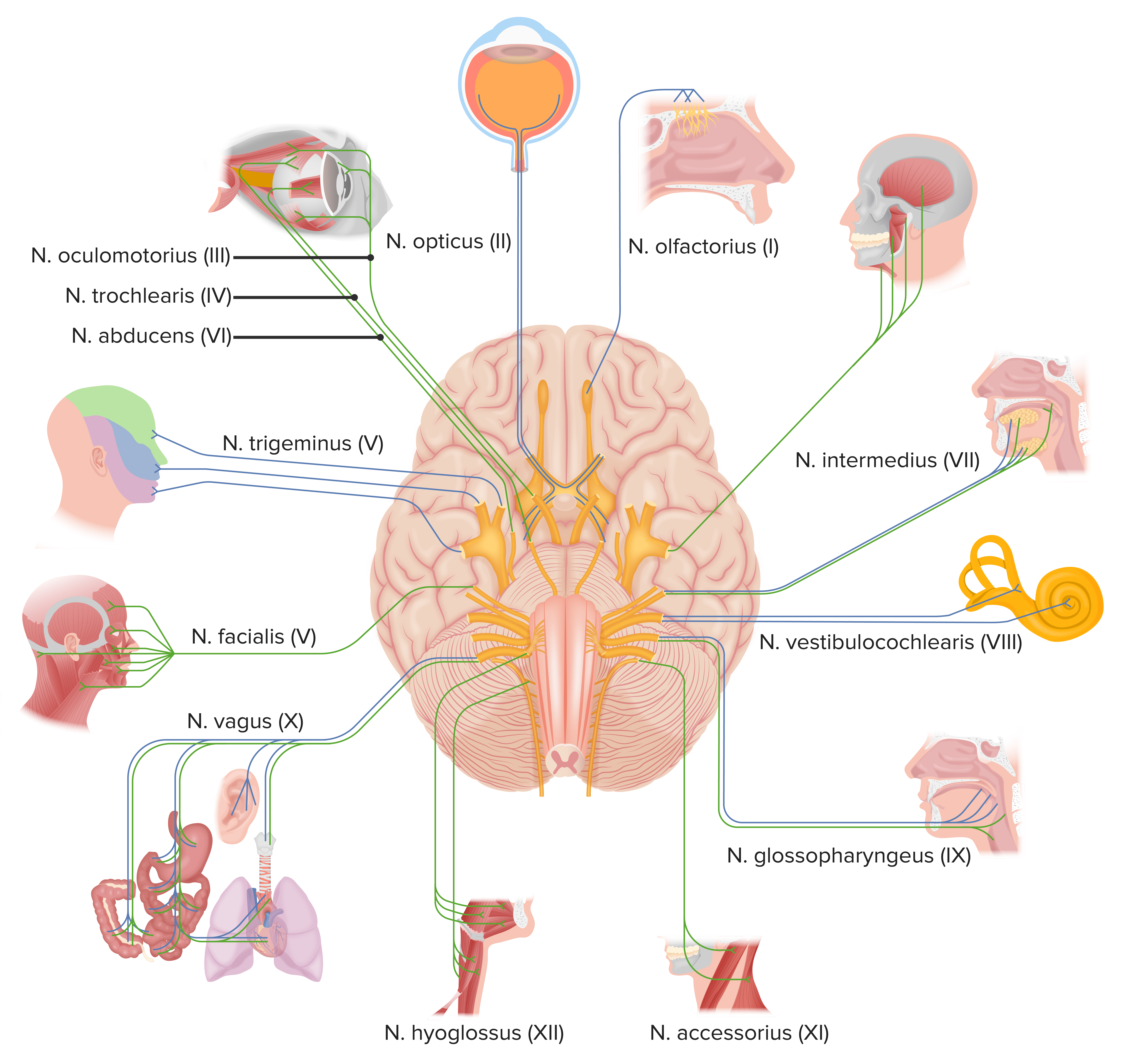
Cranial nerves
Image by Lecturio.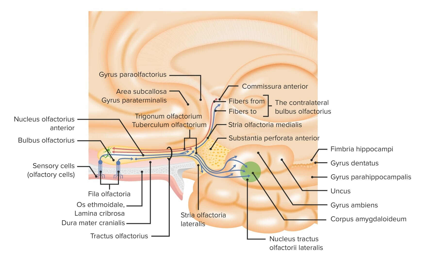
Overview of the peripheral and central components of the olfactory system
Image by Lecturio.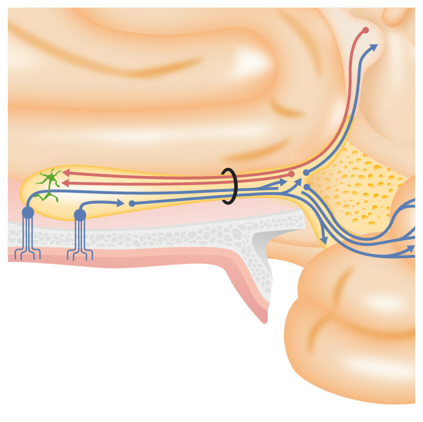
A closer look at the olfactory sensory cells in the olfactory mucosa, passing through the cribriform plate and synapsing with the olfactory tract at the olfactory bulbs
Image by Lecturio.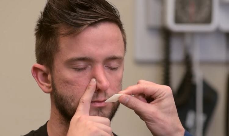
Examination of cranial nerve I: presenting the subject with a familiar olfactory stimulus
Image by Lecturio.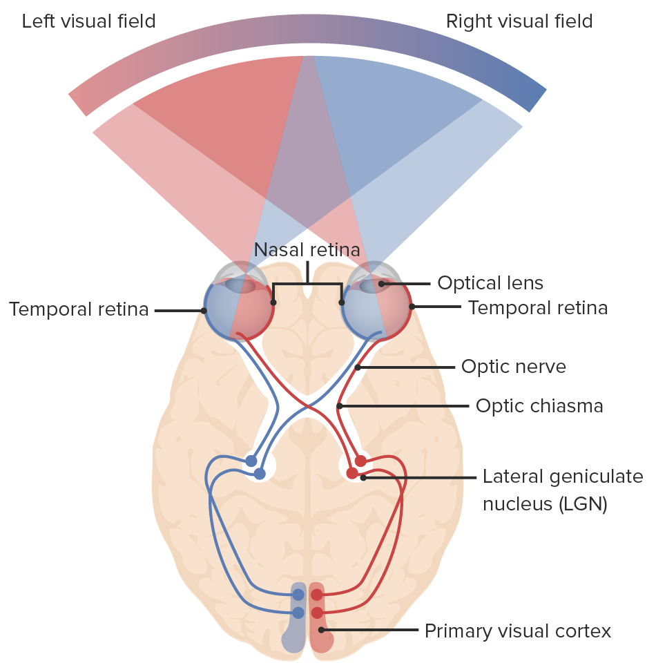
Diagram of the visual pathway and the visual fields: Light enters the eye, sending signals to the retina and through the optic nerve. The nasal fibers of each eye decussate at the optic chiasm, continuing to the optic tract with the temporal fibers. The right nasal fibers join the left temporal fibers (blue lines) and the left nasal fibers join the right temporal fibers (red lines). Neurons synapse at the lateral geniculate nucleus. Optic radiations connect the lateral geniculate nucleus to the primary visual cortex of the occipital lobe where visual information is processed.
Image by Lecturio.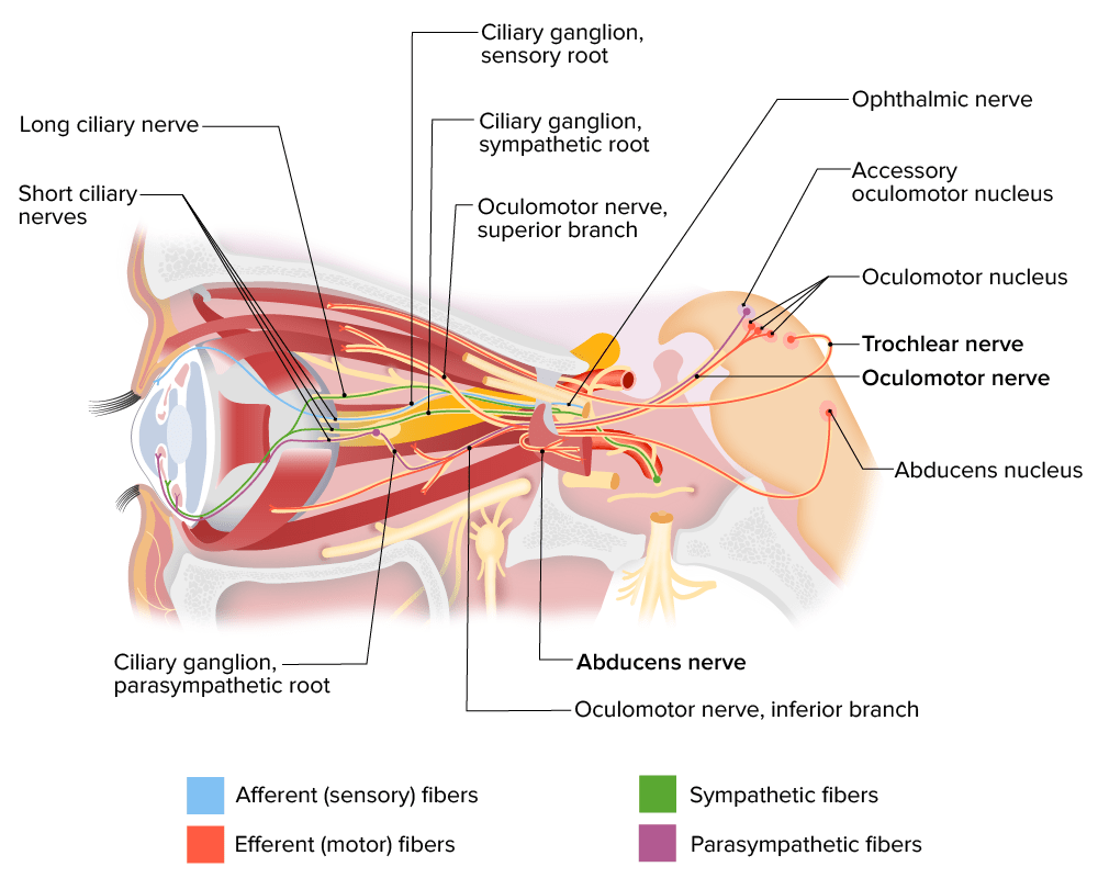
Innervation of ocular muscles by cranial nerves III, IV, and VI
Image by Lecturio.
Pupil fixed and dilated in a position of lateral and downward (down-and-out) gaze
Image: “Oculomotor nerve palsy” by Hakim W, Sherman R, Rezk T, Pannu K. License: CC BY 3.0Urgent:
Nonurgent:
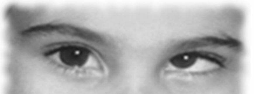
Left trochlear nerve palsy:
The subject was asked to look right after looking left. The left eye shows extortion.
| Site of damage | Clinical findings | Other findings | Common causes |
|---|---|---|---|
| Sensory Sensory Neurons which conduct nerve impulses to the central nervous system. Nervous System: Histology cortex |
|
|
|
| Internal capsule Capsule An envelope of loose gel surrounding a bacterial cell which is associated with the virulence of pathogenic bacteria. Some capsules have a well-defined border, whereas others form a slime layer that trails off into the medium. Most capsules consist of relatively simple polysaccharides but there are some bacteria whose capsules are made of polypeptides. Bacteroides | Hemifacial sensory Sensory Neurons which conduct nerve impulses to the central nervous system. Nervous System: Histology loss | Hemiparesis Hemiparesis The term hemiparesis refers to mild to moderate weakness involving one side of the body. Epidural Hemorrhage of the arm Arm The arm, or “upper arm” in common usage, is the region of the upper limb that extends from the shoulder to the elbow joint and connects inferiorly to the forearm through the cubital fossa. It is divided into 2 fascial compartments (anterior and posterior). Arm: Anatomy |
|
| Corona radiata | Central 7th cranial nerve paresis Paresis A general term referring to a mild to moderate degree of muscular weakness, occasionally used as a synonym for paralysis (severe or complete loss of motor function). In the older literature, paresis often referred specifically to paretic neurosyphilis. ‘general paresis’ and ‘general paralysis’ may still carry that connotation. Bilateral lower extremity paresis is referred to as paraparesis. Spinal Disk Herniation | ||
| VPM thalamus Thalamus The thalamus is a large, ovoid structure in the dorsal part of the diencephalon that is located between the cerebral cortex and midbrain. It consists of several interconnected nuclei of grey matter separated by the laminae of white matter. The thalamus is the main conductor of information that passes between the cerebral cortex and the periphery, spinal cord, or brain stem. Thalamus: Anatomy |
|
|
|
| Midbrain Midbrain The middle of the three primitive cerebral vesicles of the embryonic brain. Without further subdivision, midbrain develops into a short, constricted portion connecting the pons and the diencephalon. Midbrain contains two major parts, the dorsal tectum mesencephali and the ventral tegmentum mesencephali, housing components of auditory, visual, and other sensorimotor systems. Brain Stem: Anatomy |
|
Ophthalmoparesis |
|
| Site of damage | Clinical findings | Other findings | Common causes |
|---|---|---|---|
| Pons Pons The front part of the hindbrain (rhombencephalon) that lies between the medulla and the midbrain (mesencephalon) ventral to the cerebellum. It is composed of two parts, the dorsal and the ventral. The pons serves as a relay station for neural pathways between the cerebellum to the cerebrum. Brain Stem: Anatomy |
|
|
|
| Medulla |
|
|
|
| Site of damage | Clinical findings | Other findings | Common causes |
|---|---|---|---|
| Cerebellopontine angle Cerebellopontine angle Junction between the cerebellum and the pons. Acoustic Neuroma | Facial numbness |
|
|
| Gasserian ganglion | Facial numbness and weakness |
|
|
| Skull Skull The skull (cranium) is the skeletal structure of the head supporting the face and forming a protective cavity for the brain. The skull consists of 22 bones divided into the viscerocranium (facial skeleton) and the neurocranium. Skull: Anatomy base | Facial numbness and weakness |
|
Meningitis Meningitis Meningitis is inflammation of the meninges, the protective membranes of the brain, and spinal cord. The causes of meningitis are varied, with the most common being bacterial or viral infection. The classic presentation of meningitis is a triad of fever, altered mental status, and nuchal rigidity. Meningitis (bacterial, TB TB Tuberculosis (TB) is an infectious disease caused by Mycobacterium tuberculosis complex bacteria. The bacteria usually attack the lungs but can also damage other parts of the body. Approximately 30% of people around the world are infected with this pathogen, with the majority harboring a latent infection. Tuberculosis spreads through the air when a person with active pulmonary infection coughs or sneezes. Tuberculosis, cancer, sarcoid) |
| Site of damage | Clinical findings | Other findings | Common causes |
|---|---|---|---|
| V1: cavernous sinus |
|
|
|
| V1: carotid-cavernous fistula Fistula Abnormal communication most commonly seen between two internal organs, or between an internal organ and the surface of the body. Anal Fistula | Facial numbness |
|
Trauma |
| V2: maxillary region |
|
|
|
| V3: mandibular region |
|
|
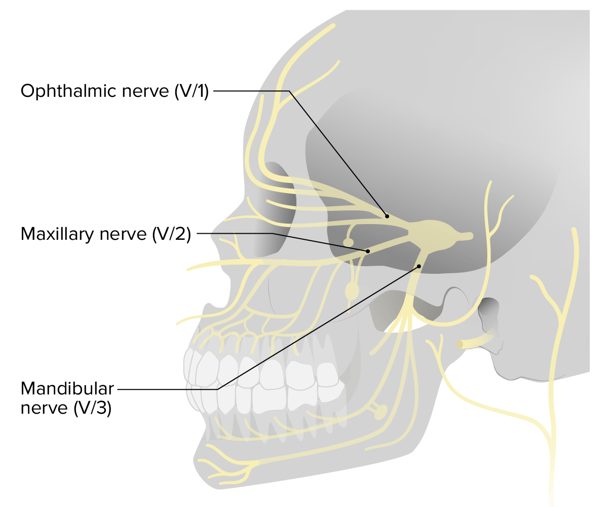
Distribution of cranial nerve V (trigeminal), with its branches labeled
Image by Lecturio.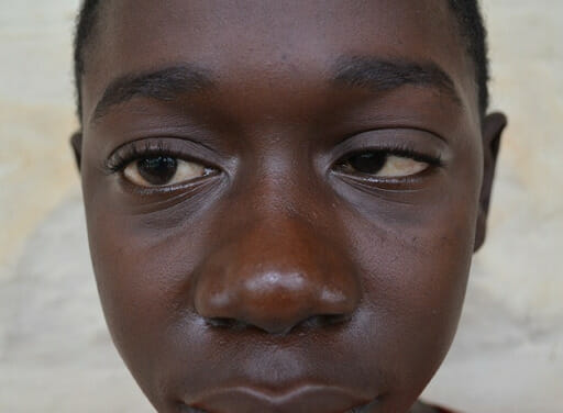
A child with nephrotic syndrome demonstrating typical nephrotic facial puffiness and right cranial nerve VI palsy: The patient is unable to abduct the right eye.
Image: “F0001” by Shalinee Bhoobun et al. License: CC BY 2.0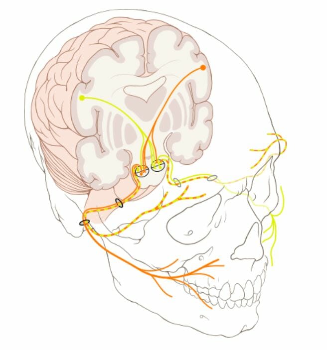
The nuclei of the facial nerve are in the brainstem.
Orange: nerves coming from the left hemisphere of the brain
Yellow: nerves coming from the right hemisphere of the brain
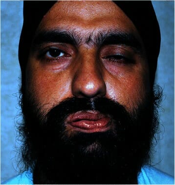
Individual with Bell palsy (lower motor lesion):
Drooped mouth and the inability to close the left eye are noted.
| Grade | Definition |
|---|---|
| I | Normal symmetrical Symmetrical Dermatologic Examination function throughout |
| II |
|
| III |
|
| IV |
|
| V |
|
| VI |
|
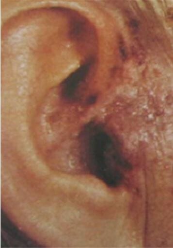
Ear vesicles seen in Ramsay-Hunt syndrome
Image: “Erythème et vésicules cutanées de la zone de Ramsay Hunt droite” by Mahfoudhi M, Lahiani R. License: CC BY 2.0General physiology:
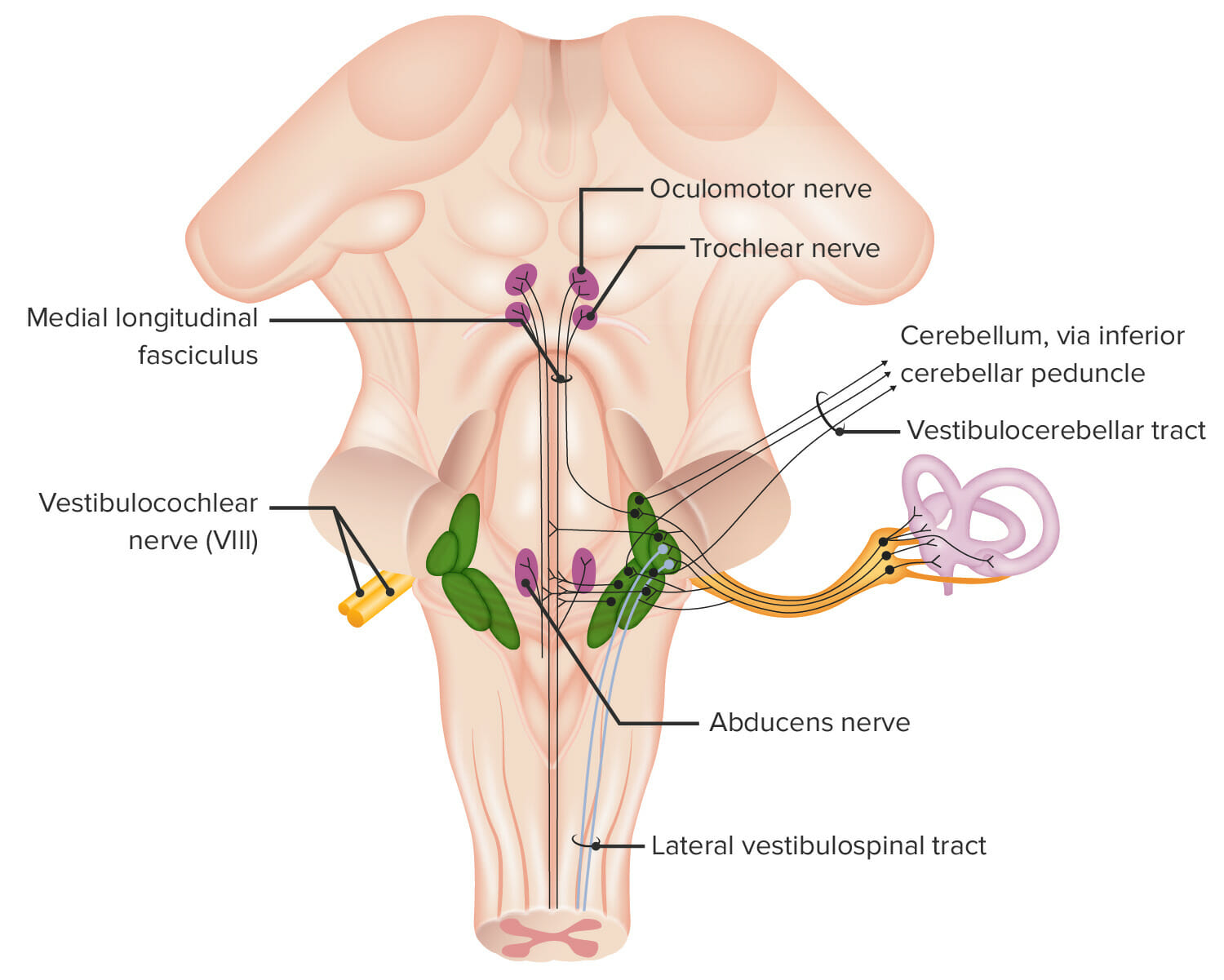
Structures associated with cranial nerve VIII (vestibulocochlear).
Image by Lecturio.Cochlear nerve:
The cochlear nerve transmits sound stimuli from the inner ear Inner ear The essential part of the hearing organ consists of two labyrinthine compartments: the bony labyrinthine and the membranous labyrinth. Ear: Anatomy (fluid-filled cochlea Cochlea The part of the inner ear (labyrinth) that is concerned with hearing. It forms the anterior part of the labyrinth, as a snail-like structure that is situated almost horizontally anterior to the vestibular labyrinth. Ear: Anatomy) to the cochlear nucleus Nucleus Within a eukaryotic cell, a membrane-limited body which contains chromosomes and one or more nucleoli (cell nucleolus). The nuclear membrane consists of a double unit-type membrane which is perforated by a number of pores; the outermost membrane is continuous with the endoplasmic reticulum. A cell may contain more than one nucleus. The Cell: Organelles (brainstem) and then to the primary auditory cortex Auditory cortex The region of the cerebral cortex that receives the auditory radiation from the medial geniculate body. Auditory and Vestibular Pathways: Anatomy ( temporal lobe Temporal lobe Lower lateral part of the cerebral hemisphere responsible for auditory, olfactory, and semantic processing. It is located inferior to the lateral fissure and anterior to the occipital lobe. Cerebral Cortex: Anatomy).
Vestibular nerve:
Vestibulocochlear nerve Vestibulocochlear nerve The 8th cranial nerve. The vestibulocochlear nerve has a cochlear part (cochlear nerve) which is concerned with hearing and a vestibular part (vestibular nerve) which mediates the sense of balance and head position. The fibers of the cochlear nerve originate from neurons of the spiral ganglion and project to the cochlear nuclei (cochlear nucleus). The fibers of the vestibular nerve arise from neurons of scarpa’s ganglion and project to the vestibular nuclei. The 12 Cranial Nerves: Overview and Functions injuries:
Injuries to the cochlear nerve often occur in either:
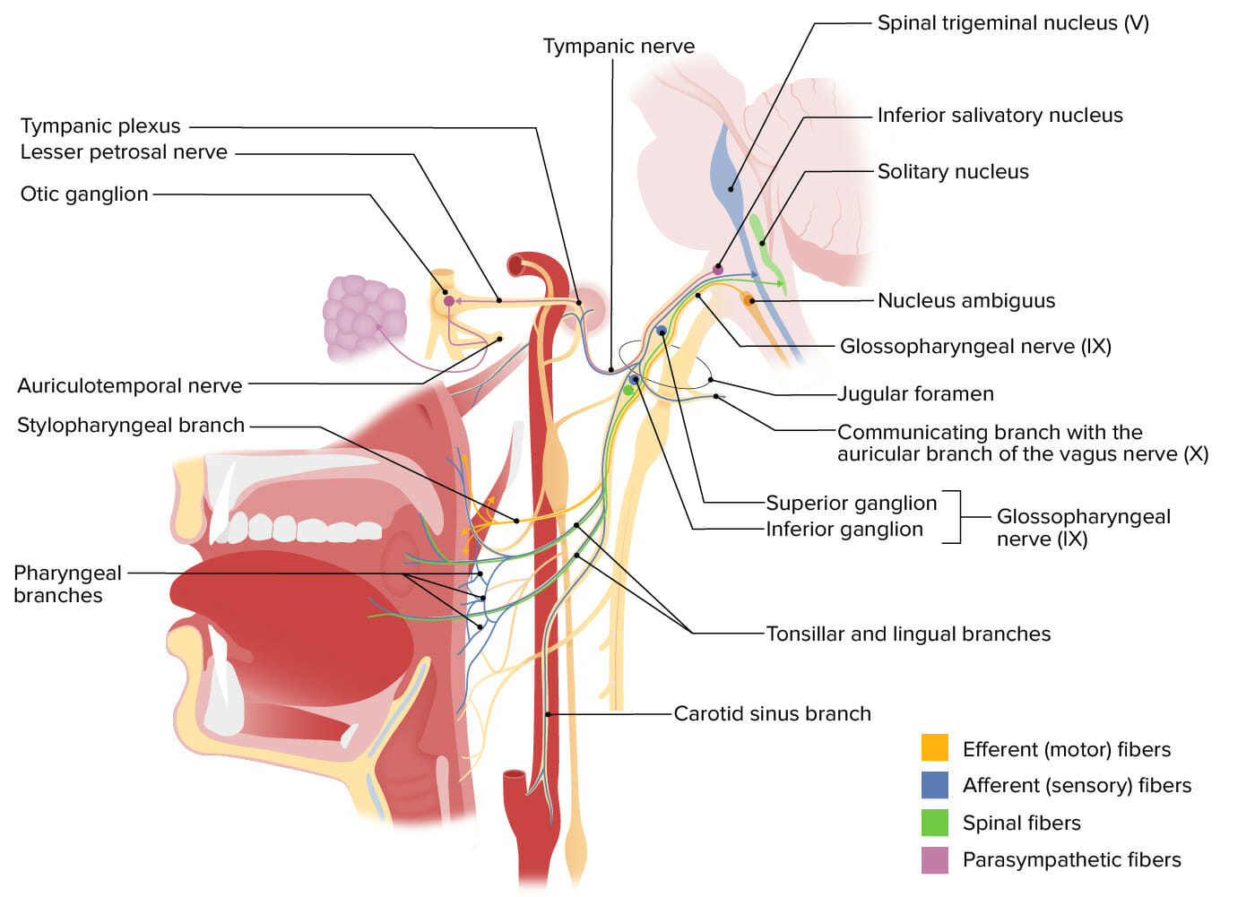
Structures innervated by cranial nerve IX (glossopharyngeal).
Image by Lecturio.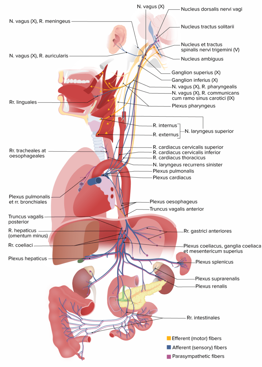
Structures innervated by cranial nerve X (vagus)
N. = nerve
R. = ramus
Rr. = rami
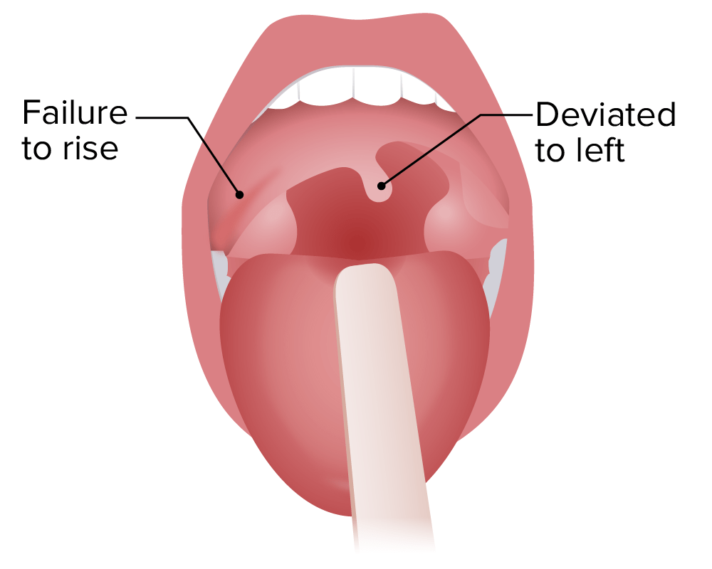
Right-sided vagal palsy: dropped arch of the soft palate on the right side, deviated uvula on the left
Image by Lecturio.
Innervation of the sternocleidomastoid and trapezius muscles by CN XI (spinal accessory nerve)
Image by Lecturio.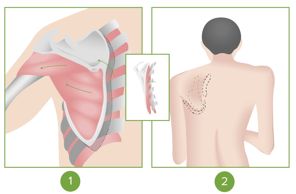
Positive scapular flip sign seen in cranial nerve XI (accessory) palsy
Image by Lecturio.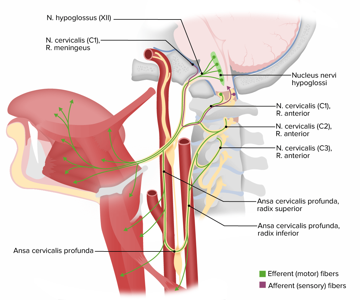
Structures innervated by cranial nerve XII (hypoglossal)
N. = nerve
R. = ramus
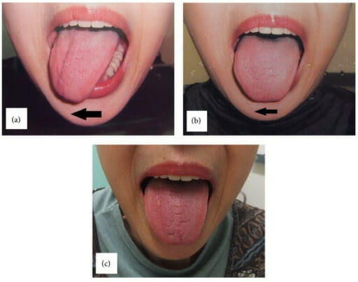
Unilateral hypoglossal nerve palsy after using the LMA device:
(a): On postoperative day 1, the tongue deviated to the right side on protrusion, demonstrating right hypoglossal nerve palsy.
(b): The deviation slightly improved 3 months later.
(c): The deviation disappeared 5 months later for complete recovery of hypoglossal nerve function.
LMA: laryngeal mask airway