Pregnancy is the time period between fertilizationFertilizationTo undergo fertilization, the sperm enters the uterus, travels towards the ampulla of the fallopian tube, and encounters the oocyte. The zona pellucida (the outer layer of the oocyte) deteriorates along with the zygote, which travels towards the uterus and eventually forms a blastocyst, allowing for implantation to occur. Fertilization and First Week of an oocyte and delivery of a fetus approximately 9 months later. The 1st sign of pregnancy is typically a missed menstrual period, after which, pregnancy should be confirmed clinically based on a positive β-hCG test (typically a qualitative urine test) and pelvic ultrasound. There are numerous maternal adaptations to pregnancy, both anatomic and physiologic, which occur to help support the developing fetus and prepare the mother's body for ultimate delivery. Pregnancy is not a pathologic condition, but good routine prenatal carePrenatal carePrenatal care is a systematic and periodic assessment of pregnant women during gestation to assure the best health outcome for the mother and her fetus. Prenatal care prevents and identifies maternal and fetal problems that adversely affect the pregnancy outcome. Prenatal Care can help achieve the best outcomes for both the mother and infant. Prenatal carePrenatal carePrenatal care is a systematic and periodic assessment of pregnant women during gestation to assure the best health outcome for the mother and her fetus. Prenatal care prevents and identifies maternal and fetal problems that adversely affect the pregnancy outcome. Prenatal Care includes appropriate lab and ultrasound testing, anticipatory guidance, and offering solutions or advice for common pregnancy discomforts.
Pregnancy is defined as the time period between fertilizationFertilizationTo undergo fertilization, the sperm enters the uterus, travels towards the ampulla of the fallopian tube, and encounters the oocyte. The zona pellucida (the outer layer of the oocyte) deteriorates along with the zygote, which travels towards the uterus and eventually forms a blastocyst, allowing for implantation to occur. Fertilization and First Week of an oocyte and delivery of a fetus approximately 9 months later.
Terminology
Gravidity: the number of times a woman has been pregnant
Parity:
The total number of deliveries
More specifically, the total number of pregnancies reaching the age of viability regardless of the outcome (live birth, stillborn, cesarean deliveryCesarean DeliveryCesarean delivery (CD) is the operative delivery of ≥ 1 infants through a surgical incision in the maternal abdomen and uterus. Cesarean deliveries may be indicated for a number of either maternal or fetal reasons, most commonly including fetal intolerance to labor, arrest of labor, a history of prior uterine surgery, fetal malpresentation, and placental abnormalities. Cesarean Delivery, etcETCThe electron transport chain (ETC) sends electrons through a series of proteins, which generate an electrochemical proton gradient that produces energy in the form of adenosine triphosphate (ATP).Electron Transport Chain (ETC).)
AbortionAbortionExpulsion of the product of fertilization before completing the term of gestation and without deliberate interference.Spontaneous Abortion:
Number of lost pregnancies prior to the age of viability
Includes both spontaneous abortions (i.e., miscarriages) and induced terminations of pregnancy
Last menstrual period (LMP): the 1st day of a woman’s LMP
Gestational age: the age of pregnancy calculated from the LMP
Embryonic age: the age of pregnancy calculated from the day of fertilizationFertilizationTo undergo fertilization, the sperm enters the uterus, travels towards the ampulla of the fallopian tube, and encounters the oocyte. The zona pellucida (the outer layer of the oocyte) deteriorates along with the zygote, which travels towards the uterus and eventually forms a blastocyst, allowing for implantation to occur. Fertilization and First Week (not used in obstetric clinical practice)
Estimated date of delivery (EDD): also known as the estimated date of confinement
Sequence of events
FertilizationFertilizationTo undergo fertilization, the sperm enters the uterus, travels towards the ampulla of the fallopian tube, and encounters the oocyte. The zona pellucida (the outer layer of the oocyte) deteriorates along with the zygote, which travels towards the uterus and eventually forms a blastocyst, allowing for implantation to occur. Fertilization and First Week of the oocyte by a sperm → embryoEmbryoThe entity of a developing mammal, generally from the cleavage of a zygote to the end of embryonic differentiation of basic structures. For the human embryo, this represents the first two months of intrauterine development preceding the stages of the fetus.Fertilization and First Week
ImplantationImplantationEndometrial implantation of embryo, mammalian at the blastocyst stage.Fertilization and First Week of the early embryoEmbryoThe entity of a developing mammal, generally from the cleavage of a zygote to the end of embryonic differentiation of basic structures. For the human embryo, this represents the first two months of intrauterine development preceding the stages of the fetus.Fertilization and First Week into the uterine wall
Fetal and placental differentiation, growth, and development
Concurrent changes occur in the mother’s body to support the developing fetus and prepare for delivery.
Labor and delivery of the infant
Puerperium: return of the mother’s body to the prepregnant state
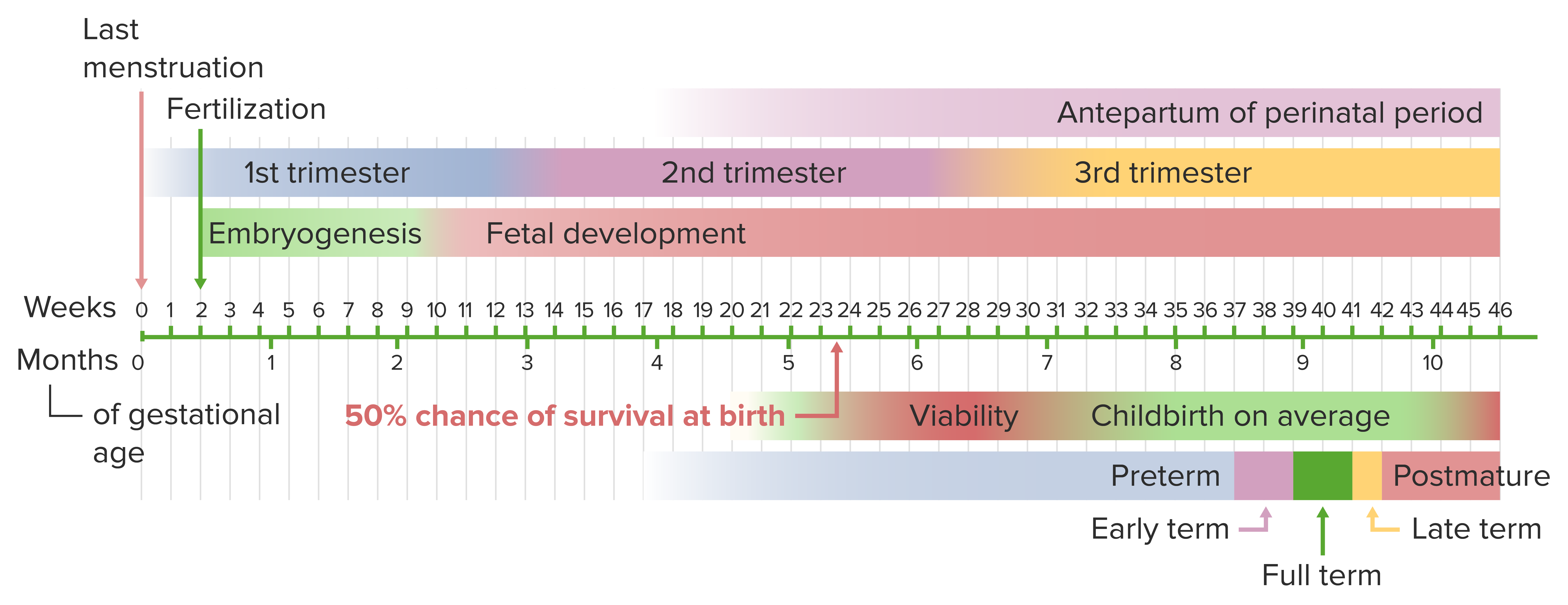
Pregnancy duration
Pregnancy is counted by completed weeks + completed days of the current week since the LMP:
Known as weeks gestational age (wga)
E.g., 35 + 4 wga would indicate that an infant is 35 weeks and 4 days gestational age
Duration of normal pregnancy:
Full-term pregnancy: 37–42 wga
Preterm pregnancy: < 37 wga
Post-term pregnancy: > 42 wga
Notes: Only about 5% of women deliver on their EDD.
Classified into trimesters:
1st trimester: 0–13 + 6 wga
2nd trimester: 14 + 0 to 27 + 6 wga
3rd trimester: 28 + 0 wga through delivery
Timeline of pregnancy from the day of last menstruation to delivery
Image by Lecturio.
Clinical Presentation
Individuals trying to get pregnant will typically present with a positive home pregnancy test. Many others may not know they are pregnant and will present with symptoms of early pregnancy, which may include:
Irregular bleeding (especially in cases of ectopic pregnancyEctopic pregnancyEctopic pregnancy refers to the implantation of a fertilized egg (embryo) outside the uterine cavity. The main cause is disruption of the normal anatomy of the fallopian tube. Ectopic Pregnancy and/or miscarriageMiscarriageSpontaneous abortion, also known as miscarriage, is the loss of a pregnancy before 20 weeks’ gestation. However, the layperson use of the term “abortion” is often intended to refer to induced termination of a pregnancy, whereas “miscarriage” is preferred for spontaneous loss.Spontaneous Abortion)
NauseaNauseaAn unpleasant sensation in the stomach usually accompanied by the urge to vomit. Common causes are early pregnancy, sea and motion sickness, emotional stress, intense pain, food poisoning, and various enteroviruses.Antiemetics and vomitingVomitingThe forcible expulsion of the contents of the stomach through the mouth.Hypokalemia
FatigueFatigueThe state of weariness following a period of exertion, mental or physical, characterized by a decreased capacity for work and reduced efficiency to respond to stimuli.Fibromyalgia
Frequent urination (typically later in pregnancy)
Diagnosis of Pregnancy and Establishing the EDD
Pregnancy is confirmed based on lab tests and obstetric ultrasound imaging.
Laboratory
The major analyteAnalyteThe molecule of interest (antigen)Immunoassays used to establish pregnancy is β-hCG.
β-hCG is a hormone produced early by the developing embryoEmbryoThe entity of a developing mammal, generally from the cleavage of a zygote to the end of embryonic differentiation of basic structures. For the human embryo, this represents the first two months of intrauterine development preceding the stages of the fetus.Fertilization and First Week.
The presence of β-hCG indicates pregnancy.
β-hCG tests may be:
Qualitative: to detect the presence or absence of β-hCG
Urine tests (available as over-the-counter kits) or a test at a medical lab
Reliable approximately 2 weeks after fertilizationFertilizationTo undergo fertilization, the sperm enters the uterus, travels towards the ampulla of the fallopian tube, and encounters the oocyte. The zona pellucida (the outer layer of the oocyte) deteriorates along with the zygote, which travels towards the uterus and eventually forms a blastocyst, allowing for implantation to occur. Fertilization and First Week
Quantitative: to determine serum β-hCG levels
Serum tests
More sensitive, reliable 6–10 days after fertilizationFertilizationTo undergo fertilization, the sperm enters the uterus, travels towards the ampulla of the fallopian tube, and encounters the oocyte. The zona pellucida (the outer layer of the oocyte) deteriorates along with the zygote, which travels towards the uterus and eventually forms a blastocyst, allowing for implantation to occur. Fertilization and First Week
Can be used to track β-hCG levels when there is a concern for an abnormal pregnancy (e.g., ectopic pregnancyEctopic pregnancyEctopic pregnancy refers to the implantation of a fertilized egg (embryo) outside the uterine cavity. The main cause is disruption of the normal anatomy of the fallopian tube. Ectopic Pregnancy or miscarriageMiscarriageSpontaneous abortion, also known as miscarriage, is the loss of a pregnancy before 20 weeks’ gestation. However, the layperson use of the term “abortion” is often intended to refer to induced termination of a pregnancy, whereas “miscarriage” is preferred for spontaneous loss.Spontaneous Abortion)
Levels should roughly double every 48–72 hours during the 1st month.
Imaging
Ultrasound is the obstetric imagingObstetric imagingObstetric imaging refers to imaging of the female reproductive tract and developing fetus during pregnancy. Ultrasonography is the 1st-line imaging modality during pregnancy as it does not emit radiation; thus, it is the safest option for the developing fetus. Obstetric Imaging modality of choice to diagnose and date a pregnancy.
Purpose of early ultrasounds:
Viability: to establish if a viable pregnancy is present
To determine the number of fetuses
To establish the location of the pregnancy (e.g., rule out ectopic pregnancyEctopic pregnancyEctopic pregnancy refers to the implantation of a fertilized egg (embryo) outside the uterine cavity. The main cause is disruption of the normal anatomy of the fallopian tube. Ectopic Pregnancy)
Dating
1st-trimester findings:
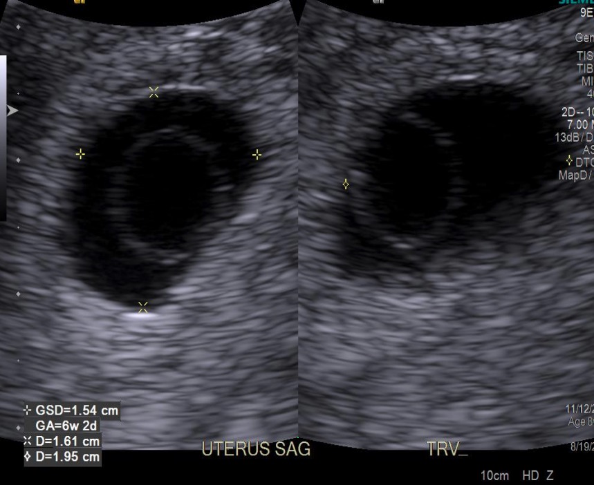
Presence of a gestational sac:
1st visible finding of pregnancy is seen around 4.5–5 wga.
A hypoechoicHypoechoicA structure that produces a low-amplitude echo (darker grays)Ultrasound (Sonography) circle within the uterine cavity, surrounded by hyperechoicHyperechoicA structure that produces a high-amplitude echo (lighter grays and white)Ultrasound (Sonography)endometriumEndometriumThe mucous membrane lining of the uterine cavity that is hormonally responsive during the menstrual cycle and pregnancy. The endometrium undergoes cyclic changes that characterize menstruation. After successful fertilization, it serves to sustain the developing embryo.Embryoblast and Trophoblast Development
Should be visible in the uterusUterusThe uterus, cervix, and fallopian tubes are part of the internal female reproductive system. The uterus has a thick wall made of smooth muscle (the myometrium) and an inner mucosal layer (the endometrium). The most inferior portion of the uterus is the cervix, which connects the uterine cavity to the vagina.Uterus, Cervix, and Fallopian Tubes: Anatomy if quantitative serum β-hCG is > 2,000
Presence of a yolk sacYolk SacThe first of four extra-embryonic membranes to form during embryogenesis. In reptiles and birds, it arises from endoderm and mesoderm to incorporate the egg yolk into the digestive tract for nourishing the embryo. In placental mammals, its nutritional function is vestigial; however, it is the source of intestinal mucosa; blood cells; and germ cells. It is sometimes called the vitelline sac, which should not be confused with the vitelline membrane of the egg.Embryoblast and Trophoblast Development:
A thin hyperechoicHyperechoicA structure that produces a high-amplitude echo (lighter grays and white)Ultrasound (Sonography) ring within the gestational sac
1st seen approximately 5–6 wga and disappears around 10 wga
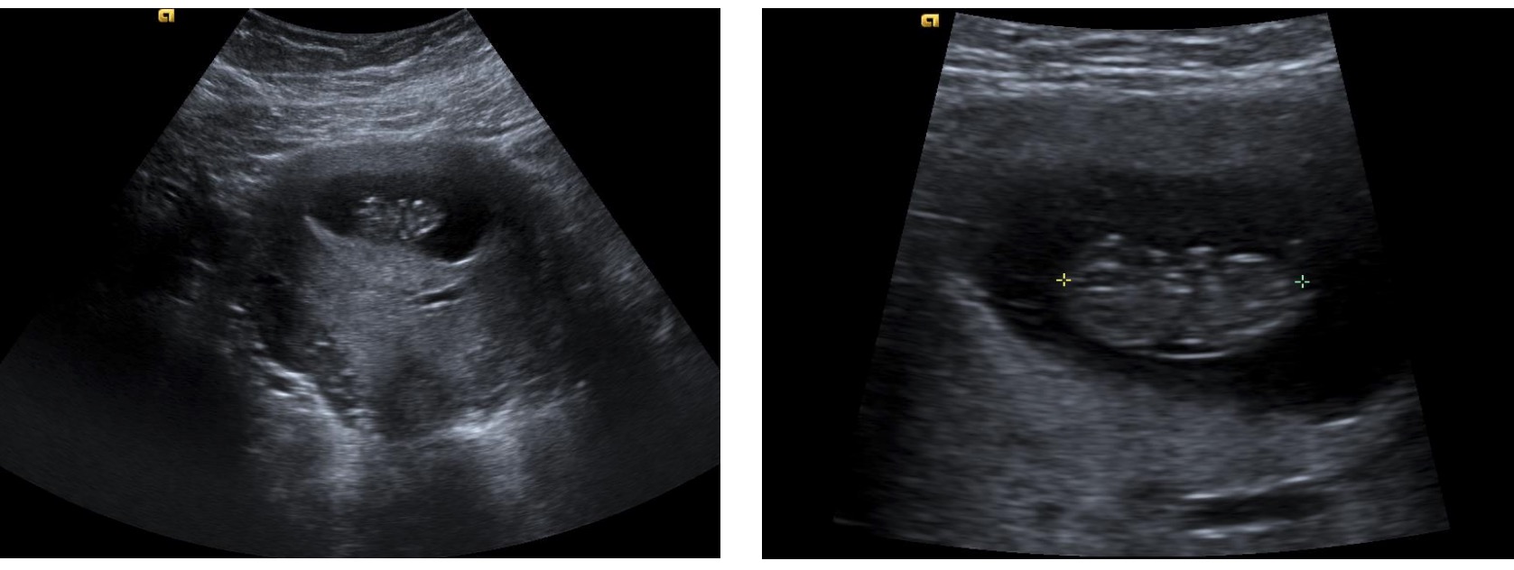
Presence of a fetal pole with a heartbeat: seen around 5.5–6 wga
2nd and 3rd trimesters: calculated using a formula by considering measurements of biparietal diameter, abdominal circumference, and femur length
Yolk sac inside a gestational sac: The yolk sac is the “white circle,” which is inside the gestational sac (the “black circle”). GSD: gestational sac diameter GA: gestational age (based on GSD measurement) D: diameter
Intrauterine pregnancy within a gestational sac: The image on the right shows the measurement of the crown-rump length. The fetal head is on the left and the rump is on the right. Early limb buds are also present. The placenta is on the bottom of the gestational sac in these images.
Establishing the EDD is one of the most important factors to accomplish after diagnosing a pregnancy. Dating a pregnancy is usually done by calculating the EDD from the LMP and comparing that date with the EDD obtained from early ultrasound measurements.
Calculating the EDD from the LMP:
The date that falls exactly 40 weeks after the LMP
Calculated by adding 280 days (or 9 months and 7 days) to the LMP
Dating by ultrasound:
Measure the crown-rump lengthCrown-Rump LengthObstetric Imaging and look up the associated date in a table (most ultrasound machines will show this along with the measurement).
Ultrasound dating is most accurate in the 1st trimester before genetic variation and the effects of intrauterine environment begin to have greater effects on fetal growth.
Establishing the final EDD:
Use the EDD obtained from the LMP if the EDD obtained from the crown-rump lengthCrown-Rump LengthObstetric Imaging measurement is within:
5 days of the LMP-EDD for pregnancies < 9 wga
7 days of the LMP-EDD for pregnancies 9–13 + 6 wga
Approximately 2 weeks in the 2nd trimester
Approximately 3 weeks in the 3rd trimester
Use the EDD obtained from ultrasound if the EDDs are further apart from each other than the dates listed above.
Calculating the EDD from the LMP is the most accurate method to date a pregnancy if that EDD is consistent with the dates obtained from the ultrasound.
If the LMP is unknown, a 1st-trimester ultrasound is the next most accurate way to date a pregnancy.
To support fetal growth and development and prepare the mother’s body for eventual delivery, numerous anatomic and physiologic changes occur within a woman’s body during pregnancy.
Reproductive tract
UterusUterusThe uterus, cervix, and fallopian tubes are part of the internal female reproductive system. The uterus has a thick wall made of smooth muscle (the myometrium) and an inner mucosal layer (the endometrium). The most inferior portion of the uterus is the cervix, which connects the uterine cavity to the vagina.Uterus, Cervix, and Fallopian Tubes: Anatomy:
Increased uterine size:
MassMassThree-dimensional lesion that occupies a space within the breastImaging of the Breast ↑ from approximately 70 grams to 1100 grams
Volume capacity ↑ from approximately 10 mL to 5 L
HypertrophyHypertrophyGeneral increase in bulk of a part or organ due to cell enlargement and accumulation of fluids and secretions, not due to tumor formation, nor to an increase in the number of cells (hyperplasia).Cellular Adaptation of the uterine wall with an accumulation of fibrousFibrousFibrocystic Change and elasticElasticConnective Tissue: Histology tissue
Growth is initiated through ↑ estrogenEstrogenCompounds that interact with estrogen receptors in target tissues to bring about the effects similar to those of estradiol. Estrogens stimulate the female reproductive organs, and the development of secondary female sex characteristics. Estrogenic chemicals include natural, synthetic, steroidal, or non-steroidal compounds.Ovaries: Anatomy levels
By 28 wga, uterine growth slows and the uterusUterusThe uterus, cervix, and fallopian tubes are part of the internal female reproductive system. The uterus has a thick wall made of smooth muscle (the myometrium) and an inner mucosal layer (the endometrium). The most inferior portion of the uterus is the cervix, which connects the uterine cavity to the vagina.Uterus, Cervix, and Fallopian Tubes: Anatomy continues to stretch and become thinner.
Blood flowBlood flowBlood flow refers to the movement of a certain volume of blood through the vasculature over a given unit of time (e.g., mL per minute).Vascular Resistance, Flow, and Mean Arterial Pressure: ↑ from 50 mL/min to 450–750 mL/min at term
Muscle contraction:
UterusUterusThe uterus, cervix, and fallopian tubes are part of the internal female reproductive system. The uterus has a thick wall made of smooth muscle (the myometrium) and an inner mucosal layer (the endometrium). The most inferior portion of the uterus is the cervix, which connects the uterine cavity to the vagina.Uterus, Cervix, and Fallopian Tubes: Anatomy is maintained in a passive noncontractile state through ↑ levels of progesteroneProgesteroneThe major progestational steroid that is secreted primarily by the corpus luteum and the placenta. Progesterone acts on the uterus, the mammary glands and the brain. It is required in embryo implantation; pregnancy maintenance, and the development of mammary tissue for milk production. Progesterone, converted from pregnenolone, also serves as an intermediate in the biosynthesis of gonadal steroid hormones and adrenal corticosteroids.Gonadal Hormones (a smooth muscle relaxant)
Braxton-Hicks contractions:
Irregular contractions that do not cause cervical change
More noticeable as the pregnancy progresses
Uterine involution: return of the uterusUterusThe uterus, cervix, and fallopian tubes are part of the internal female reproductive system. The uterus has a thick wall made of smooth muscle (the myometrium) and an inner mucosal layer (the endometrium). The most inferior portion of the uterus is the cervix, which connects the uterine cavity to the vagina.Uterus, Cervix, and Fallopian Tubes: Anatomy to its pre-pregnant state in the 1st several weeks postpartum
CervixCervixThe uterus, cervix, and fallopian tubes are part of the internal female reproductive system. The most inferior portion of the uterus is the cervix, which connects the uterine cavity to the vagina. Externally, the cervix is lined by stratified squamous cells; however, the cervical canal is lined by columnar epithelium.Uterus, Cervix, and Fallopian Tubes: Anatomy:
CervixCervixThe uterus, cervix, and fallopian tubes are part of the internal female reproductive system. The most inferior portion of the uterus is the cervix, which connects the uterine cavity to the vagina. Externally, the cervix is lined by stratified squamous cells; however, the cervical canal is lined by columnar epithelium.Uterus, Cervix, and Fallopian Tubes: Anatomysoftens and can become bluish due to:
EdemaEdemaEdema is a condition in which excess serous fluid accumulates in the body cavity or interstitial space of connective tissues. Edema is a symptom observed in several medical conditions. It can be categorized into 2 types, namely, peripheral (in the extremities) and internal (in an organ or body cavity). Edema
Increased vascularization
HypertrophyHypertrophyGeneral increase in bulk of a part or organ due to cell enlargement and accumulation of fluids and secretions, not due to tumor formation, nor to an increase in the number of cells (hyperplasia).Cellular Adaptation and hyperplasiaHyperplasiaAn increase in the number of cells in a tissue or organ without tumor formation. It differs from hypertrophy, which is an increase in bulk without an increase in the number of cells.Cellular Adaptation of the cervical glands
Glandular cells normally lining the cervical canalCervical canalUterus, Cervix, and Fallopian Tubes: Anatomy become visible on the surface of the cervixCervixThe uterus, cervix, and fallopian tubes are part of the internal female reproductive system. The most inferior portion of the uterus is the cervix, which connects the uterine cavity to the vagina. Externally, the cervix is lined by stratified squamous cells; however, the cervical canal is lined by columnar epithelium.Uterus, Cervix, and Fallopian Tubes: Anatomy.
Can cause benignBenignFibroadenoma bleeding (all bleeding in pregnancy should be fully worked up)
Endocervical mucosal cells produce a mucus plug, an immunological barrier for uterine contents.
OvariesOvariesOvaries are the paired gonads of the female reproductive system that contain haploid gametes known as oocytes. The ovaries are located intraperitoneally in the pelvis, just posterior to the broad ligament, and are connected to the pelvic sidewall and to the uterus by ligaments. These organs function to secrete hormones (estrogen and progesterone) and to produce the female germ cells (oocytes).Ovaries: Anatomy:
OvulationOvulationThe discharge of an ovum from a rupturing follicle in the ovary.Menstrual Cycle and follicle development are suppressed by ↑ estrogenEstrogenCompounds that interact with estrogen receptors in target tissues to bring about the effects similar to those of estradiol. Estrogens stimulate the female reproductive organs, and the development of secondary female sex characteristics. Estrogenic chemicals include natural, synthetic, steroidal, or non-steroidal compounds.Ovaries: Anatomy levels
The corpus luteumCorpus LuteumThe yellow body derived from the ruptured ovarian follicle after ovulation. The process of corpus luteum formation, luteinization, is regulated by luteinizing hormone.Ovaries: Anatomy supplies progesteroneProgesteroneThe major progestational steroid that is secreted primarily by the corpus luteum and the placenta. Progesterone acts on the uterus, the mammary glands and the brain. It is required in embryo implantation; pregnancy maintenance, and the development of mammary tissue for milk production. Progesterone, converted from pregnenolone, also serves as an intermediate in the biosynthesis of gonadal steroid hormones and adrenal corticosteroids.Gonadal Hormonesduring the 1st part of pregnancy until the placentaPlacentaA highly vascularized mammalian fetal-maternal organ and major site of transport of oxygen, nutrients, and fetal waste products. It includes a fetal portion (chorionic villi) derived from trophoblasts and a maternal portion (decidua) derived from the uterine endometrium. The placenta produces an array of steroid, protein and peptide hormones (placental hormones).Placenta, Umbilical Cord, and Amniotic Cavity is developed enough to take over this function.
Anatomic and physiologic changes in pregnancy by system
Table: Anatomic and physiologic changes in pregnancy
System
Parameters that ↑ in pregnancy
Parameters that ↓ in pregnancy
Symptoms and anatomic changes
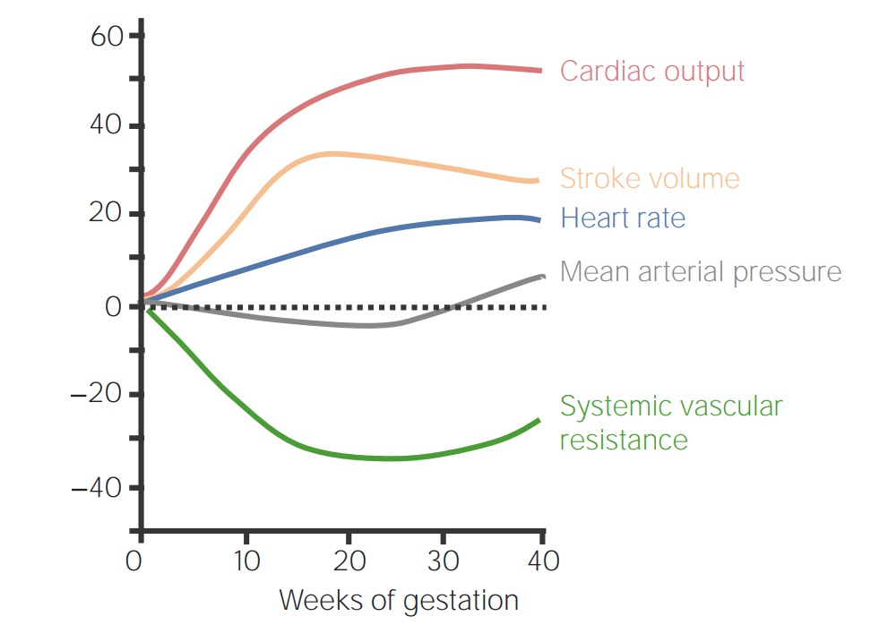
Cardiovascular system
CO
HR
Stroke volumeStroke volumeThe amount of blood pumped out of the heart per beat, not to be confused with cardiac output (volume/time). It is calculated as the difference between the end-diastolic volume and the end-systolic volume.Cardiac Cycle
Venous pressure
Peripheral vascular resistanceResistancePhysiologically, the opposition to flow of air caused by the forces of friction. As a part of pulmonary function testing, it is the ratio of driving pressure to the rate of air flow.Ventilation: Mechanics of Breathing
HemorrhoidsHemorrhoidsHemorrhoids are normal vascular cushions in the anal canal composed of dilated vascular tissue, smooth muscle, and connective tissue. They do not cause issues unless they are enlarged, inflamed, thrombosed, or prolapsed. Patients often present with rectal bleeding of bright red blood, or they may have pain, perianal pruritus, or a palpable mass. Hemorrhoids
Supine hypotensive syndrome
Increased risk of heart failureHeart FailureA heterogeneous condition in which the heart is unable to pump out sufficient blood to meet the metabolic need of the body. Heart failure can be caused by structural defects, functional abnormalities (ventricular dysfunction), or a sudden overload beyond its capacity. Chronic heart failure is more common than acute heart failure which results from sudden insult to cardiac function, such as myocardial infarction.Total Anomalous Pulmonary Venous Return (TAPVR) in individuals with underlying cardiac disease
Hematologic system
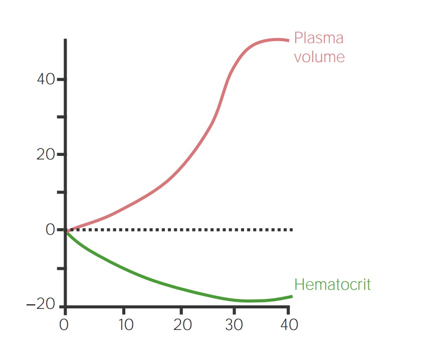
PlasmaPlasmaThe residual portion of blood that is left after removal of blood cells by centrifugation without prior blood coagulation.Transfusion Products volume (↑ 40%‒50% due to water retention)
RBC massMassThree-dimensional lesion that occupies a space within the breastImaging of the Breast (↑ 15%‒30%)
WBC count:
Up to 29,000/µL can be physiologic
Work-up if > 20,000/µL
Coagulation factorsCoagulation factorsEndogenous substances, usually proteins, that are involved in the blood coagulation process.Hemostasis: II, VII, VIII, X, and XII
FibrinogenFibrinogenPlasma glycoprotein clotted by thrombin, composed of a dimer of three non-identical pairs of polypeptide chains (alpha, beta, gamma) held together by disulfide bonds. Fibrinogen clotting is a sol-gel change involving complex molecular arrangements: whereas fibrinogen is cleaved by thrombin to form polypeptides a and b, the proteolytic action of other enzymes yields different fibrinogen degradation products.Hemostasis
Hemoglobin and hematocritHematocritThe volume of packed red blood cells in a blood specimen. The volume is measured by centrifugation in a tube with graduated markings, or with automated blood cell counters. It is an indicator of erythrocyte status in disease. For example, anemia shows a low value; polycythemia, a high value.Neonatal Polycythemia concentrations
PlateletsPlateletsPlatelets are small cell fragments involved in hemostasis. Thrombopoiesis takes place primarily in the bone marrow through a series of cell differentiation and is influenced by several cytokines. Platelets are formed after fragmentation of the megakaryocyte cytoplasm. Platelets: Histology
AnticoagulantsAnticoagulantsAnticoagulants are drugs that retard or interrupt the coagulation cascade. The primary classes of available anticoagulants include heparins, vitamin K-dependent antagonists (e.g., warfarin), direct thrombin inhibitors, and factor Xa inhibitors. Anticoagulants: protein SProtein SProtein S augments the activity of protein C.Hemostasis
Fibrinolysis
Blood viscosityBlood viscosityThe internal resistance of the blood to shear forces. The in vitro measure of whole blood viscosity is of limited clinical utility because it bears little relationship to the actual viscosity within the circulation, but an increase in the viscosity of circulating blood can contribute to morbidity in patients suffering from disorders such as sickle cell anemia and polycythemia.Vascular Resistance, Flow, and Mean Arterial Pressure (improves placental perfusion)
PT and aPTT may be slightly ↓
HypercoagulabilityHypercoagulabilityHypercoagulable States → risk of DVTDVTDeep vein thrombosis (DVT) usually occurs in the deep veins of the lower extremities. The affected veins include the femoral, popliteal, iliofemoral, and pelvic veins. Proximal DVT is more likely to cause a pulmonary embolism (PE) and is generally considered more serious. Deep Vein Thrombosis/PE
Dilutional anemiaAnemiaAnemia is a condition in which individuals have low Hb levels, which can arise from various causes. Anemia is accompanied by a reduced number of RBCs and may manifest with fatigue, shortness of breath, pallor, and weakness. Subtypes are classified by the size of RBCs, chronicity, and etiology. Anemia: Overview and Types → fatigueFatigueThe state of weariness following a period of exertion, mental or physical, characterized by a decreased capacity for work and reduced efficiency to respond to stimuli.Fibromyalgia, shortness of breathShortness of breathDyspnea is the subjective sensation of breathing discomfort. Dyspnea is a normal manifestation of heavy physical or psychological exertion, but also may be caused by underlying conditions (both pulmonary and extrapulmonary).Dyspnea
EdemaEdemaEdema is a condition in which excess serous fluid accumulates in the body cavity or interstitial space of connective tissues. Edema is a symptom observed in several medical conditions. It can be categorized into 2 types, namely, peripheral (in the extremities) and internal (in an organ or body cavity). Edema
Respiratory system
Tidal volumeTidal volumeThe volume of air inspired or expired during each normal, quiet respiratory cycle. Common abbreviations are tv or V with subscript t.Ventilation: Mechanics of Breathing
Pulmonary vascular resistanceResistancePhysiologically, the opposition to flow of air caused by the forces of friction. As a part of pulmonary function testing, it is the ratio of driving pressure to the rate of air flow.Ventilation: Mechanics of Breathing
DiaphragmDiaphragmThe diaphragm is a large, dome-shaped muscle that separates the thoracic cavity from the abdominal cavity. The diaphragm consists of muscle fibers and a large central tendon, which is divided into right and left parts. As the primary muscle of inspiration, the diaphragm contributes 75% of the total inspiratory muscle force.Diaphragm: Anatomy rises by approximately 4 cm due to uterine expansion.
Physiologic respiratory alkalosisAlkalosisA pathological condition that removes acid or adds base to the body fluids.Respiratory Alkalosis
GI system
Intraabdominal pressure
GI motilityGI MotilityThe primary functions of the GI tract are digestion and absorption, which require coordinated contractions of the smooth muscles present in the GI tract. Peristaltic waves, segmentation contractions, and the migrating motor complex are all important contraction patterns that help to mix contents, get them in contact with the intestinal walls, and propel material down the tract at appropriate times and in appropriate amounts.Gastrointestinal Motility (delayed emptying)
StomachStomachThe stomach is a muscular sac in the upper left portion of the abdomen that plays a critical role in digestion. The stomach develops from the foregut and connects the esophagus with the duodenum. Structurally, the stomach is C-shaped and forms a greater and lesser curvature and is divided grossly into regions: the cardia, fundus, body, and pylorus. Stomach: Anatomy and intestines are displaced upward.
ConstipationConstipationConstipation is common and may be due to a variety of causes. Constipation is generally defined as bowel movement frequency < 3 times per week. Patients who are constipated often strain to pass hard stools. The condition is classified as primary (also known as idiopathic or functional constipation) or secondary, and as acute or chronic. Constipation
NauseaNauseaAn unpleasant sensation in the stomach usually accompanied by the urge to vomit. Common causes are early pregnancy, sea and motion sickness, emotional stress, intense pain, food poisoning, and various enteroviruses.Antiemetics and vomitingVomitingThe forcible expulsion of the contents of the stomach through the mouth.Hypokalemia
Renal system
Kidney size (due to ↑ blood volume)
GFRGFRThe volume of water filtered out of plasma through glomerular capillary walls into Bowman’s capsules per unit of time. It is considered to be equivalent to inulin clearance.Kidney Function Tests (↑ approximately 50%)
Renal plasmaPlasmaThe residual portion of blood that is left after removal of blood cells by centrifugation without prior blood coagulation.Transfusion ProductsflowFlowBlood flows through the heart, arteries, capillaries, and veins in a closed, continuous circuit. Flow is the movement of volume per unit of time. Flow is affected by the pressure gradient and the resistance fluid encounters between 2 points. Vascular resistance is the opposition to flow, which is caused primarily by blood friction against vessel walls.Vascular Resistance, Flow, and Mean Arterial Pressure
BicarbonateBicarbonateInorganic salts that contain the -HCO3 radical. They are an important factor in determining the ph of the blood and the concentration of bicarbonate ions is regulated by the kidney. Levels in the blood are an index of the alkali reserve or buffering capacity.Electrolytes excretion (compensatory mechanism for respiratory alkalosisAlkalosisA pathological condition that removes acid or adds base to the body fluids.Respiratory Alkalosis)
Serum creatinine (should be < 0.8 mg/dL in pregnancy)
Serum BUN
Serum sodiumSodiumA member of the alkali group of metals. It has the atomic symbol na, atomic number 11, and atomic weight 23.Hyponatremia
GlucoseGlucoseA primary source of energy for living organisms. It is naturally occurring and is found in fruits and other parts of plants in its free state. It is used therapeutically in fluid and nutrient replacement.Lactose Intolerance reabsorption
Urinary incontinenceUrinary incontinenceUrinary incontinence (UI) is involuntary loss of bladder control or unintentional voiding, which represents a hygienic or social problem to the patient. Urinary incontinence is a symptom, a sign, and a disorder. The 5 types of UI include stress, urge, mixed, overflow, and functional.Urinary Incontinence
HydronephrosisHydronephrosisHydronephrosis is dilation of the renal collecting system as a result of the obstruction of urine outflow. Hydronephrosis can be unilateral or bilateral. Nephrolithiasis is the most common cause of hydronephrosis in young adults, while prostatic hyperplasia and neoplasm are seen in older patients. Hydronephrosis in the 3rd trimester (due to ureteral compressionCompressionBlunt Chest Trauma)
↑ Risk for UTIUTIUrinary tract infections (UTIs) represent a wide spectrum of diseases, from self-limiting simple cystitis to severe pyelonephritis that can result in sepsis and death. Urinary tract infections are most commonly caused by Escherichia coli, but may also be caused by other bacteria and fungi. Urinary Tract Infections (UTIs)
Compensatory metabolic acidosisAcidosisA pathologic condition of acid accumulation or depletion of base in the body. The two main types are respiratory acidosis and metabolic acidosis, due to metabolic acid build up.Respiratory Acidosis
Endocrine and metabolic systems
Basal metabolic rate
Total T3T3A T3 thyroid hormone normally synthesized and secreted by the thyroid gland in much smaller quantities than thyroxine (T4). Most T3 is derived from peripheral monodeiodination of T4 at the 5′ position of the outer ring of the iodothyronine nucleus. The hormone finally delivered and used by the tissues is mainly t3.Thyroid Hormones and T4T4The major hormone derived from the thyroid gland. Thyroxine is synthesized via the iodination of tyrosines (monoiodotyrosine) and the coupling of iodotyrosines (diiodotyrosine) in the thyroglobulin. Thyroxine is released from thyroglobulin by proteolysis and secreted into the blood. Thyroxine is peripherally deiodinated to form triiodothyronine which exerts a broad spectrum of stimulatory effects on cell metabolism.Thyroid Hormones (thyroidThyroidThe thyroid gland is one of the largest endocrine glands in the human body. The thyroid gland is a highly vascular, brownish-red gland located in the visceral compartment of the anterior region of the neck.Thyroid Gland: AnatomyhormonesHormonesHormones are messenger molecules that are synthesized in one part of the body and move through the bloodstream to exert specific regulatory effects on another part of the body. Hormones play critical roles in coordinating cellular activities throughout the body in response to the constant changes in both the internal and external environments. Hormones: Overview and Types
GlucoseGlucoseA primary source of energy for living organisms. It is naturally occurring and is found in fruits and other parts of plants in its free state. It is used therapeutically in fluid and nutrient replacement.Lactose Intolerance intolerance
Anterior pituitaryPituitaryA small, unpaired gland situated in the sella turcica. It is connected to the hypothalamus by a short stalk which is called the infundibulum.Hormones: Overview and Types volume
ProgesteroneProgesteroneThe major progestational steroid that is secreted primarily by the corpus luteum and the placenta. Progesterone acts on the uterus, the mammary glands and the brain. It is required in embryo implantation; pregnancy maintenance, and the development of mammary tissue for milk production. Progesterone, converted from pregnenolone, also serves as an intermediate in the biosynthesis of gonadal steroid hormones and adrenal corticosteroids.Gonadal Hormones and estrogenEstrogenCompounds that interact with estrogen receptors in target tissues to bring about the effects similar to those of estradiol. Estrogens stimulate the female reproductive organs, and the development of secondary female sex characteristics. Estrogenic chemicals include natural, synthetic, steroidal, or non-steroidal compounds.Ovaries: Anatomy
ProlactinProlactinA lactogenic hormone secreted by the adenohypophysis. It is a polypeptide of approximately 23 kd. Besides its major action on lactation, in some species prolactin exerts effects on reproduction, maternal behavior, fat metabolism, immunomodulation and osmoregulation.Breasts: Anatomy
Oxytocin
Relaxin
ReninReninA highly specific (leu-leu) endopeptidase that generates angiotensin I from its precursor angiotensinogen, leading to a cascade of reactions which elevate blood pressure and increase sodium retention by the kidney in the renin-angiotensin system.Renal Sodium and Water Regulation and aldosteroneAldosteroneA hormone secreted by the adrenal cortex that regulates electrolyte and water balance by increasing the renal retention of sodium and the excretion of potassium.Hyperkalemia
ErythropoietinErythropoietinGlycoprotein hormone, secreted chiefly by the kidney in the adult and the liver in the fetus, that acts on erythroid stem cells of the bone marrow to stimulate proliferation and differentiation.Erythrocytes: Histology
TSH
FSHFSHA major gonadotropin secreted by the adenohypophysis. Follicle-stimulating hormone stimulates gametogenesis and the supporting cells such as the ovarian granulosa cells, the testicular sertoli cells, and leydig cells. Fsh consists of two noncovalently linked subunits, alpha and beta. Within a species, the alpha subunit is common in the three pituitary glycoprotein hormones (TSH, LH, and FSH), but the beta subunit is unique and confers its biological specificity.Menstrual Cycle and LHLHA major gonadotropin secreted by the adenohypophysis. Luteinizing hormone regulates steroid production by the interstitial cells of the testis and the ovary. The preovulatory luteinizing hormone surge in females induces ovulation, and subsequent luteinization of the follicle. Luteinizing hormone consists of two noncovalently linked subunits, alpha and beta. Within a species, the alpha subunit is common in the three pituitary glycoprotein hormones (TSH, LH, and FSH), but the beta subunit is unique and confers its biological specificity.Menstrual Cycle
↑ Caloric needs
↑ Volume of the anterior pituitaryPituitaryA small, unpaired gland situated in the sella turcica. It is connected to the hypothalamus by a short stalk which is called the infundibulum.Hormones: Overview and Types
Breast enlargement and tenderness
PainPainAn unpleasant sensation induced by noxious stimuli which are detected by nerve endings of nociceptive neurons.Pain: Types and Pathways from stretching of ligaments (e.g., pelvic painPainAn unpleasant sensation induced by noxious stimuli which are detected by nerve endings of nociceptive neurons.Pain: Types and Pathways, round ligamentRound ligamentA fibromuscular band that attaches to the uterus and then passes along the broad ligament, out through the inguinal ring, and into the labium majus.Uterus, Cervix, and Fallopian Tubes: AnatomypainPainAn unpleasant sensation induced by noxious stimuli which are detected by nerve endings of nociceptive neurons.Pain: Types and Pathways)
SkinSkinThe skin, also referred to as the integumentary system, is the largest organ of the body. The skin is primarily composed of the epidermis (outer layer) and dermis (deep layer). The epidermis is primarily composed of keratinocytes that undergo rapid turnover, while the dermis contains dense layers of connective tissue.Skin: Structure and Functions changes
Stretch marks
HyperpigmentationHyperpigmentationExcessive pigmentation of the skin, usually as a result of increased epidermal or dermal melanin pigmentation, hypermelanosis. Hyperpigmentation can be localized or generalized. The condition may arise from exposure to light, chemicals or other substances, or from a primary metabolic imbalance.Malassezia Fungi of:
Face (known as melasmaMelasmaMelasma is a benign skin condition characterized by hyperpigmentation of sun-exposed regions due to excess melanin production and deposition. The condition mainly affects women during their reproductive years, particularly those with darker skin tones. Melasma, or the “mask of pregnancy”)
Nipples
PerineumPerineumThe body region lying between the genital area and the anus on the surface of the trunk, and to the shallow compartment lying deep to this area that is inferior to the pelvic diaphragm. The surface area is between the vulva and the anus in the female, and between the scrotum and the anus in the male.Vagina, Vulva, and Pelvic Floor: Anatomy
The typical schedule of prenatal visits for low-risk individuals:
Every 4 weeks up through 28 wga
Every 2 weeks from 28–36 wga
Every week from 36 wga until delivery
Prenatal visits
Parameters to measure/monitor for healthy, uncomplicated individuals at routine prenatal visits:
1st visit:
Ultrasound to confirm the estimated date of confinement (either in-office or ordered)
Full physical exam, including a pelvic exam
Recommend supplements:
Folic acid (best if started prior to pregnancy)
Multivitamins with ironIronA metallic element with atomic symbol fe, atomic number 26, and atomic weight 55. 85. It is an essential constituent of hemoglobins; cytochromes; and iron-binding proteins. It plays a role in cellular redox reactions and in the transport of oxygen.Trace Elements
All visits:
Weight
Blood pressure
Fetal HR (using dopplerDopplerUltrasonography applying the doppler effect, with frequency-shifted ultrasound reflections produced by moving targets (usually red blood cells) in the bloodstream along the ultrasound axis in direct proportion to the velocity of movement of the targets, to determine both direction and velocity of blood flow.Ultrasound (Sonography) auscultation)
Should experience 10 movements in a 2-hour period at least once daily
Individuals who report decreased fetal movement should be evaluated.
TDaP immunization (recommended between 27–36 wga)
Rh immunoglobulin to Rh-negative women
Starting at 34–36 wga: Assess fetal presentationFetal presentationFetal presentation describes which part of the fetus will enter through the cervix first, while position is the orientation of the fetus compared to the maternal bony pelvis. Presentations include vertex (the fetal occiput will present through the cervix first), face, brow, shoulder, and breech. Fetal Malpresentation and Malposition (vertex or breech).
Routine pregnancy laboratory and imaging studies
All pregnant individuals should have certain labs done at different points during their pregnancy. These include:
At their 1st obstetrics appointment:
CBC
Blood type and screen (may indicate future compatibility issues with fetal blood type)
UrinalysisUrinalysisExamination of urine by chemical, physical, or microscopic means. Routine urinalysis usually includes performing chemical screening tests, determining specific gravity, observing any unusual color or odor, screening for bacteriuria, and examining the sediment microscopically.Urinary Tract Infections (UTIs) in Children
RubellaRubellaAn acute infectious disease caused by the rubella virus. The virus enters the respiratory tract via airborne droplet and spreads to the lymphatic system.Rubella Virus immunity status
Hepatitis B surface antigenHepatitis B surface antigenThose hepatitis B antigens found on the surface of the dane particle and on the 20 nm spherical and tubular particles. Several subspecificities of the surface antigen are known. These were formerly called the Australia antigen.Hepatitis B Virus
Rapid plasma reagin testRapid plasma reagin testTreponema for syphilisSyphilisSyphilis is a bacterial infection caused by the spirochete Treponema pallidum pallidum (T. p. pallidum), which is usually spread through sexual contact. Syphilis has 4 clinical stages: primary, secondary, latent, and tertiary. Syphilis
GonorrheaGonorrheaGonorrhea is a sexually transmitted infection (STI) caused by the gram-negative bacteria Neisseria gonorrhoeae (N. gonorrhoeae). Gonorrhea may be asymptomatic but commonly manifests as cervicitis or urethritis with less common presentations such as proctitis, conjunctivitis, or pharyngitis. Gonorrhea and chlamydiaChlamydiaChlamydiae are obligate intracellular gram-negative bacteria. They lack a peptidoglycan layer and are best visualized using Giemsa stain. The family of Chlamydiaceae comprises 3 pathogens that can infect humans: Chlamydia trachomatis, Chlamydia psittaci, and Chlamydia pneumoniae.Chlamydia testing
ScreeningScreeningPreoperative Care for fetal aneuploidy (e.g., trisomy 21Trisomy 21Down syndrome, or trisomy 21, is the most common chromosomal aberration and the most frequent genetic cause of developmental delay. Both boys and girls are affected and have characteristic craniofacial and musculoskeletal features, as well as multiple medical anomalies involving the cardiac, gastrointestinal, ocular, and auditory systems.Down syndrome (Trisomy 21)):
Multiple options available
Options include different combinations of multiple serum analytes and ultrasound.
A common option used is a test assessing the cell-free fetal DNADNAA deoxyribonucleotide polymer that is the primary genetic material of all cells. Eukaryotic and prokaryotic organisms normally contain DNA in a double-stranded state, yet several important biological processes transiently involve single-stranded regions. DNA, which consists of a polysugar-phosphate backbone possessing projections of purines (adenine and guanine) and pyrimidines (thymine and cytosine), forms a double helix that is held together by hydrogen bonds between these purines and pyrimidines (adenine to thymine and guanine to cytosine).DNA Types and Structure found in maternal serum (often referred to as noninvasive prenatal testing (NIPT)).
Other tests:
Full anatomic assessment of the fetus, placentaPlacentaA highly vascularized mammalian fetal-maternal organ and major site of transport of oxygen, nutrients, and fetal waste products. It includes a fetal portion (chorionic villi) derived from trophoblasts and a maternal portion (decidua) derived from the uterine endometrium. The placenta produces an array of steroid, protein and peptide hormones (placental hormones).Placenta, Umbilical Cord, and Amniotic Cavity, and uterusUterusThe uterus, cervix, and fallopian tubes are part of the internal female reproductive system. The uterus has a thick wall made of smooth muscle (the myometrium) and an inner mucosal layer (the endometrium). The most inferior portion of the uterus is the cervix, which connects the uterine cavity to the vagina.Uterus, Cervix, and Fallopian Tubes: Anatomy/cervixCervixThe uterus, cervix, and fallopian tubes are part of the internal female reproductive system. The most inferior portion of the uterus is the cervix, which connects the uterine cavity to the vagina. Externally, the cervix is lined by stratified squamous cells; however, the cervical canal is lined by columnar epithelium.Uterus, Cervix, and Fallopian Tubes: Anatomy at 18–22 wga
1-hour glucoseGlucoseA primary source of energy for living organisms. It is naturally occurring and is found in fruits and other parts of plants in its free state. It is used therapeutically in fluid and nutrient replacement.Lactose IntolerancetoleranceTolerancePharmacokinetics and Pharmacodynamics test (GTT) at 24–28 wga
CBC is often repeated with GTT.
Group B StreptococcusStreptococcusStreptococcus is one of the two medically important genera of gram-positive cocci, the other being Staphylococcus. Streptococci are identified as different species on blood agar on the basis of their hemolytic pattern and sensitivity to optochin and bacitracin. There are many pathogenic species of streptococci, including S. pyogenes, S. agalactiae, S. pneumoniae, and the viridans streptococci.Streptococcus culture at 36–37 wga
Bedside ultrasound to check fetal presentationFetal presentationFetal presentation describes which part of the fetus will enter through the cervix first, while position is the orientation of the fetus compared to the maternal bony pelvis. Presentations include vertex (the fetal occiput will present through the cervix first), face, brow, shoulder, and breech. Fetal Malpresentation and Malposition (vertex/head down, breech/buttocks down) around 35 wga
Diet, exercise, and weight gain
Table: Safe and unsafe diets in pregnancy
Safe
Unsafe
ModeratecaffeineCaffeineA methylxanthine naturally occurring in some beverages and also used as a pharmacological agent. Caffeine’s most notable pharmacological effect is as a central nervous system stimulant, increasing alertness and producing agitation. Several cellular actions of caffeine have been observed, but it is not entirely clear how each contributes to its pharmacological profile. Among the most important are inhibition of cyclic nucleotide phosphodiesterases, antagonism of adenosine receptors, and modulation of intracellular calcium handling.Stimulants intake
Artificial sweeteners
FishFISHA type of in situ hybridization in which target sequences are stained with fluorescent dye so their location and size can be determined using fluorescence microscopy. This staining is sufficiently distinct that the hybridization signal can be seen both in metaphase spreads and in interphase nuclei.Chromosome Testing: limitLimitA value (e.g., pressure or time) that should not be exceeded and which is specified by the operator to protect the lungInvasive Mechanical Ventilation to < 12 oz/week
Excess caffeineCaffeineA methylxanthine naturally occurring in some beverages and also used as a pharmacological agent. Caffeine’s most notable pharmacological effect is as a central nervous system stimulant, increasing alertness and producing agitation. Several cellular actions of caffeine have been observed, but it is not entirely clear how each contributes to its pharmacological profile. Among the most important are inhibition of cyclic nucleotide phosphodiesterases, antagonism of adenosine receptors, and modulation of intracellular calcium handling.Stimulants intake
Saccharine
Unpasteurized foods, especially dairy (risk of listeriaListeriaListeria spp. are motile, flagellated, gram-positive, facultative intracellular bacilli. The major pathogenic species is Listeria monocytogenes. Listeria are part of the normal gastrointestinal flora of domestic mammals and poultry and are transmitted to humans through the ingestion of contaminated food, especially unpasteurized dairy products. Listeria Monocytogenes/Listeriosis)
Swordfish, shark, king mackerel, or raw fishFISHA type of in situ hybridization in which target sequences are stained with fluorescent dye so their location and size can be determined using fluorescence microscopy. This staining is sufficiently distinct that the hybridization signal can be seen both in metaphase spreads and in interphase nuclei.Chromosome Testing (risk of mercuryMercuryA silver metallic element that exists as a liquid at room temperature. It has the atomic symbol Hg (from hydrargyrum, liquid silver), atomic number 80, and atomic weight 200. 59. Mercury is used in many industrial applications and its salts have been employed therapeutically as purgatives, antisyphilitics, disinfectants, and astringents. It can be absorbed through the skin and mucous membranes which leads to mercury poisoning. Because of its toxicity, the clinical use of mercury and mercurials is diminishing.Renal Tubular Acidosis poisoning)
Weight gain:
The recommended weight gain during pregnancy is based on the individual’s prepregnancy BMIBMIAn indicator of body density as determined by the relationship of body weight to body height. Bmi=weight (kg)/height squared (m2). Bmi correlates with body fat (adipose tissue). Their relationship varies with age and gender. For adults, bmi falls into these categories: below 18. 5 (underweight); 18. 5-24. 9 (normal); 25. 0-29. 9 (overweight); 30. 0 and above (obese).Obesity. Normal weight-gain recommendations:
Underweight (BMIBMIAn indicator of body density as determined by the relationship of body weight to body height. Bmi=weight (kg)/height squared (m2). Bmi correlates with body fat (adipose tissue). Their relationship varies with age and gender. For adults, bmi falls into these categories: below 18. 5 (underweight); 18. 5-24. 9 (normal); 25. 0-29. 9 (overweight); 30. 0 and above (obese).Obesity < 18.5): 28–40 lbs
Normal weight (BMIBMIAn indicator of body density as determined by the relationship of body weight to body height. Bmi=weight (kg)/height squared (m2). Bmi correlates with body fat (adipose tissue). Their relationship varies with age and gender. For adults, bmi falls into these categories: below 18. 5 (underweight); 18. 5-24. 9 (normal); 25. 0-29. 9 (overweight); 30. 0 and above (obese).Obesity 18.5–24.9): 25–35 lbs
Overweight (BMIBMIAn indicator of body density as determined by the relationship of body weight to body height. Bmi=weight (kg)/height squared (m2). Bmi correlates with body fat (adipose tissue). Their relationship varies with age and gender. For adults, bmi falls into these categories: below 18. 5 (underweight); 18. 5-24. 9 (normal); 25. 0-29. 9 (overweight); 30. 0 and above (obese).Obesity 25–29.9): 15–25 lbs
Obese (BMIBMIAn indicator of body density as determined by the relationship of body weight to body height. Bmi=weight (kg)/height squared (m2). Bmi correlates with body fat (adipose tissue). Their relationship varies with age and gender. For adults, bmi falls into these categories: below 18. 5 (underweight); 18. 5-24. 9 (normal); 25. 0-29. 9 (overweight); 30. 0 and above (obese).Obesity > 30): 11–20 lbs
Note: Weight lossWeight lossDecrease in existing body weight.Bariatric Surgery is not recommended during pregnancy.
Exercise:
Purpose: controls weight gain, improves delivery, improves weight lossWeight lossDecrease in existing body weight.Bariatric Surgery after pregnancy
Recommendation: moderate exercise for 30 minutes on most days of the week
In general, women can continue performing exercises they were doing prior to pregnancy at the same level of intensity (goal: maintain fitness level rather than increasing exercise intensity).
Avoid contact sports and/or activities associated with the risk of falling or abdominal trauma (e.g., soccer, horseback riding, downhill skiing).
Avoid exercising in hot weather.
Safety
Air travel is safe for up to 36 weeks (after which, the risk of labor or complications on board ↑).
Precautions to prevent deep vein thrombosisThrombosisFormation and development of a thrombus or blood clot in the blood vessel.Epidemic Typhus during long trips (both flights and road trips):
Frequent ambulation in the airplane or at rest stops (every 1–2 hours)
Frequent handwashing
Avoid kitty litter (to ↓ risk of toxoplasmosisToxoplasmosisToxoplasmosis is an infectious disease caused by Toxoplasma gondii, an obligate intracellular protozoan parasite. Felines are the definitive host, but transmission to humans can occur through contact with cat feces or the consumption of contaminated foods. The clinical presentation and complications depend on the host’s immune status. Toxoplasma/Toxoplasmosis).
PainPainAn unpleasant sensation induced by noxious stimuli which are detected by nerve endings of nociceptive neurons.Pain: Types and Pathways
Pregnancy hormonesHormonesHormones are messenger molecules that are synthesized in one part of the body and move through the bloodstream to exert specific regulatory effects on another part of the body. Hormones play critical roles in coordinating cellular activities throughout the body in response to the constant changes in both the internal and external environments. Hormones: Overview and Types (e.g., progesteroneProgesteroneThe major progestational steroid that is secreted primarily by the corpus luteum and the placenta. Progesterone acts on the uterus, the mammary glands and the brain. It is required in embryo implantation; pregnancy maintenance, and the development of mammary tissue for milk production. Progesterone, converted from pregnenolone, also serves as an intermediate in the biosynthesis of gonadal steroid hormones and adrenal corticosteroids.Gonadal Hormones, relaxin) cause ligaments to stretch more easily and ↑ water retention
Common pains:
Pelvic painPainAn unpleasant sensation induced by noxious stimuli which are detected by nerve endings of nociceptive neurons.Pain: Types and Pathways from stretching at the pubic symphysisPubic SymphysisA slightly movable cartilaginous joint which occurs between the pubic bones.Vagina, Vulva, and Pelvic Floor: Anatomy and sacroiliac joints → maternity support belts can help
Round ligamentRound ligamentA fibromuscular band that attaches to the uterus and then passes along the broad ligament, out through the inguinal ring, and into the labium majus.Uterus, Cervix, and Fallopian Tubes: AnatomypainPainAn unpleasant sensation induced by noxious stimuli which are detected by nerve endings of nociceptive neurons.Pain: Types and Pathways (described subsequently)
FootFootThe foot is the terminal portion of the lower limb, whose primary function is to bear weight and facilitate locomotion. The foot comprises 26 bones, including the tarsal bones, metatarsal bones, and phalanges. The bones of the foot form longitudinal and transverse arches and are supported by various muscles, ligaments, and tendons.Foot: AnatomypainPainAn unpleasant sensation induced by noxious stimuli which are detected by nerve endings of nociceptive neurons.Pain: Types and Pathways
Low back painPainAn unpleasant sensation induced by noxious stimuli which are detected by nerve endings of nociceptive neurons.Pain: Types and Pathways (from shifting center of gravity)
Carpal tunnelCarpal TunnelThe carpal tunnel is formed by the transverse carpal ligament (flexor retinaculum) superiorly and the carpal bones inferiorly.Carpal Tunnel Syndrome syndrome
Round ligamentRound ligamentA fibromuscular band that attaches to the uterus and then passes along the broad ligament, out through the inguinal ring, and into the labium majus.Uterus, Cervix, and Fallopian Tubes: AnatomypainPainAn unpleasant sensation induced by noxious stimuli which are detected by nerve endings of nociceptive neurons.Pain: Types and Pathways:
Round ligaments attach the uterusUterusThe uterus, cervix, and fallopian tubes are part of the internal female reproductive system. The uterus has a thick wall made of smooth muscle (the myometrium) and an inner mucosal layer (the endometrium). The most inferior portion of the uterus is the cervix, which connects the uterine cavity to the vagina.Uterus, Cervix, and Fallopian Tubes: Anatomy to the pelvic sidewall.
As the uterusUterusThe uterus, cervix, and fallopian tubes are part of the internal female reproductive system. The uterus has a thick wall made of smooth muscle (the myometrium) and an inner mucosal layer (the endometrium). The most inferior portion of the uterus is the cervix, which connects the uterine cavity to the vagina.Uterus, Cervix, and Fallopian Tubes: Anatomy grows, the round ligaments can stretch and cause painPainAn unpleasant sensation induced by noxious stimuli which are detected by nerve endings of nociceptive neurons.Pain: Types and Pathways.
Differentiating round ligamentRound ligamentA fibromuscular band that attaches to the uterus and then passes along the broad ligament, out through the inguinal ring, and into the labium majus.Uterus, Cervix, and Fallopian Tubes: AnatomypainPainAn unpleasant sensation induced by noxious stimuli which are detected by nerve endings of nociceptive neurons.Pain: Types and Pathways (benignBenignFibroadenoma) from more concerning painPainAn unpleasant sensation induced by noxious stimuli which are detected by nerve endings of nociceptive neurons.Pain: Types and Pathways:
Round ligamentRound ligamentA fibromuscular band that attaches to the uterus and then passes along the broad ligament, out through the inguinal ring, and into the labium majus.Uterus, Cervix, and Fallopian Tubes: AnatomypainPainAn unpleasant sensation induced by noxious stimuli which are detected by nerve endings of nociceptive neurons.Pain: Types and Pathways is often unilateral.
On exam, push the uterusUterusThe uterus, cervix, and fallopian tubes are part of the internal female reproductive system. The uterus has a thick wall made of smooth muscle (the myometrium) and an inner mucosal layer (the endometrium). The most inferior portion of the uterus is the cervix, which connects the uterine cavity to the vagina.Uterus, Cervix, and Fallopian Tubes: Anatomytoward the painful side; if this maneuver relieves painPainAn unpleasant sensation induced by noxious stimuli which are detected by nerve endings of nociceptive neurons.Pain: Types and Pathways, it is likely round ligamentRound ligamentA fibromuscular band that attaches to the uterus and then passes along the broad ligament, out through the inguinal ring, and into the labium majus.Uterus, Cervix, and Fallopian Tubes: AnatomypainPainAn unpleasant sensation induced by noxious stimuli which are detected by nerve endings of nociceptive neurons.Pain: Types and Pathways.
Analgesics:
AcetaminophenAcetaminophenAcetaminophen is an over-the-counter nonopioid analgesic and antipyretic medication and the most commonly used analgesic worldwide. Despite the widespread use of acetaminophen, its mechanism of action is not entirely understood.Acetaminophen isthe safest analgesic.
Try to avoid NSAIDsNSAIDSPrimary vs Secondary Headaches due to their effects on the fetal kidneysKidneysThe kidneys are a pair of bean-shaped organs located retroperitoneally against the posterior wall of the abdomen on either side of the spine. As part of the urinary tract, the kidneys are responsible for blood filtration and excretion of water-soluble waste in the urine.Kidneys: Anatomy.
Gastrointestinal symptoms
NauseaNauseaAn unpleasant sensation in the stomach usually accompanied by the urge to vomit. Common causes are early pregnancy, sea and motion sickness, emotional stress, intense pain, food poisoning, and various enteroviruses.Antiemetics and vomitingVomitingThe forcible expulsion of the contents of the stomach through the mouth.Hypokalemia:
Common especially in early pregnancy (colloquially, “morning sickness”)
More common in the mornings but may occur throughout the day
Often improves in the early 2nd trimester
Management:
Vitamin B6Vitamin B6Vitamin B 6 refers to several picolines (especially pyridoxine; pyridoxal; & pyridoxamine) that are efficiently converted by the body to pyridoxal phosphate which is a coenzyme for synthesis of amino acids, neurotransmitters (serotonin, norepinephrine), sphingolipids, and aminolevulinic acid. During transamination of amino acids, pyridoxal phosphate is transiently converted into pyridoxamine phosphate. Although pyridoxine and vitamin B 6 are still frequently used as synonyms, especially by medical researchers, this practice is erroneous and sometimes misleading. Most of vitamin b6 is eventually degraded to pyridoxic acid and excreted in the urine.Water-soluble Vitamins and their Deficiencies supplementation
Dietary changes: eat 1st thing in the morning. Consume smaller meals more frequently.
ConstipationConstipationConstipation is common and may be due to a variety of causes. Constipation is generally defined as bowel movement frequency < 3 times per week. Patients who are constipated often strain to pass hard stools. The condition is classified as primary (also known as idiopathic or functional constipation) or secondary, and as acute or chronic. Constipation
The primary symptom of pregnancy is a “missed” menstrual cycleMenstrual cycleThe menstrual cycle is the cyclic pattern of hormonal and tissular activity that prepares a suitable uterine environment for the fertilization and implantation of an ovum. The menstrual cycle involves both an endometrial and ovarian cycle that are dependent on one another for proper functioning. There are 2 phases of the ovarian cycle and 3 phases of the endometrial cycle.Menstrual Cycle. Pregnancy must always be ruled out using a simple urine-pregnancy test in reproductive-aged women presenting with abnormal bleeding. Other common causes of abnormal uterine bleedingAbnormal Uterine BleedingAbnormal uterine bleeding is the medical term for abnormalities in the frequency, volume, duration, and regularity of the menstrual cycle. Abnormal uterine bleeding is classified using the acronym PALM-COEIN, with PALM representing the structural causes and COEIN indicating the non-structural causes. Abnormal Uterine Bleeding include:
Polycystic ovarian syndromePolycystic ovarian syndromePolycystic ovarian syndrome (PCOS) is the most common endocrine disorder of reproductive-age women, affecting nearly 5%-10% of women in the age group. It is characterized by hyperandrogenism, chronic anovulation leading to oligomenorrhea (or amenorrhea), and metabolic dysfunction.Polycystic Ovarian Syndrome: a metabolic condition resulting in abnormally elevated androgen levels that can suppress menstruationMenstruationThe periodic shedding of the endometrium and associated menstrual bleeding in the menstrual cycle of humans and primates. Menstruation is due to the decline in circulating progesterone, and occurs at the late luteal phase when luteolysis of the corpus luteum takes place.Menstrual Cycle. Polycystic ovarian syndromePolycystic ovarian syndromePolycystic ovarian syndrome (PCOS) is the most common endocrine disorder of reproductive-age women, affecting nearly 5%-10% of women in the age group. It is characterized by hyperandrogenism, chronic anovulation leading to oligomenorrhea (or amenorrhea), and metabolic dysfunction.Polycystic Ovarian Syndrome is typically treated with oral contraceptiveOral contraceptiveCompounds, usually hormonal, taken orally in order to block ovulation and prevent the occurrence of pregnancy. The hormones are generally estrogen or progesterone or both.Benign Liver Tumors pills and lifestyle modifications to promote weight lossWeight lossDecrease in existing body weight.Bariatric Surgery.
Hypothalamic amenorrheaAmenorrheaAbsence of menstruation.Congenital Malformations of the Female Reproductive System: a condition in which the hypothalamusHypothalamusThe hypothalamus is a collection of various nuclei within the diencephalon in the center of the brain. The hypothalamus plays a vital role in endocrine regulation as the primary regulator of the pituitary gland, and it is the major point of integration between the central nervous and endocrine systems.Hypothalamus decreases gonadotropin-releasing hormoneGonadotropin-releasing hormoneA decapeptide that stimulates the synthesis and secretion of both pituitary gonadotropins, luteinizing hormone and follicle stimulating hormone. Gnrh is produced by neurons in the septum preoptic area of the hypothalamus and released into the pituitary portal blood, leading to stimulation of gonadotrophs in the anterior pituitary gland.Puberty (GnRH) secretionSecretionCoagulation Studies, which in turn decreases the release of follicle-stimulating hormone (FSHFSHA major gonadotropin secreted by the adenohypophysis. Follicle-stimulating hormone stimulates gametogenesis and the supporting cells such as the ovarian granulosa cells, the testicular sertoli cells, and leydig cells. Fsh consists of two noncovalently linked subunits, alpha and beta. Within a species, the alpha subunit is common in the three pituitary glycoprotein hormones (TSH, LH, and FSH), but the beta subunit is unique and confers its biological specificity.Menstrual Cycle), resulting in the suppressionSuppressionDefense Mechanisms of ovulationOvulationThe discharge of an ovum from a rupturing follicle in the ovary.Menstrual Cycle and menstruationMenstruationThe periodic shedding of the endometrium and associated menstrual bleeding in the menstrual cycle of humans and primates. Menstruation is due to the decline in circulating progesterone, and occurs at the late luteal phase when luteolysis of the corpus luteum takes place.Menstrual Cycle. Hypothalamic amenorrheaAmenorrheaAbsence of menstruation.Congenital Malformations of the Female Reproductive System most commonly arises in the setting of eating disorders and/or women athletes. Management typically involves nutritional support and psychotherapyPsychotherapyPsychotherapy is interpersonal treatment based on the understanding of psychological principles and mechanisms of mental disease. The treatment approach is often individualized, depending on the psychiatric condition(s) or circumstance. Psychotherapy.
PrematurePrematureChildbirth before 37 weeks of pregnancy (259 days from the first day of the mother’s last menstrual period, or 245 days after fertilization).Necrotizing Enterocolitis ovarian insufficiency: a condition of prematurePrematureChildbirth before 37 weeks of pregnancy (259 days from the first day of the mother’s last menstrual period, or 245 days after fertilization).Necrotizing EnterocolitismenopauseMenopauseMenopause is a physiologic process in women characterized by the permanent cessation of menstruation that occurs after the loss of ovarian activity. Menopause can only be diagnosed retrospectively, after 12 months without menstrual bleeding. Menopause in which the ovarian follicles fail to ovulate starting at an abnormally young age. Management involves hormone replacement therapyHormone Replacement TherapyHormone replacement therapy (HRT) is used to treat symptoms associated with female menopause and in combination to suppress ovulation. Risks and side effects include uterine bleeding, predisposition to cancer, breast tenderness, hyperpigmentation, migraine headaches, hypertension, bloating, and mood changes.Noncontraceptive Estrogen and Progestins and fertility treatments as desired.
Pelvic painPainAn unpleasant sensation induced by noxious stimuli which are detected by nerve endings of nociceptive neurons.Pain: Types and Pathways
Some individuals may present with pelvic painPainAn unpleasant sensation induced by noxious stimuli which are detected by nerve endings of nociceptive neurons.Pain: Types and Pathways and/or bleeding, which are symptoms that are more concerning for an abnormal pregnancy. Again, pregnancy should always be tested for in these individuals using a urine-pregnancy test. If the test is positive, the differential diagnosis includes:
Ectopic pregnancyEctopic pregnancyEctopic pregnancy refers to the implantation of a fertilized egg (embryo) outside the uterine cavity. The main cause is disruption of the normal anatomy of the fallopian tube. Ectopic Pregnancy:implantationImplantationEndometrial implantation of embryo, mammalian at the blastocyst stage.Fertilization and First Week of an embryoEmbryoThe entity of a developing mammal, generally from the cleavage of a zygote to the end of embryonic differentiation of basic structures. For the human embryo, this represents the first two months of intrauterine development preceding the stages of the fetus.Fertilization and First Week outside of the uterine cavity, most commonly in the fallopian tubeFallopian TubeA pair of highly specialized canals extending from the uterus to its corresponding ovary. They provide the means for ovum transport from the ovaries and they are the site of the ovum’s final maturation and fertilization. The fallopian tube consists of an interstitium, an isthmus, an ampulla, an infundibulum, and fimbriae. Its wall consists of three layers: serous, muscular, and an internal mucosal layer lined with both ciliated and secretory cells.Uterus, Cervix, and Fallopian Tubes: Anatomy. The growing embryoEmbryoThe entity of a developing mammal, generally from the cleavage of a zygote to the end of embryonic differentiation of basic structures. For the human embryo, this represents the first two months of intrauterine development preceding the stages of the fetus.Fertilization and First Week can lead to tubal rupture and internal hemorrhage, which is associated with a high rate of maternal morbidityMorbidityThe proportion of patients with a particular disease during a given year per given unit of population.Measures of Health Status and mortalityMortalityAll deaths reported in a given population.Measures of Health Status when not treated promptly.
Threatened, spontaneous, incomplete, or missed abortionMissed abortionThe retention in the uterus of a dead fetus two months or more after its death.Spontaneous Abortion (i.e., miscarriageMiscarriageSpontaneous abortion, also known as miscarriage, is the loss of a pregnancy before 20 weeks’ gestation. However, the layperson use of the term “abortion” is often intended to refer to induced termination of a pregnancy, whereas “miscarriage” is preferred for spontaneous loss.Spontaneous Abortion): Spontaneous abortionAbortionExpulsion of the product of fertilization before completing the term of gestation and without deliberate interference.Spontaneous Abortion (SAB) refers to the spontaneous loss of a pregnancy with complete expulsion of the fetal tissue. A threatened abortionThreatened abortionUterine bleeding from a gestation of less than 20 weeks without any cervical dilatation. It is characterized by vaginal bleeding, lower back discomfort, or midline pelvic cramping and a risk factor for miscarriage.Spontaneous Abortion means that symptoms are present (pelvic painPainAn unpleasant sensation induced by noxious stimuli which are detected by nerve endings of nociceptive neurons.Pain: Types and Pathways and bleeding), suggesting an SAB is possible. A missed abortionMissed abortionThe retention in the uterus of a dead fetus two months or more after its death.Spontaneous Abortion refers to a fetus that has died in utero but has not yet been expelled. An incomplete abortionIncomplete abortionPremature loss of pregnancy in which not all the products of conception have been expelled.Spontaneous Abortion refers to a fetus that has died but only a part of the fetal tissue has been expelled. Individuals are diagnosed and followed up with serial ultrasounds and, sometimes, by monitoring β-hCG levels.