Ectopic pregnancy refers to the implantation of a fertilized egg (embryo) outside the uterine cavity. The main cause is disruption of the normal anatomy of the fallopian tube. Affected patients may suffer from acute abdominal pain as the developing embryo increases in size and/or from vaginal bleeding; if the pregnancy ruptures, internal bleeding can be significant and lead to hemodynamic instability and hemorrhagic shock. Diagnosis involves measures of serum human chorionic gonadotropin (hCG) levels and transvaginal ultrasonography; often, serial assessments are required over several days to establish the correct diagnosis. Management can be expectant, medical, or surgical depending on the clinical situation. Severe cases involving rupture of the fallopian tube and hemorrhage are considered a medical emergency and require immediate surgery.
Diagnosed in approximately 10% of patientsPatientsIndividuals participating in the health care system for the purpose of receiving therapeutic, diagnostic, or preventive procedures.Clinician–Patient Relationship presenting with vaginal bleeding and abdominal painAbdominal PainAcute Abdomen in early pregnancyPregnancyThe status during which female mammals carry their developing young (embryos or fetuses) in utero before birth, beginning from fertilization to birth.Pregnancy: Diagnosis, Physiology, and Care
Etiology
Ectopic pregnancyPregnancyThe status during which female mammals carry their developing young (embryos or fetuses) in utero before birth, beginning from fertilization to birth.Pregnancy: Diagnosis, Physiology, and Care (EP) can occur when the fertilized egg does not enter the uterine cavity by way of the fallopian tubeFallopian TubeA pair of highly specialized canals extending from the uterus to its corresponding ovary. They provide the means for ovum transport from the ovaries and they are the site of the ovum’s final maturation and fertilization. The fallopian tube consists of an interstitium, an isthmus, an ampulla, an infundibulum, and fimbriae. Its wall consists of three layers: serous, muscular, and an internal mucosal layer lined with both ciliated and secretory cells.Uterus, Cervix, and Fallopian Tubes: Anatomy by the 5th to 6th day of gestation.
Caused by:
Abnormal passage of the embryoEmbryoThe entity of a developing mammal, generally from the cleavage of a zygote to the end of embryonic differentiation of basic structures. For the human embryo, this represents the first two months of intrauterine development preceding the stages of the fetus.Fertilization and First Week through the fallopian tubeFallopian TubeA pair of highly specialized canals extending from the uterus to its corresponding ovary. They provide the means for ovum transport from the ovaries and they are the site of the ovum’s final maturation and fertilization. The fallopian tube consists of an interstitium, an isthmus, an ampulla, an infundibulum, and fimbriae. Its wall consists of three layers: serous, muscular, and an internal mucosal layer lined with both ciliated and secretory cells.Uterus, Cervix, and Fallopian Tubes: Anatomy
Pelvic inflammatory diseasePelvic inflammatory diseasePelvic inflammatory disease (PID) is defined as a polymicrobial infection of the upper female reproductive system. The disease can affect the uterus, fallopian tubes, ovaries, and adjacent structures. Pelvic inflammatory disease is closely linked with sexually transmitted diseases, most commonly caused by Chlamydia trachomatis, Neisseria gonorrhoeae, and Gardnerella vaginalis. Pelvic Inflammatory Disease (50% of cases, increases risk 3-fold)
Adhesions after tubal surgery (25% of cases)
Assisted reproduction (e.g., in vitro fertilizationIn vitro fertilizationAn assisted reproductive technique that includes the direct handling and manipulation of oocytes and sperm to achieve fertilization in vitro.Infertility (IVF))
Prior EP
Abnormal endometriumEndometriumThe mucous membrane lining of the uterine cavity that is hormonally responsive during the menstrual cycle and pregnancy. The endometrium undergoes cyclic changes that characterize menstruation. After successful fertilization, it serves to sustain the developing embryo.Embryoblast and Trophoblast Development (e.g., endometriosisEndometriosisEndometriosis is a common disease in which patients have endometrial tissue implanted outside of the uterus. Endometrial implants can occur anywhere in the pelvis, including the ovaries, the broad and uterosacral ligaments, the pelvic peritoneum, and the urinary and gastrointestinal tracts.Endometriosis or fibroidsFibroidsA benign tumor derived from smooth muscle tissue, also known as a fibroid tumor. They rarely occur outside of the uterus and the gastrointestinal tract but can occur in the skin and subcutaneous tissue, probably arising from the smooth muscle of small blood vessels in these tissues.Infertility)
SmokingSmokingWillful or deliberate act of inhaling and exhaling smoke from burning substances or agents held by hand.Interstitial Lung Diseases
Advanced age (> 35 years old)
Intrauterine device/oral contraceptives (if pregnancyPregnancyThe status during which female mammals carry their developing young (embryos or fetuses) in utero before birth, beginning from fertilization to birth.Pregnancy: Diagnosis, Physiology, and Care occurs despite their use)
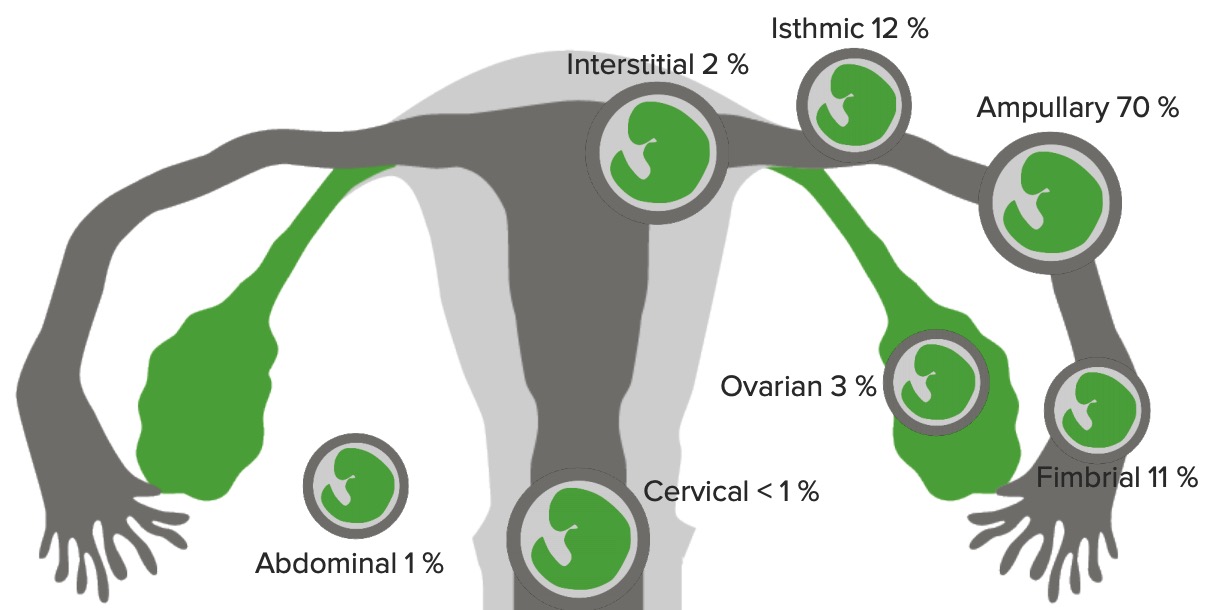
90%‒95% within the fallopian tubeFallopian TubeA pair of highly specialized canals extending from the uterus to its corresponding ovary. They provide the means for ovum transport from the ovaries and they are the site of the ovum’s final maturation and fertilization. The fallopian tube consists of an interstitium, an isthmus, an ampulla, an infundibulum, and fimbriae. Its wall consists of three layers: serous, muscular, and an internal mucosal layer lined with both ciliated and secretory cells.Uterus, Cervix, and Fallopian Tubes: Anatomy
3% in the ovary
1% in the peritoneal cavityPeritoneal CavityThe space enclosed by the peritoneum. It is divided into two portions, the greater sac and the lesser sac or omental bursa, which lies behind the stomach. The two sacs are connected by the foramen of winslow, or epiploic foramen.Peritoneum: Anatomy (abdominal)
< 1% in the cervixCervixThe uterus, cervix, and fallopian tubes are part of the internal female reproductive system. The most inferior portion of the uterus is the cervix, which connects the uterine cavity to the vagina. Externally, the cervix is lined by stratified squamous cells; however, the cervical canal is lined by columnar epithelium.Uterus, Cervix, and Fallopian Tubes: Anatomy
Ectopic pregnancyPregnancyThe status during which female mammals carry their developing young (embryos or fetuses) in utero before birth, beginning from fertilization to birth.Pregnancy: Diagnosis, Physiology, and Care can present before, during, or after rupture. Early on (prior to rupturing), symptoms can be relatively mild, such as light bleeding or cramping. If the pregnancyPregnancyThe status during which female mammals carry their developing young (embryos or fetuses) in utero before birth, beginning from fertilization to birth.Pregnancy: Diagnosis, Physiology, and Care does not spontaneously abort, it will eventually rupture, leading to a potentially massive internal hemorrhage.
Common presentation: vaginal bleeding and/or pelvic painPainAn unpleasant sensation induced by noxious stimuli which are detected by nerve endings of nociceptive neurons.Pain: Types and Pathways in the setting of a positive pregnancyPregnancyThe status during which female mammals carry their developing young (embryos or fetuses) in utero before birth, beginning from fertilization to birth.Pregnancy: Diagnosis, Physiology, and Care test (early in pregnancyPregnancyThe status during which female mammals carry their developing young (embryos or fetuses) in utero before birth, beginning from fertilization to birth.Pregnancy: Diagnosis, Physiology, and Care)
Bleeding is often light.
PainPainAn unpleasant sensation induced by noxious stimuli which are detected by nerve endings of nociceptive neurons.Pain: Types and Pathways:
Typically menstrual-like cramping
May be unilateral or diffuse
Signs of a ruptured ectopic pregnancyPregnancyThe status during which female mammals carry their developing young (embryos or fetuses) in utero before birth, beginning from fertilization to birth.Pregnancy: Diagnosis, Physiology, and Care:
Acute abdomenAcute AbdomenAcute abdomen, which is in many cases a surgical emergency, is the sudden onset of abdominal pain that may be caused by inflammation, infection, perforation, ischemia, or obstruction. The location of the pain, its characteristics, and associated symptoms (e.g., jaundice) are important tools that help narrow the differential diagnosis.Acute Abdomen:
Severe lower abdominal or pelvic painPainAn unpleasant sensation induced by noxious stimuli which are detected by nerve endings of nociceptive neurons.Pain: Types and Pathways
PainPainAn unpleasant sensation induced by noxious stimuli which are detected by nerve endings of nociceptive neurons.Pain: Types and Pathways may be more diffuse if there is blood in the abdominal cavity.
Hemodynamic instability: Rupture causes significant internal bleeding and may lead to hypovolemic shockHypovolemic ShockTypes of Shock.
General pregnancyPregnancyThe status during which female mammals carry their developing young (embryos or fetuses) in utero before birth, beginning from fertilization to birth.Pregnancy: Diagnosis, Physiology, and Care symptoms:
Closed cervixCervixThe uterus, cervix, and fallopian tubes are part of the internal female reproductive system. The most inferior portion of the uterus is the cervix, which connects the uterine cavity to the vagina. Externally, the cervix is lined by stratified squamous cells; however, the cervical canal is lined by columnar epithelium.Uterus, Cervix, and Fallopian Tubes: Anatomy
Adnexal tenderness
An adnexal massMassThree-dimensional lesion that occupies a space within the breastImaging of the Breast may be felt in 10%–20% of cases.
Very important: Perform a pregnancyPregnancyThe status during which female mammals carry their developing young (embryos or fetuses) in utero before birth, beginning from fertilization to birth.Pregnancy: Diagnosis, Physiology, and Care test on all women of reproductive age who present with abdominal painAbdominal PainAcute Abdomen!
Typically start with a rapid urine hCG test.
If the rapid urine hCG test is positive and the individual has symptoms (e.g., bleeding, cramping), order a quantitative serum hCG
Complete blood count (CBC): to look for anemiaAnemiaAnemia is a condition in which individuals have low Hb levels, which can arise from various causes. Anemia is accompanied by a reduced number of RBCs and may manifest with fatigue, shortness of breath, pallor, and weakness. Subtypes are classified by the size of RBCs, chronicity, and etiology. Anemia: Overview and Types/evidence of hemorrhage
Liver function testsLiver function testsLiver function tests, also known as hepatic function panels, are one of the most commonly performed screening blood tests. Such tests are also used to detect, evaluate, and monitor acute and chronic liver diseases.Liver Function Tests (LFTs), basic metabolic panelBasic Metabolic PanelPrimary vs Secondary Headaches (BMPBMPPrimary vs Secondary Headaches), urinalysisUrinalysisExamination of urine by chemical, physical, or microscopic means. Routine urinalysis usually includes performing chemical screening tests, determining specific gravity, observing any unusual color or odor, screening for bacteriuria, and examining the sediment microscopically.Urinary Tract Infections (UTIs) in Children (UA): to evaluate for other causes of acute abdomenAcute AbdomenAcute abdomen, which is in many cases a surgical emergency, is the sudden onset of abdominal pain that may be caused by inflammation, infection, perforation, ischemia, or obstruction. The location of the pain, its characteristics, and associated symptoms (e.g., jaundice) are important tools that help narrow the differential diagnosis.Acute Abdomen and look for contraindicationsContraindicationsA condition or factor associated with a recipient that makes the use of a drug, procedure, or physical agent improper or inadvisable. Contraindications may be absolute (life threatening) or relative (higher risk of complications in which benefits may outweigh risks).Noninvasive Ventilation to methotrexateMethotrexateAn antineoplastic antimetabolite with immunosuppressant properties. It is an inhibitor of tetrahydrofolate dehydrogenase and prevents the formation of tetrahydrofolate, necessary for synthesis of thymidylate, an essential component of DNA.Antimetabolite Chemotherapy (a treatment for EP)
After a positive urine pregnancyPregnancyThe status during which female mammals carry their developing young (embryos or fetuses) in utero before birth, beginning from fertilization to birth.Pregnancy: Diagnosis, Physiology, and Care test, the 1st step is to determine the gestational ageGestational ageThe age of the conceptus, beginning from the time of fertilization. In clinical obstetrics, the gestational age is often estimated as the time from the last day of the last menstruation which is about 2 weeks before ovulation and fertilization.Pregnancy: Diagnosis, Physiology, and Care based on the LMPLMPThe 1st day of a woman’s LMPPregnancy: Diagnosis, Physiology, and Care.
Frequently done using a digital obstetric calculator.
Knowing the approximate gestational ageGestational ageThe age of the conceptus, beginning from the time of fertilization. In clinical obstetrics, the gestational age is often estimated as the time from the last day of the last menstruation which is about 2 weeks before ovulation and fertilization.Pregnancy: Diagnosis, Physiology, and Care is important when interpreting an ultrasound
Diagnostic studies
Ultrasonography is the imaging method of choice and the gold standard for evaluating early pregnancies.
Ultrasonography:
Should be performed immediately in all individuals with a positive pregnancyPregnancyThe status during which female mammals carry their developing young (embryos or fetuses) in utero before birth, beginning from fertilization to birth.Pregnancy: Diagnosis, Physiology, and Care test and symptoms (e.g., bleeding, cramping) to determine the location and viability of the pregnancyPregnancyThe status during which female mammals carry their developing young (embryos or fetuses) in utero before birth, beginning from fertilization to birth.Pregnancy: Diagnosis, Physiology, and Care.
Normal pregnancyPregnancyThe status during which female mammals carry their developing young (embryos or fetuses) in utero before birth, beginning from fertilization to birth.Pregnancy: Diagnosis, Physiology, and Care:
At 5–6 weeks’ gestation, a gestational sac and yolk sacYolk SacThe first of four extra-embryonic membranes to form during embryogenesis. In reptiles and birds, it arises from endoderm and mesoderm to incorporate the egg yolk into the digestive tract for nourishing the embryo. In placental mammals, its nutritional function is vestigial; however, it is the source of intestinal mucosa; blood cells; and germ cells. It is sometimes called the vitelline sac, which should not be confused with the vitelline membrane of the egg.Embryoblast and Trophoblast Development are present within the uterusUterusThe uterus, cervix, and fallopian tubes are part of the internal female reproductive system. The uterus has a thick wall made of smooth muscle (the myometrium) and an inner mucosal layer (the endometrium). The most inferior portion of the uterus is the cervix, which connects the uterine cavity to the vagina.Uterus, Cervix, and Fallopian Tubes: Anatomy.
Presence of a fetal pole with a heartbeat: seen around 5.5–6 weeks’ gestation age
EP findings:
An empty uterine cavity without an amniotic sac or with a pseudo-gestational sac (a collection of fluid within the uterusUterusThe uterus, cervix, and fallopian tubes are part of the internal female reproductive system. The uterus has a thick wall made of smooth muscle (the myometrium) and an inner mucosal layer (the endometrium). The most inferior portion of the uterus is the cervix, which connects the uterine cavity to the vagina.Uterus, Cervix, and Fallopian Tubes: Anatomy that appears becaue of the hormonal milieu of an ectopic pregnancyPregnancyThe status during which female mammals carry their developing young (embryos or fetuses) in utero before birth, beginning from fertilization to birth.Pregnancy: Diagnosis, Physiology, and Care; will never contain a yolk sacYolk SacThe first of four extra-embryonic membranes to form during embryogenesis. In reptiles and birds, it arises from endoderm and mesoderm to incorporate the egg yolk into the digestive tract for nourishing the embryo. In placental mammals, its nutritional function is vestigial; however, it is the source of intestinal mucosa; blood cells; and germ cells. It is sometimes called the vitelline sac, which should not be confused with the vitelline membrane of the egg.Embryoblast and Trophoblast Development, which is an embryonic structure)
Adnexal massMassThree-dimensional lesion that occupies a space within the breastImaging of the Breast, especially when it contains a hypoechoicHypoechoicA structure that produces a low-amplitude echo (darker grays)Ultrasound (Sonography) area and is separate from the ovary (Note: Fallopian tubesFallopian tubesThe uterus, cervix, and fallopian tubes are part of the internal female reproductive system. The fallopian tubes receive an ovum after ovulation and help move it and/or a fertilized embryo toward the uterus via ciliated cells lining the tubes and peristaltic movements of its smooth muscle. Uterus, Cervix, and Fallopian Tubes: Anatomy are not normally visible on an ultrasound.)
Definitive diagnosis: visualization of a gestational sac with a yolk sacYolk SacThe first of four extra-embryonic membranes to form during embryogenesis. In reptiles and birds, it arises from endoderm and mesoderm to incorporate the egg yolk into the digestive tract for nourishing the embryo. In placental mammals, its nutritional function is vestigial; however, it is the source of intestinal mucosa; blood cells; and germ cells. It is sometimes called the vitelline sac, which should not be confused with the vitelline membrane of the egg.Embryoblast and Trophoblast Development, embryoEmbryoThe entity of a developing mammal, generally from the cleavage of a zygote to the end of embryonic differentiation of basic structures. For the human embryo, this represents the first two months of intrauterine development preceding the stages of the fetus.Fertilization and First Week, or both outside the uterusUterusThe uterus, cervix, and fallopian tubes are part of the internal female reproductive system. The uterus has a thick wall made of smooth muscle (the myometrium) and an inner mucosal layer (the endometrium). The most inferior portion of the uterus is the cervix, which connects the uterine cavity to the vagina.Uterus, Cervix, and Fallopian Tubes: Anatomy (most do not progress to this stage)
In the case of tubal rupture, free fluid (blood) is present in the pouch of DouglasPouch of DouglasA sac or recess formed by a fold of the peritoneum.Ovaries: Anatomy.
Possible to determine if the embryoEmbryoThe entity of a developing mammal, generally from the cleavage of a zygote to the end of embryonic differentiation of basic structures. For the human embryo, this represents the first two months of intrauterine development preceding the stages of the fetus.Fertilization and First Week is alive by the detection of a fetal heartbeat
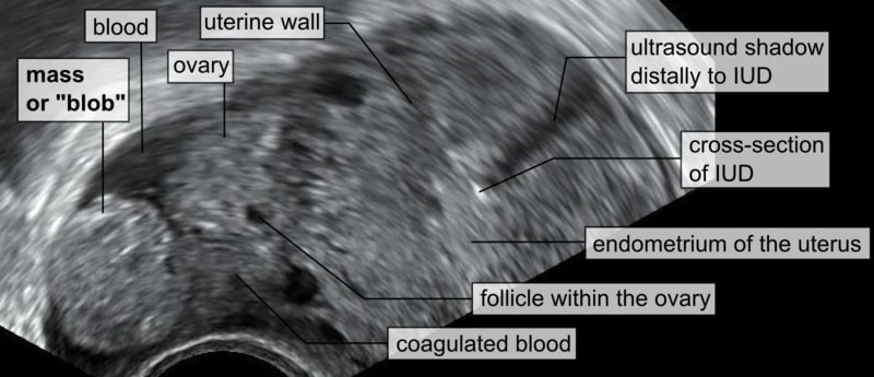
Transvaginal ultrasound showing an intrauterine device (IUD) with its respective distal shadow, blood in the abdominal cavity, no visible intrauterine gestational sac, and a spherical mass (or “blob”) close to the right ovary representing the EP
Image: “Blob sign” by Mikael Häggström. License: CC0
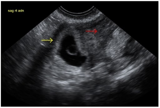
Transvaginal ultrasound showing an endometrial stripe of 2.4 cm (red arrow), no visible intrauterine pregnancy, and a right adnexal mass, suggestive of an EP (yellow arrow)
Image: “Transvaginal ultrasound” by Department of Obstetrics and Gynecology, George Washington University, Washington, DC, USA. License: CC BY 4.0
An EP must be closely monitored. The management of an EP can be expectant, medical, or surgical depending on the patient’s condition, hCG trend, and additional maternal and fetal factors.
Emergency management
In cases where the individual is hemodynamically unstable owing to a ruptured ectopic pregnancyPregnancyThe status during which female mammals carry their developing young (embryos or fetuses) in utero before birth, beginning from fertilization to birth.Pregnancy: Diagnosis, Physiology, and Care:
Give intravenous fluidsIntravenous FluidsIntravenous fluids are one of the most common interventions administered in medicine to approximate physiologic bodily fluids. Intravenous fluids are divided into 2 categories: crystalloid and colloid solutions. Intravenous fluids have a wide variety of indications, including intravascular volume expansion, electrolyte manipulation, and maintenance fluids. Intravenous Fluids to compensate for blood loss.
Reserved for hemodynamically stable patientsHemodynamically Stable PatientsBlunt Chest Trauma with an unruptured massMassThree-dimensional lesion that occupies a space within the breastImaging of the Breast and without any contraindicationsContraindicationsA condition or factor associated with a recipient that makes the use of a drug, procedure, or physical agent improper or inadvisable. Contraindications may be absolute (life threatening) or relative (higher risk of complications in which benefits may outweigh risks).Noninvasive Ventilation
MethotrexateMethotrexateAn antineoplastic antimetabolite with immunosuppressant properties. It is an inhibitor of tetrahydrofolate dehydrogenase and prevents the formation of tetrahydrofolate, necessary for synthesis of thymidylate, an essential component of DNA.Antimetabolite Chemotherapy (IM or, occasionally, injected directly into the EP) to halt trophoblastic growth and cause resolution of the pregnancyPregnancyThe status during which female mammals carry their developing young (embryos or fetuses) in utero before birth, beginning from fertilization to birth.Pregnancy: Diagnosis, Physiology, and Care
Must monitor the patient’s hCG levels to 0
Adverse effects of methotrexateMethotrexateAn antineoplastic antimetabolite with immunosuppressant properties. It is an inhibitor of tetrahydrofolate dehydrogenase and prevents the formation of tetrahydrofolate, necessary for synthesis of thymidylate, an essential component of DNA.Antimetabolite Chemotherapy:
Bone marrowBone marrowThe soft tissue filling the cavities of bones. Bone marrow exists in two types, yellow and red. Yellow marrow is found in the large cavities of large bones and consists mostly of fat cells and a few primitive blood cells. Red marrow is a hematopoietic tissue and is the site of production of erythrocytes and granular leukocytes. Bone marrow is made up of a framework of connective tissue containing branching fibers with the frame being filled with marrow cells.Bone Marrow: Composition and HematopoiesissuppressionSuppressionDefense Mechanisms
Pulmonary fibrosisFibrosisAny pathological condition where fibrous connective tissue invades any organ, usually as a consequence of inflammation or other injury.Bronchiolitis Obliterans
Renal failureRenal failureConditions in which the kidneys perform below the normal level in the ability to remove wastes, concentrate urine, and maintain electrolyte balance; blood pressure; and calcium metabolism. Renal insufficiency can be classified by the degree of kidney damage (as measured by the level of proteinuria) and reduction in glomerular filtration rate.Crush Syndrome
Gastric ulcers
Surgical management
Indications:
Hemodynamic instability
Symptoms of a ruptured ectopic pregnancyPregnancyThe status during which female mammals carry their developing young (embryos or fetuses) in utero before birth, beginning from fertilization to birth.Pregnancy: Diagnosis, Physiology, and Care
ContraindicationsContraindicationsA condition or factor associated with a recipient that makes the use of a drug, procedure, or physical agent improper or inadvisable. Contraindications may be absolute (life threatening) or relative (higher risk of complications in which benefits may outweigh risks).Noninvasive Ventilation to methotrexateMethotrexateAn antineoplastic antimetabolite with immunosuppressant properties. It is an inhibitor of tetrahydrofolate dehydrogenase and prevents the formation of tetrahydrofolate, necessary for synthesis of thymidylate, an essential component of DNA.Antimetabolite Chemotherapy
Failure of methotrexateMethotrexateAn antineoplastic antimetabolite with immunosuppressant properties. It is an inhibitor of tetrahydrofolate dehydrogenase and prevents the formation of tetrahydrofolate, necessary for synthesis of thymidylate, an essential component of DNA.Antimetabolite Chemotherapy
Patient preference for surgery over methotrexateMethotrexateAn antineoplastic antimetabolite with immunosuppressant properties. It is an inhibitor of tetrahydrofolate dehydrogenase and prevents the formation of tetrahydrofolate, necessary for synthesis of thymidylate, an essential component of DNA.Antimetabolite Chemotherapy
Indications for concurrent surgical procedure (e.g., individual desires tubal sterilizationTubal sterilizationProcedures that render the female sterile by interrupting the flow in the fallopian tube. These procedures generally are surgical, and may also use chemicals or physical means.Reproductive Ethical Issues)
Laparoscopic approach is preferred (over open).
Options include:
Salpingectomy:
Removal of the entire tube, including the EP
Definitive treatment
Salpingotomy:
Opening the tube and removing only the pregnancyPregnancyThe status during which female mammals carry their developing young (embryos or fetuses) in utero before birth, beginning from fertilization to birth.Pregnancy: Diagnosis, Physiology, and Care
Possible for trophoblastic tissue to persist → hCG should be followed to 0
Discuss pros/consCoNSStaphylococcus of salpingectomy vs salpingotomy with individual prior to surgery noting that it may be impossible to save the tube.
Expectant management
Only for patientsPatientsIndividuals participating in the health care system for the purpose of receiving therapeutic, diagnostic, or preventive procedures.Clinician–Patient Relationship who meet the following criteria:
Asymptomatic
No evidence of extrauterine sac/massMassThree-dimensional lesion that occupies a space within the breastImaging of the Breast on ultrasound
Low and decreasing serum hCG (evidence of spontaneous resolution)
Agreeable to close follow-up
Steps include:
Monitoring the pattern of serial hCG levels → follow levels all the way to 0
Giving strict return-for-followup instructions to ensure that hCG is monitored
Abandoning expectant management if significant abdominal painAbdominal PainAcute Abdomen develops or hCG increases or fails to decline
Since EPs typically present with bleeding and/or cramping in early pregnancyPregnancyThe status during which female mammals carry their developing young (embryos or fetuses) in utero before birth, beginning from fertilization to birth.Pregnancy: Diagnosis, Physiology, and Care, it is critical to differentiate an EP from other types of abnormal pregnancyPregnancyThe status during which female mammals carry their developing young (embryos or fetuses) in utero before birth, beginning from fertilization to birth.Pregnancy: Diagnosis, Physiology, and Care events, most notably threatened, missed, inevitable, incomplete, and complete spontaneous miscarriages. In addition, bleeding may be due to a molar pregnancyPregnancyThe status during which female mammals carry their developing young (embryos or fetuses) in utero before birth, beginning from fertilization to birth.Pregnancy: Diagnosis, Physiology, and Care or non-obstetric causes. PainPainAn unpleasant sensation induced by noxious stimuli which are detected by nerve endings of nociceptive neurons.Pain: Types and Pathways can also be due to non-obstetric causes.
Obstetric and gynecologic causes of vaginal bleeding in pregnancyPregnancyThe status during which female mammals carry their developing young (embryos or fetuses) in utero before birth, beginning from fertilization to birth.Pregnancy: Diagnosis, Physiology, and Care
The most notable causes of vaginal bleeding in early pregnancyPregnancyThe status during which female mammals carry their developing young (embryos or fetuses) in utero before birth, beginning from fertilization to birth.Pregnancy: Diagnosis, Physiology, and Care include:
Ectopic pregnancyPregnancyThe status during which female mammals carry their developing young (embryos or fetuses) in utero before birth, beginning from fertilization to birth.Pregnancy: Diagnosis, Physiology, and Care
Failed/failing pregnancies:
Threatened miscarriageMiscarriageSpontaneous abortion, also known as miscarriage, is the loss of a pregnancy before 20 weeks’ gestation. However, the layperson use of the term “abortion” is often intended to refer to induced termination of a pregnancy, whereas “miscarriage” is preferred for spontaneous loss.Spontaneous Abortion
Missed miscarriageMiscarriageSpontaneous abortion, also known as miscarriage, is the loss of a pregnancy before 20 weeks’ gestation. However, the layperson use of the term “abortion” is often intended to refer to induced termination of a pregnancy, whereas “miscarriage” is preferred for spontaneous loss.Spontaneous Abortion
Inevitable miscarriageMiscarriageSpontaneous abortion, also known as miscarriage, is the loss of a pregnancy before 20 weeks’ gestation. However, the layperson use of the term “abortion” is often intended to refer to induced termination of a pregnancy, whereas “miscarriage” is preferred for spontaneous loss.Spontaneous Abortion
Incomplete miscarriageMiscarriageSpontaneous abortion, also known as miscarriage, is the loss of a pregnancy before 20 weeks’ gestation. However, the layperson use of the term “abortion” is often intended to refer to induced termination of a pregnancy, whereas “miscarriage” is preferred for spontaneous loss.Spontaneous Abortion
Complete spontaneous miscarriageMiscarriageSpontaneous abortion, also known as miscarriage, is the loss of a pregnancy before 20 weeks’ gestation. However, the layperson use of the term “abortion” is often intended to refer to induced termination of a pregnancy, whereas “miscarriage” is preferred for spontaneous loss.Spontaneous Abortion
Molar pregnancyPregnancyThe status during which female mammals carry their developing young (embryos or fetuses) in utero before birth, beginning from fertilization to birth.Pregnancy: Diagnosis, Physiology, and Care
Non-obstetric causes of bleeding (e.g., cervical polyps)
Table: Diagnosing vaginal bleeding in early pregnancyPregnancyThe status during which female mammals carry their developing young (embryos or fetuses) in utero before birth, beginning from fertilization to birth.Pregnancy: Diagnosis, Physiology, and Care
Diagnosis
Typical bleeding pattern
CervixCervixThe uterus, cervix, and fallopian tubes are part of the internal female reproductive system. The most inferior portion of the uterus is the cervix, which connects the uterine cavity to the vagina. Externally, the cervix is lined by stratified squamous cells; however, the cervical canal is lined by columnar epithelium.Uterus, Cervix, and Fallopian Tubes: Anatomy
Cramping, pelvic painPainAn unpleasant sensation induced by noxious stimuli which are detected by nerve endings of nociceptive neurons.Pain: Types and Pathways
Products of conception (POC)
Threatened miscarriageMiscarriageSpontaneous abortion, also known as miscarriage, is the loss of a pregnancy before 20 weeks’ gestation. However, the layperson use of the term “abortion” is often intended to refer to induced termination of a pregnancy, whereas “miscarriage” is preferred for spontaneous loss.Spontaneous Abortion
Light
Closed
Yes
POC visible in uterusUterusThe uterus, cervix, and fallopian tubes are part of the internal female reproductive system. The uterus has a thick wall made of smooth muscle (the myometrium) and an inner mucosal layer (the endometrium). The most inferior portion of the uterus is the cervix, which connects the uterine cavity to the vagina.Uterus, Cervix, and Fallopian Tubes: Anatomy on ultrasound (depending on dates)
Missed miscarriageMiscarriageSpontaneous abortion, also known as miscarriage, is the loss of a pregnancy before 20 weeks’ gestation. However, the layperson use of the term “abortion” is often intended to refer to induced termination of a pregnancy, whereas “miscarriage” is preferred for spontaneous loss.Spontaneous Abortion
None or light
Closed
Possible
POC visible on ultrasound without a fetal heartbeat
Inevitable miscarriageMiscarriageSpontaneous abortion, also known as miscarriage, is the loss of a pregnancy before 20 weeks’ gestation. However, the layperson use of the term “abortion” is often intended to refer to induced termination of a pregnancy, whereas “miscarriage” is preferred for spontaneous loss.Spontaneous Abortion
Heavy
Dilated
Yes
POC visible in uterusUterusThe uterus, cervix, and fallopian tubes are part of the internal female reproductive system. The uterus has a thick wall made of smooth muscle (the myometrium) and an inner mucosal layer (the endometrium). The most inferior portion of the uterus is the cervix, which connects the uterine cavity to the vagina.Uterus, Cervix, and Fallopian Tubes: Anatomy or cervixCervixThe uterus, cervix, and fallopian tubes are part of the internal female reproductive system. The most inferior portion of the uterus is the cervix, which connects the uterine cavity to the vagina. Externally, the cervix is lined by stratified squamous cells; however, the cervical canal is lined by columnar epithelium.Uterus, Cervix, and Fallopian Tubes: Anatomy on ultrasound (depending on dates)
Incomplete miscarriageMiscarriageSpontaneous abortion, also known as miscarriage, is the loss of a pregnancy before 20 weeks’ gestation. However, the layperson use of the term “abortion” is often intended to refer to induced termination of a pregnancy, whereas “miscarriage” is preferred for spontaneous loss.Spontaneous Abortion
Heavy
Dilated
Yes
Partial expulsion of POC; POC may be visible at os
Complete miscarriageMiscarriageSpontaneous abortion, also known as miscarriage, is the loss of a pregnancy before 20 weeks’ gestation. However, the layperson use of the term “abortion” is often intended to refer to induced termination of a pregnancy, whereas “miscarriage” is preferred for spontaneous loss.Spontaneous Abortion
Light
Closed
Yes
History of POC expulsion
No POC visible in the uterusUterusThe uterus, cervix, and fallopian tubes are part of the internal female reproductive system. The uterus has a thick wall made of smooth muscle (the myometrium) and an inner mucosal layer (the endometrium). The most inferior portion of the uterus is the cervix, which connects the uterine cavity to the vagina.Uterus, Cervix, and Fallopian Tubes: Anatomy on ultrasound
Ectopic pregnancyPregnancyThe status during which female mammals carry their developing young (embryos or fetuses) in utero before birth, beginning from fertilization to birth.Pregnancy: Diagnosis, Physiology, and Care
Light
Closed
Yes
No intrauterine POC (though a pseudogestational sac may be present)
Tender adnexal massMassThree-dimensional lesion that occupies a space within the breastImaging of the Breast, which may show a yolk sacYolk SacThe first of four extra-embryonic membranes to form during embryogenesis. In reptiles and birds, it arises from endoderm and mesoderm to incorporate the egg yolk into the digestive tract for nourishing the embryo. In placental mammals, its nutritional function is vestigial; however, it is the source of intestinal mucosa; blood cells; and germ cells. It is sometimes called the vitelline sac, which should not be confused with the vitelline membrane of the egg.Embryoblast and Trophoblast Development and possibly a fetal pole with or without cardiac activity
Bleeding molar pregnancyPregnancyThe status during which female mammals carry their developing young (embryos or fetuses) in utero before birth, beginning from fertilization to birth.Pregnancy: Diagnosis, Physiology, and Care
Heavy
Dilated
Yes
Partial expulsion of POC which resembles grapes
No evidence of a fetus
Non-obstetric causes of vaginal bleeding
VariableVariableVariables represent information about something that can change. The design of the measurement scales, or of the methods for obtaining information, will determine the data gathered and the characteristics of that data. As a result, a variable can be qualitative or quantitative, and may be further classified into subgroups.Types of Variables
Closed
Often no
Ultrasound findings show normal embryologic/fetal findings appropriate for gestational ageGestational ageThe age of the conceptus, beginning from the time of fertilization. In clinical obstetrics, the gestational age is often estimated as the time from the last day of the last menstruation which is about 2 weeks before ovulation and fertilization.Pregnancy: Diagnosis, Physiology, and Care
Other cervical, vaginal, or uterine pathology may be identified (e.g., cervical polyps)
Note that threatened miscarriages will ultimately resolve or progress to another diagnosis:
May progress to complete spontaneous miscarriageMiscarriageSpontaneous abortion, also known as miscarriage, is the loss of a pregnancy before 20 weeks’ gestation. However, the layperson use of the term “abortion” is often intended to refer to induced termination of a pregnancy, whereas “miscarriage” is preferred for spontaneous loss.Spontaneous Abortion
A subchorionic hematomaHematomaA collection of blood outside the blood vessels. Hematoma can be localized in an organ, space, or tissue.Intussusception may subsequently be identified on an ultrasound (pregnancyPregnancyThe status during which female mammals carry their developing young (embryos or fetuses) in utero before birth, beginning from fertilization to birth.Pregnancy: Diagnosis, Physiology, and Care may still continue normally).
Bleeding that resolves spontaneously with no other abnormal findings is typically attributed to bleeding from a physiologic cause, such as implantationImplantationEndometrial implantation of embryo, mammalian at the blastocyst stage.Fertilization and First Week bleeding.
Management of failed or failing intrauterine pregnancies (missed, inevitable, incomplete, or complete spontaneous miscarriages):
Expectant (i.e., await spontaneous passage of POC)
Medical management (e.g., misoprostolMisoprostolA synthetic analog of natural prostaglandin e1. It produces a dose-related inhibition of gastric acid and pepsin secretion, and enhances mucosal resistance to injury. It is an effective anti-ulcer agent and also has oxytocic properties.Eicosanoids to induce passage of POC)
Surgical management, with uterine evacuation (e.g., suction dilatation and curettageCurettageA scraping, usually of the interior of a cavity or tract, for removal of new growth or other abnormal tissue, or to obtain material for tissue diagnosis. It is performed with a curet (curette), a spoon-shaped instrument designed for that purpose.Benign Bone Tumors)
Non-obstetric causes of pelvic painPainAn unpleasant sensation induced by noxious stimuli which are detected by nerve endings of nociceptive neurons.Pain: Types and Pathways/cramping in pregnancyPregnancyThe status during which female mammals carry their developing young (embryos or fetuses) in utero before birth, beginning from fertilization to birth.Pregnancy: Diagnosis, Physiology, and Care
AppendicitisAppendicitisAppendicitis is the acute inflammation of the vermiform appendix and the most common abdominal surgical emergency globally. The condition has a lifetime risk of 8%. Characteristic features include periumbilical abdominal pain that migrates to the right lower quadrant, fever, anorexia, nausea, and vomiting.Appendicitis: inflammationInflammationInflammation is a complex set of responses to infection and injury involving leukocytes as the principal cellular mediators in the body’s defense against pathogenic organisms. Inflammation is also seen as a response to tissue injury in the process of wound healing. The 5 cardinal signs of inflammation are pain, heat, redness, swelling, and loss of function. Inflammation of the appendixAppendixA worm-like blind tube extension from the cecum.Colon, Cecum, and Appendix: Anatomy caused by obstruction (e.g., by fecaliths or infection). Produces symptoms such as abdominal painAbdominal PainAcute Abdomen, vomitingVomitingThe forcible expulsion of the contents of the stomach through the mouth.Hypokalemia, and malaiseMalaiseTick-borne Encephalitis Virus. Diagnostic findings may include an elevated WBC count and a thickened appendiceal wall on CT scan. Surgery is the recommended treatment, although some patientsPatientsIndividuals participating in the health care system for the purpose of receiving therapeutic, diagnostic, or preventive procedures.Clinician–Patient Relationship respond to antibiotics. Unlike in EP, US may reveal an enlarged appendiceal diameter and hCG levels will rise appropriately.
Kidney stonesKidney stonesNephrolithiasis is the formation of a stone, or calculus, anywhere along the urinary tract caused by precipitations of solutes in the urine. The most common type of kidney stone is the calcium oxalate stone, but other types include calcium phosphate, struvite (ammonium magnesium phosphate), uric acid, and cystine stones.Nephrolithiasis: urine can become supersaturated with soluble substances (e.g., calciumCalciumA basic element found in nearly all tissues. It is a member of the alkaline earth family of metals with the atomic symbol ca, atomic number 20, and atomic weight 40. Calcium is the most abundant mineral in the body and combines with phosphorus to form calcium phosphate in the bones and teeth. It is essential for the normal functioning of nerves and muscles and plays a role in blood coagulation (as factor IV) and in many enzymatic processes.Electrolytes oxalate), which crystallize and form stones that deposit throughout the urinary tractUrinary tractThe urinary tract is located in the abdomen and pelvis and consists of the kidneys, ureters, urinary bladder, and urethra. The structures permit the excretion of urine from the body. Urine flows from the kidneys through the ureters to the urinary bladder and out through the urethra.Urinary Tract: Anatomy. PatientsPatientsIndividuals participating in the health care system for the purpose of receiving therapeutic, diagnostic, or preventive procedures.Clinician–Patient Relationship may present with colicky flank or abdominal painAbdominal PainAcute Abdomen. Diagnostic studies reveal hematuriaHematuriaPresence of blood in the urine.Renal Cell Carcinoma, urinary stones on CT scan, and/or hydronephrosisHydronephrosisHydronephrosis is dilation of the renal collecting system as a result of the obstruction of urine outflow. Hydronephrosis can be unilateral or bilateral. Nephrolithiasis is the most common cause of hydronephrosis in young adults, while prostatic hyperplasia and neoplasm are seen in older patients. Hydronephrosis on US. Management consists of painPainAn unpleasant sensation induced by noxious stimuli which are detected by nerve endings of nociceptive neurons.Pain: Types and Pathways control and varies depending on the chemical nature of the stones. hCG levels will rise appropriately and an early pregnancyPregnancyThe status during which female mammals carry their developing young (embryos or fetuses) in utero before birth, beginning from fertilization to birth.Pregnancy: Diagnosis, Physiology, and Care should otherwise progress normally on ultrasound.
Sherwood, L. Human Physiology: From Cells to Systems. (9th ed., pp. 753, 756). Cengage Learning.
Marion, L. L., & Meeks, G. R. (2012). Ectopic pregnancy: History, incidence, epidemiology, and risk factors. Clinical Obstetrics and Gynecology, 55(2), 376–386. Retrieved June 28, 2026, from https://doi.org/10.1097/GRF.0b013e3182516d7b
Scibetta, E. W., & Han, C. S. (2019). Ultrasound in Early Pregnancy: Viability, Unknown Locations, and Ectopic Pregnancies. Obstetrics and gynecology clinics of North America, 46(4), 783–795. Retrieved June 28, 2026, from https://doi.org/10.1016/j.ogc.2019.07.013