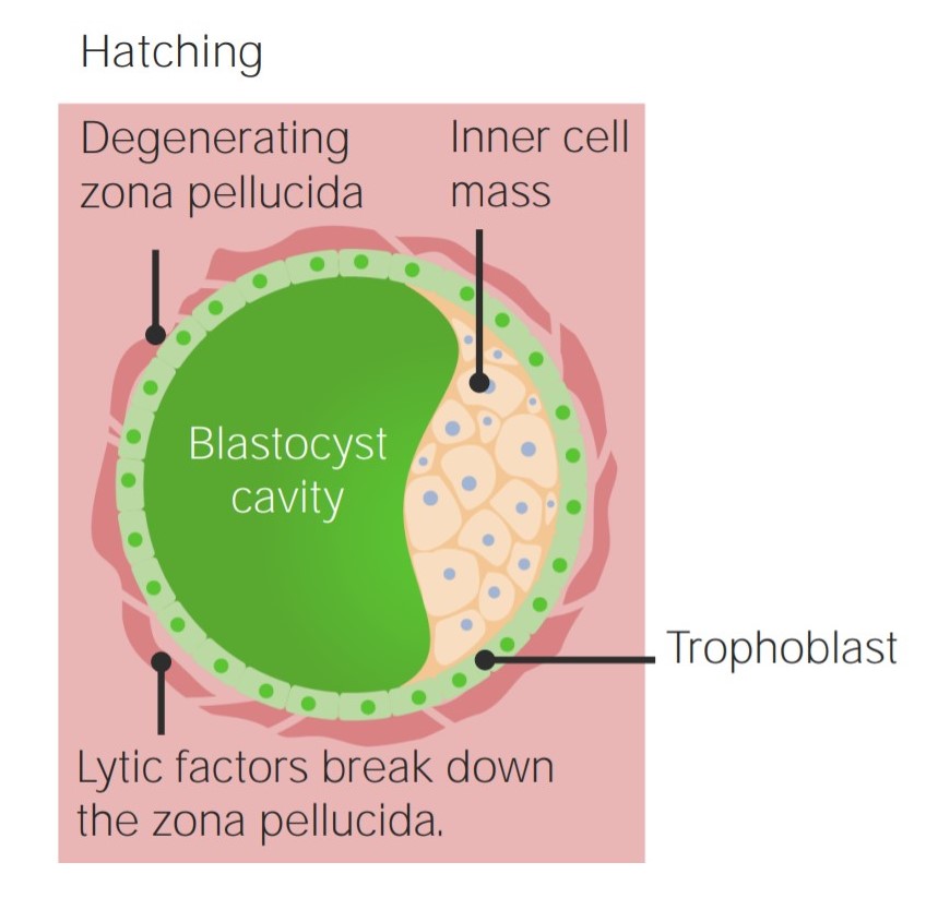
Before the developing blastocyst Blastocyst A post-morula preimplantation mammalian embryo that develops from a 32-cell stage into a fluid-filled hollow ball of over a hundred cells. A blastocyst has two distinctive tissues. The outer layer of trophoblasts gives rise to extra-embryonic tissues. The inner cell mass gives rise to the embryonic disc and eventual embryo proper. Fertilization and First Week reaches the uterine wall, it needs to undergo several stages of differentiation. After a continuous process of cleavage and compaction, the morula Morula An early embryo that is a compact mass of about 16 blastomeres. It resembles a cluster of mulberries with two types of cells, outer cells and inner cells. Morula is the stage before blastula in non-mammalian animals or a blastocyst in mammals. Fertilization and First Week gives rise to the trophoblast Trophoblast Cells lining the outside of the blastocyst. After binding to the endometrium, trophoblasts develop into two distinct layers, an inner layer of mononuclear cytotrophoblasts and an outer layer of continuous multinuclear cytoplasm, the syncytiotrophoblasts, which form the early fetal-maternal interface (placenta). Fertilization and First Week and embryoblast, which are the primary components of the blastocyst Blastocyst A post-morula preimplantation mammalian embryo that develops from a 32-cell stage into a fluid-filled hollow ball of over a hundred cells. A blastocyst has two distinctive tissues. The outer layer of trophoblasts gives rise to extra-embryonic tissues. The inner cell mass gives rise to the embryonic disc and eventual embryo proper. Fertilization and First Week. Uterine fluid passes through the zona pellucida Zona pellucida A tough transparent membrane surrounding the ovum. It is penetrated by the sperm during fertilization. Fertilization and First Week to form the blastocyst Blastocyst A post-morula preimplantation mammalian embryo that develops from a 32-cell stage into a fluid-filled hollow ball of over a hundred cells. A blastocyst has two distinctive tissues. The outer layer of trophoblasts gives rise to extra-embryonic tissues. The inner cell mass gives rise to the embryonic disc and eventual embryo proper. Fertilization and First Week cavity. When the blastocyst Blastocyst A post-morula preimplantation mammalian embryo that develops from a 32-cell stage into a fluid-filled hollow ball of over a hundred cells. A blastocyst has two distinctive tissues. The outer layer of trophoblasts gives rise to extra-embryonic tissues. The inner cell mass gives rise to the embryonic disc and eventual embryo proper. Fertilization and First Week reaches the endometrium, implantation Implantation Endometrial implantation of embryo, mammalian at the blastocyst stage. Fertilization and First Week begins by the trophoblast Trophoblast Cells lining the outside of the blastocyst. After binding to the endometrium, trophoblasts develop into two distinct layers, an inner layer of mononuclear cytotrophoblasts and an outer layer of continuous multinuclear cytoplasm, the syncytiotrophoblasts, which form the early fetal-maternal interface (placenta). Fertilization and First Week dividing into the cytotrophoblast and syncytiotrophoblast, with the syncytiotrophoblast primarily being responsible for invading the endometrium. The embryoblast divides into the epiblast and hypoblast, which are responsible for creating the amniotic cavity and yolk sac, respectively.
Last updated: Jul 10, 2026
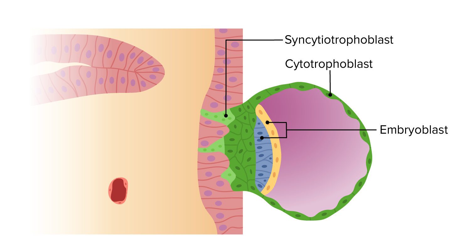
Embryoblast and trophoblast surrounded by the degenerating zona pellucida
Image by Lecturio.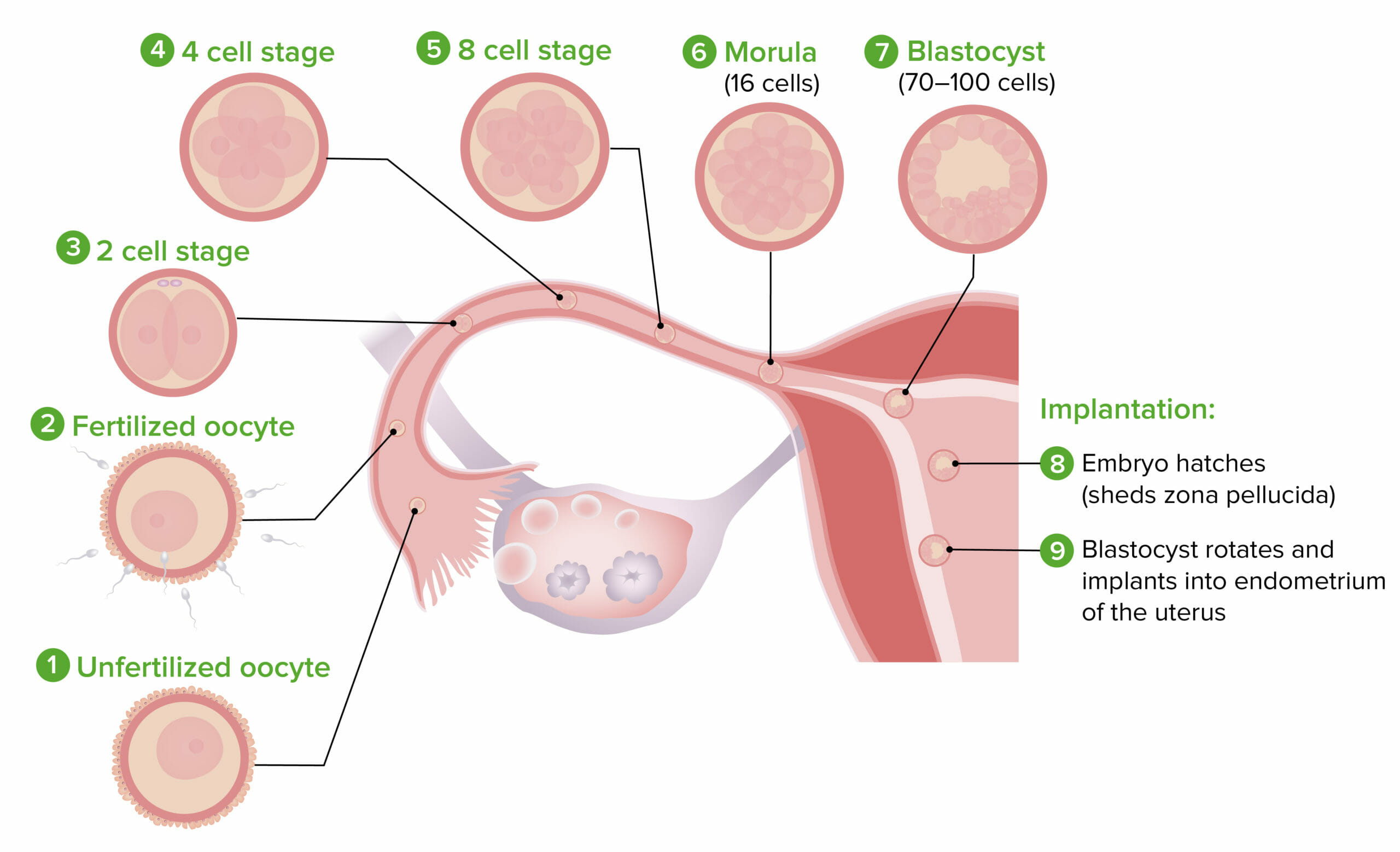
Anatomical Location of Fertilization and Early Divisions:
The blastocyst is formed around the 5th day of gestation. It is composed of a trophoblast and embryoblast.
The trophoblast Trophoblast Cells lining the outside of the blastocyst. After binding to the endometrium, trophoblasts develop into two distinct layers, an inner layer of mononuclear cytotrophoblasts and an outer layer of continuous multinuclear cytoplasm, the syncytiotrophoblasts, which form the early fetal-maternal interface (placenta). Fertilization and First Week is primarily responsible for implantation Implantation Endometrial implantation of embryo, mammalian at the blastocyst stage. Fertilization and First Week of the blastocyst Blastocyst A post-morula preimplantation mammalian embryo that develops from a 32-cell stage into a fluid-filled hollow ball of over a hundred cells. A blastocyst has two distinctive tissues. The outer layer of trophoblasts gives rise to extra-embryonic tissues. The inner cell mass gives rise to the embryonic disc and eventual embryo proper. Fertilization and First Week.
Blastocyst penetration guided by syncytiotrophoblasts
Image by Lecturio.The embryoblast gives rise to the bilaminar disk and amniotic cavity. The embryoblast differentiates into the epiblast and hypoblast. Together, they are referred to as the bilaminar disc. The process begins around the 8th day of gestation.
Hypoblast:
Epiblast:
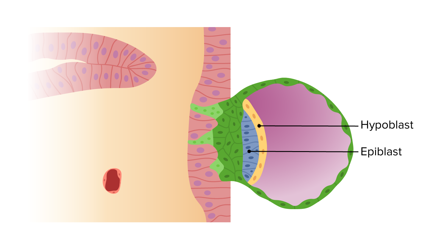
The hypoblast and epiblast form the bilaminar disc:
This begins around the 8th day of gestation. Throughout the rest of the 2nd week, a cavity will develop within both the hypoblast and the epiblast to form the primary yolk sac and amniotic cavity, respectively.
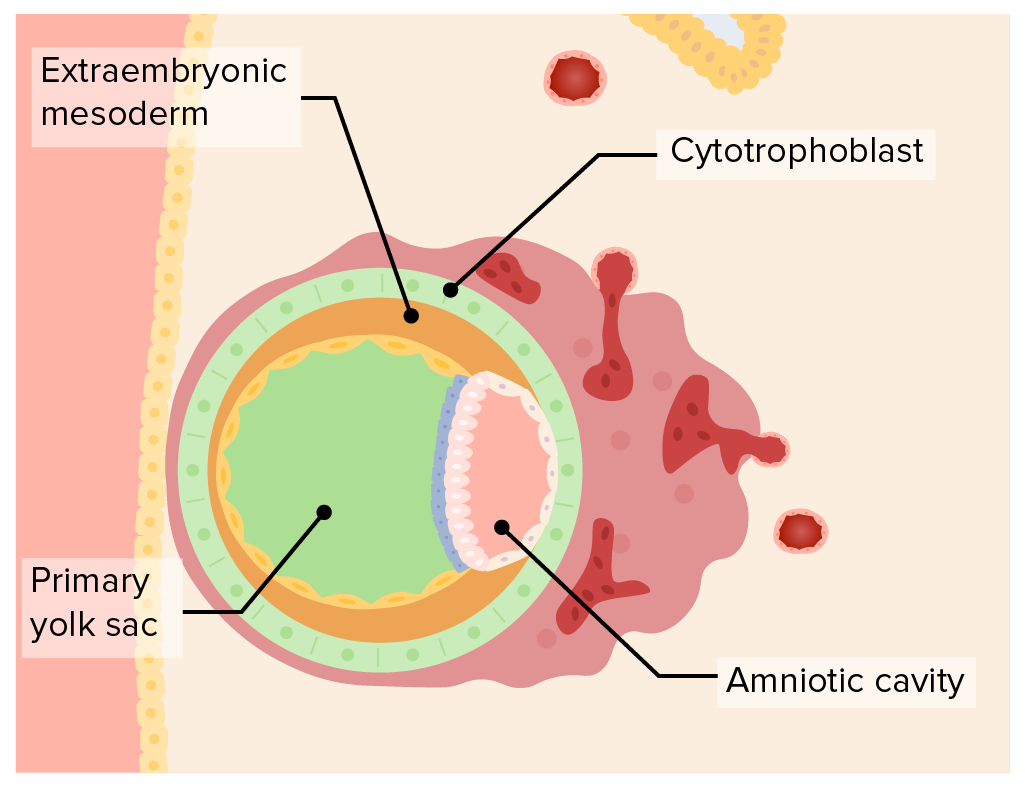
The amniotic cavity:
The amniotic cavity is contained within the epiblast while the primary yolk sac is contained within the hypoblast.
The 2nd week of development is known as the week of 2s:
Spontaneous abortion Abortion Expulsion of the product of fertilization before completing the term of gestation and without deliberate interference. Spontaneous Abortion: caused by inadequate production of estrogens and progesterone Progesterone The major progestational steroid that is secreted primarily by the corpus luteum and the placenta. Progesterone acts on the uterus, the mammary glands and the brain. It is required in embryo implantation; pregnancy maintenance, and the development of mammary tissue for milk production. Progesterone, converted from pregnenolone, also serves as an intermediate in the biosynthesis of gonadal steroid hormones and adrenal corticosteroids. Gonadal Hormones by the corpus luteum Corpus Luteum The yellow body derived from the ruptured ovarian follicle after ovulation. The process of corpus luteum formation, luteinization, is regulated by luteinizing hormone. Ovaries: Anatomy. Other causes include chromosomal abnormalities and physical trauma. The major symptom of early spontaneous abortion Abortion Expulsion of the product of fertilization before completing the term of gestation and without deliberate interference. Spontaneous Abortion is vaginal bleeding. Treatment of early spontaneous abortion Abortion Expulsion of the product of fertilization before completing the term of gestation and without deliberate interference. Spontaneous Abortion is with supportive care.