The uterus, cervix, and fallopian tubes are part of the internal female reproductive system. The fallopian tubes receive an ovum after ovulationOvulationThe discharge of an ovum from a rupturing follicle in the ovary.Menstrual Cycle and help move it and/or a fertilized embryoEmbryoThe entity of a developing mammal, generally from the cleavage of a zygote to the end of embryonic differentiation of basic structures. For the human embryo, this represents the first two months of intrauterine development preceding the stages of the fetus.Fertilization and First Week toward the uterus via ciliated cells lining the tubes and peristaltic movements of its smooth muscle. The uterus has a thick wall made of smooth muscle (the myometrium) and an inner mucosal layer (the endometriumEndometriumThe mucous membrane lining of the uterine cavity that is hormonally responsive during the menstrual cycle and pregnancy. The endometrium undergoes cyclic changes that characterize menstruation. After successful fertilization, it serves to sustain the developing embryo.Embryoblast and Trophoblast Development). The most inferior portion of the uterus is the cervix, which connects the uterine cavity to the vaginaVaginaThe vagina is the female genital canal, extending from the vulva externally to the cervix uteri internally. The structures have sexual, reproductive, and urinary functions and a rich blood supply, mainly arising from the internal iliac artery.Vagina, Vulva, and Pelvic Floor: Anatomy. Externally, the cervix is lined by stratified squamous cells; however, the cervical canal is lined by columnar epitheliumEpitheliumThe epithelium is a complex of specialized cellular organizations arranged into sheets and lining cavities and covering the surfaces of the body. The cells exhibit polarity, having an apical and a basal pole. Structures important for the epithelial integrity and function involve the basement membrane, the semipermeable sheet on which the cells rest, and interdigitations, as well as cellular junctions. Surface Epithelium: Histology. The transition point is known as the squamocolumnar junctionSquamocolumnar junctionEsophagus: Anatomy, which is the site of most cervical cancers. These organs are supplied by the uterine and ovarian arteriesArteriesArteries are tubular collections of cells that transport oxygenated blood and nutrients from the heart to the tissues of the body. The blood passes through the arteries in order of decreasing luminal diameter, starting in the largest artery (the aorta) and ending in the small arterioles. Arteries are classified into 3 types: large elastic arteries, medium muscular arteries, and small arteries and arterioles. Arteries: Histology and innervated by the autonomic nervous systemAutonomic nervous systemThe ANS is a component of the peripheral nervous system that uses both afferent (sensory) and efferent (effector) neurons, which control the functioning of the internal organs and involuntary processes via connections with the CNS. The ANS consists of the sympathetic and parasympathetic nervous systems. Autonomic Nervous System: Anatomy.
The uterus, cervix, and fallopian tubes are all important organs in the female reproductive tract.
Gross anatomy of the female reproductive system
Image by Lecturio.
Location
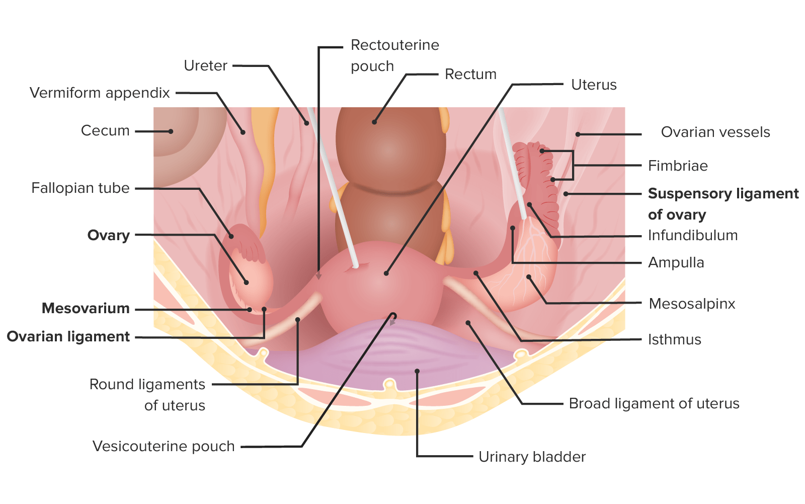
The uterus and fallopian tubes are pelvic organs.
2 fallopian tubes (left and right):
Arise from the superolateral portions of the uterus
Connect the ovariesOvariesOvaries are the paired gonads of the female reproductive system that contain haploid gametes known as oocytes. The ovaries are located intraperitoneally in the pelvis, just posterior to the broad ligament, and are connected to the pelvic sidewall and to the uterus by ligaments. These organs function to secrete hormones (estrogen and progesterone) and to produce the female germ cells (oocytes).Ovaries: Anatomy (female gonadsGonadsThe gamete-producing glands, ovary or testis.Hormones: Overview and Types) to the uterus
Uterus: in the midline, between the bladderBladderA musculomembranous sac along the urinary tract. Urine flows from the kidneys into the bladder via the ureters, and is held there until urination.Pyelonephritis and Perinephric Abscess and the rectumRectumThe rectum and anal canal are the most terminal parts of the lower GI tract/large intestine that form a functional unit and control defecation. Fecal continence is maintained by several important anatomic structures including rectal folds, anal valves, the sling-like puborectalis muscle, and internal and external anal sphincters. Rectum and Anal Canal: Anatomy
Cervix: inferior portion of the uterus, connecting the uterine cavity to the vaginaVaginaThe vagina is the female genital canal, extending from the vulva externally to the cervix uteri internally. The structures have sexual, reproductive, and urinary functions and a rich blood supply, mainly arising from the internal iliac artery.Vagina, Vulva, and Pelvic Floor: Anatomy
Relationship to peritoneal cavityPeritoneal CavityThe space enclosed by the peritoneum. It is divided into two portions, the greater sac and the lesser sac or omental bursa, which lies behind the stomach. The two sacs are connected by the foramen of winslow, or epiploic foramen.Peritoneum: Anatomy:
IntraperitonealIntraperitonealPeritoneum: Anatomy (i.e., covered by peritoneumPeritoneumThe peritoneum is a serous membrane lining the abdominopelvic cavity. This lining is formed by connective tissue and originates from the mesoderm. The membrane lines both the abdominal walls (as parietal peritoneum) and all of the visceral organs (as visceral peritoneum).Peritoneum: Anatomy):
Subperitoneal (i.e., entirely below the peritoneal cavityPeritoneal CavityThe space enclosed by the peritoneum. It is divided into two portions, the greater sac and the lesser sac or omental bursa, which lies behind the stomach. The two sacs are connected by the foramen of winslow, or epiploic foramen.Peritoneum: Anatomy): cervix
Location of the uterus and fallopian tubes in situ
Image by Lecturio.
Function
Uterus:
Site of implantationImplantationEndometrial implantation of embryo, mammalian at the blastocyst stage.Fertilization and First Week for a fertilized embryoEmbryoThe entity of a developing mammal, generally from the cleavage of a zygote to the end of embryonic differentiation of basic structures. For the human embryo, this represents the first two months of intrauterine development preceding the stages of the fetus.Fertilization and First Week
Growth and nourishment of the fetus
Able to shed its lining when pregnancyPregnancyThe status during which female mammals carry their developing young (embryos or fetuses) in utero before birth, beginning from fertilization to birth.Pregnancy: Diagnosis, Physiology, and Care does not occur (i.e., menstruationMenstruationThe periodic shedding of the endometrium and associated menstrual bleeding in the menstrual cycle of humans and primates. Menstruation is due to the decline in circulating progesterone, and occurs at the late luteal phase when luteolysis of the corpus luteum takes place.Menstrual Cycle)
Cervix:
Opening of the uterus into the vaginaVaginaThe vagina is the female genital canal, extending from the vulva externally to the cervix uteri internally. The structures have sexual, reproductive, and urinary functions and a rich blood supply, mainly arising from the internal iliac artery.Vagina, Vulva, and Pelvic Floor: Anatomy, which leads out of the body:
Allows passage of menstrual blood into the vaginaVaginaThe vagina is the female genital canal, extending from the vulva externally to the cervix uteri internally. The structures have sexual, reproductive, and urinary functions and a rich blood supply, mainly arising from the internal iliac artery.Vagina, Vulva, and Pelvic Floor: Anatomy
Allows sperm into the uterus
Secretes fluid that can promote or inhibit sperm entry into the uterus, depending on the stage of the menstrual cycleMenstrual cycleThe menstrual cycle is the cyclic pattern of hormonal and tissular activity that prepares a suitable uterine environment for the fertilization and implantation of an ovum. The menstrual cycle involves both an endometrial and ovarian cycle that are dependent on one another for proper functioning. There are 2 phases of the ovarian cycle and 3 phases of the endometrial cycle.Menstrual Cycle
In pregnancyPregnancyThe status during which female mammals carry their developing young (embryos or fetuses) in utero before birth, beginning from fertilization to birth.Pregnancy: Diagnosis, Physiology, and Care:
Keeps the uterus closed and protected during pregnancyPregnancyThe status during which female mammals carry their developing young (embryos or fetuses) in utero before birth, beginning from fertilization to birth.Pregnancy: Diagnosis, Physiology, and Care
Dilates during labor to allow delivery of the fetus
Fallopian tubes:
Accept an oocyte from the ovariesOvariesOvaries are the paired gonads of the female reproductive system that contain haploid gametes known as oocytes. The ovaries are located intraperitoneally in the pelvis, just posterior to the broad ligament, and are connected to the pelvic sidewall and to the uterus by ligaments. These organs function to secrete hormones (estrogen and progesterone) and to produce the female germ cells (oocytes).Ovaries: Anatomy upon ovulationOvulationThe discharge of an ovum from a rupturing follicle in the ovary.Menstrual Cycle
Facilitate movement of the oocyte along the tube to meet a potential sperm
Typical site of fertilizationFertilizationTo undergo fertilization, the sperm enters the uterus, travels towards the ampulla of the fallopian tube, and encounters the oocyte. The zona pellucida (the outer layer of the oocyte) deteriorates along with the zygote, which travels towards the uterus and eventually forms a blastocyst, allowing for implantation to occur. Fertilization and First Week by the sperm
Facilitate movement of a fertilized embryoEmbryoThe entity of a developing mammal, generally from the cleavage of a zygote to the end of embryonic differentiation of basic structures. For the human embryo, this represents the first two months of intrauterine development preceding the stages of the fetus.Fertilization and First Week into the uterus for implantationImplantationEndometrial implantation of embryo, mammalian at the blastocyst stage.Fertilization and First Week
Embryologic development
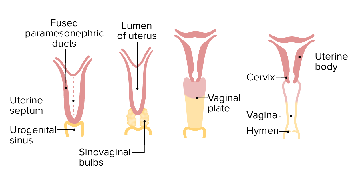
The uterus, cervix, and fallopian tubes derive from the paramesonephric ductsParamesonephric ductsA pair of ducts near the wolffian ducts in a developing embryo. In the male embryo, they degenerate with the appearance of testicular anti-mullerian hormone. In the absence of anti-mullerian hormone, mullerian ducts give rise to the female reproductive tract, including the oviducts; uterus; cervix; and vagina.Development of the Urogenital System (müllerian ducts)
At 6 weeks of embryologic life, the müllerian ducts fuse at the caudal end:
The fused medial/caudal portion gives rise to the uterus and upper vaginaVaginaThe vagina is the female genital canal, extending from the vulva externally to the cervix uteri internally. The structures have sexual, reproductive, and urinary functions and a rich blood supply, mainly arising from the internal iliac artery.Vagina, Vulva, and Pelvic Floor: Anatomy.
The unfused lateral/cranial portions give rise to the fallopian tubes.
A longitudinal midline septum exists within the uterine cavity where the müllerian ducts came together → usually regresses by week 20
The gubernaculumGubernaculumAn embryonic structure that helps guide proper descent of gonads into their final positions. It attaches the caudal end of the fetal gonads to the developing scrotum in male and the labium majorum in female. It gives rise to the caudal ligaments of the gonad: the scrotal ligament in male and the uterine round and proper ovarian ligaments in female. It includes morphofunctional equivalent structures in non-mammals.Congenital Malformations of the Female Reproductive System gives rise to the supporting ligaments (broad and round ligaments).
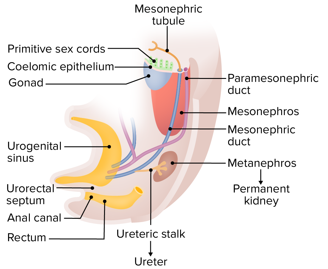
At around 6 weeks, the mesothelium of the gonads invades underlying mesoderm, forming the primitive sex cords. Paramesonephric (i.e., müllerian) ducts develop alongside the mesonephric ducts and fuse at the caudal end. At this stage, the mesonephric ducts are functioning as part of the primitive urinary system (mesonephros); however, as the definitive kidney (from the metanephros) takes over, the mesonephric ducts will begin differentiating into male genital structures in males or will regress in females.
Image by Lecturio.
Development of the uterus, cervix, and vagina (anterior view) from the paramesonephric (müllerian) ducts and the urogenital sinus. Pink structures are derived from the paramesonephric ducts; yellow structures are derived from the urogenital sinus.
Image by Lecturio.
Gross Anatomy
Structure and size of the uterus
Pear-shaped and hollow
Composed of smooth muscle
Size:
Length: 7–8 cm
Width: 4–5 cm
Thickness: 2.5–4 cm
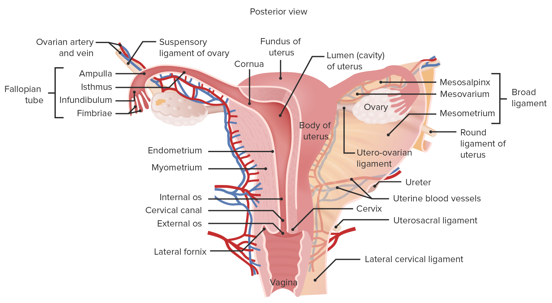
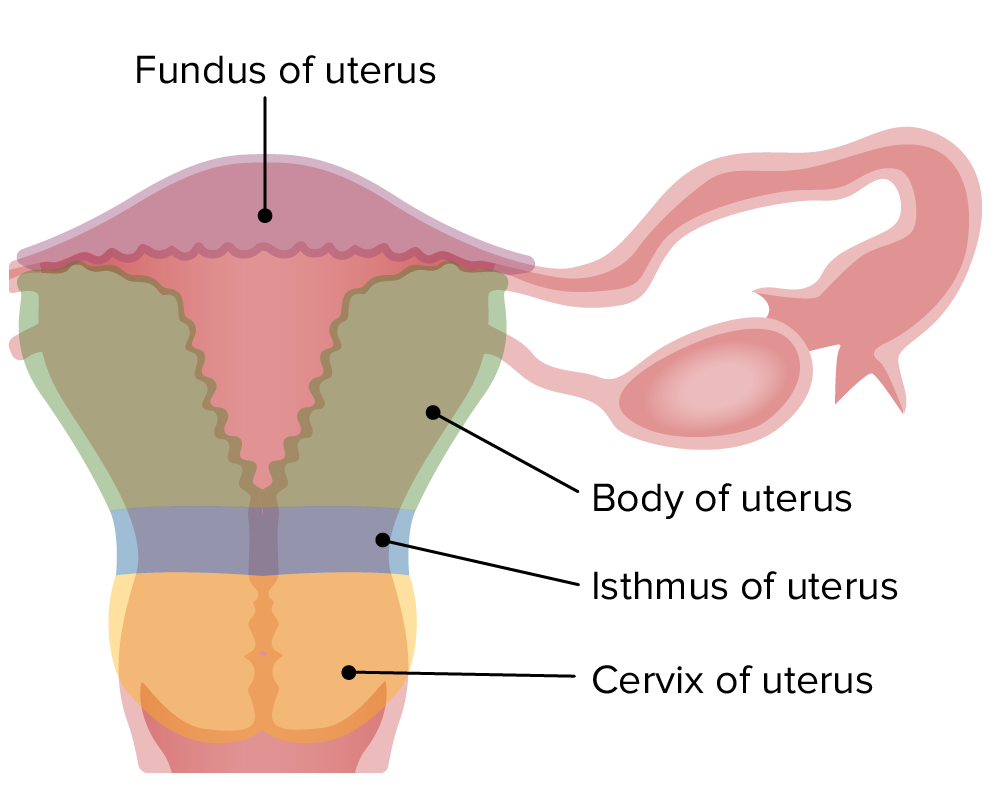
Parts of the uterus
FundusFundusThe superior portion of the body of the stomach above the level of the cardiac notch.Stomach: Anatomy: broad, superior curvature of the uterus
Body:
Main central portion
Uterine horns: superior lateral openings to the fallopian tubes
Uterine cavity:
Internal cavity
Shaped like an inverted triangle
Also referred to as the endometrial cavity
Isthmus:
Narrowing of the uterus just inferior to its body
The transition point between the uterine body and the cervix
Cervix:
FibrousFibrousFibrocystic Change, cylindrical structure; makes up the inferior portion of the uterus
Connected to and leads into the vaginaVaginaThe vagina is the female genital canal, extending from the vulva externally to the cervix uteri internally. The structures have sexual, reproductive, and urinary functions and a rich blood supply, mainly arising from the internal iliac artery.Vagina, Vulva, and Pelvic Floor: Anatomy
Cervical canal: passage leading from the uterine cavity into the vaginaVaginaThe vagina is the female genital canal, extending from the vulva externally to the cervix uteri internally. The structures have sexual, reproductive, and urinary functions and a rich blood supply, mainly arising from the internal iliac artery.Vagina, Vulva, and Pelvic Floor: Anatomy
Internal os: internal opening of the canal into the uterine body
External os: external opening of the canal into the vaginaVaginaThe vagina is the female genital canal, extending from the vulva externally to the cervix uteri internally. The structures have sexual, reproductive, and urinary functions and a rich blood supply, mainly arising from the internal iliac artery.Vagina, Vulva, and Pelvic Floor: Anatomy
Visible on speculum exam
Has an anterior and a posterior lip
Shape and size of the external os differs between nulliparous and multiparousMultiparousA woman with prior deliveriesNormal and Abnormal Labor women.
Parts of the uterus.
Image by Lecturio.
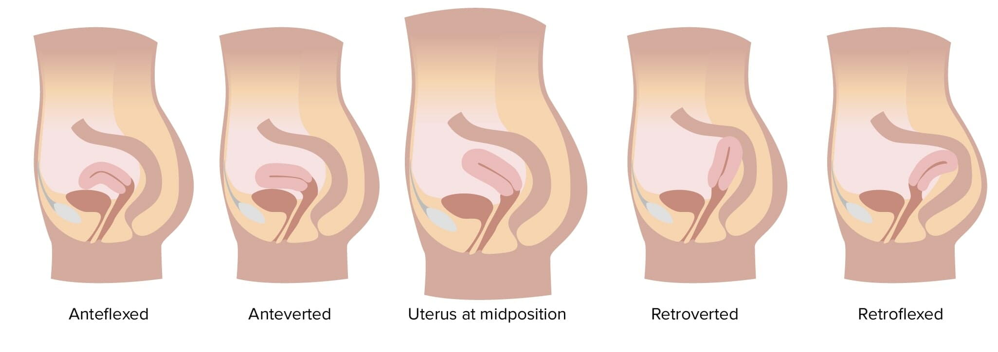
Uterine orientations
The uterus is often tilted or bent forward or backward. It is clinically important to determine the orientationOrientationAwareness of oneself in relation to time, place and person.Psychiatric Assessment of the uterus before any uterine procedure in order to minimize risks of complications (like uterine perforationPerforationA pathological hole in an organ, blood vessel or other soft part of the body, occurring in the absence of external force.Esophagitis). The 5 orientations are:
Anteverted:
The entire uterus is tilted forward, over the bladderBladderA musculomembranous sac along the urinary tract. Urine flows from the kidneys into the bladder via the ureters, and is held there until urination.Pyelonephritis and Perinephric Abscess.
Anteflexed: forward bend in the body musculature, in addition to being anteverted
Midposition: between anteverted and retroverted positions
Retroverted: tilted posteriorly, toward the rectumRectumThe rectum and anal canal are the most terminal parts of the lower GI tract/large intestine that form a functional unit and control defecation. Fecal continence is maintained by several important anatomic structures including rectal folds, anal valves, the sling-like puborectalis muscle, and internal and external anal sphincters. Rectum and Anal Canal: Anatomy
Retroflexed:backward bend in the body musculature, in addition to being retroverted
Uterine orientations
Image by Lecturio.
Anatomic relationships of the uterus
The uterus is in contact with a number of other organs and spaces:
Anteriorly:
BladderBladderA musculomembranous sac along the urinary tract. Urine flows from the kidneys into the bladder via the ureters, and is held there until urination.Pyelonephritis and Perinephric Abscess
Vesicouterine pouch: recess formed by the peritoneal fold between the uterus and bladderBladderA musculomembranous sac along the urinary tract. Urine flows from the kidneys into the bladder via the ureters, and is held there until urination.Pyelonephritis and Perinephric Abscess
Posteriorly:
RectumRectumThe rectum and anal canal are the most terminal parts of the lower GI tract/large intestine that form a functional unit and control defecation. Fecal continence is maintained by several important anatomic structures including rectal folds, anal valves, the sling-like puborectalis muscle, and internal and external anal sphincters. Rectum and Anal Canal: Anatomy
Rectouterine pouch
Also known as the Douglas pouch
The recess formed by the peritoneal fold between the rectumRectumThe rectum and anal canal are the most terminal parts of the lower GI tract/large intestine that form a functional unit and control defecation. Fecal continence is maintained by several important anatomic structures including rectal folds, anal valves, the sling-like puborectalis muscle, and internal and external anal sphincters. Rectum and Anal Canal: Anatomy and the posterior uterine wall
Lowermost point of the peritoneumPeritoneumThe peritoneum is a serous membrane lining the abdominopelvic cavity. This lining is formed by connective tissue and originates from the mesoderm. The membrane lines both the abdominal walls (as parietal peritoneum) and all of the visceral organs (as visceral peritoneum).Peritoneum: Anatomy
Laterally:
Fallopian tubes
OvariesOvariesOvaries are the paired gonads of the female reproductive system that contain haploid gametes known as oocytes. The ovaries are located intraperitoneally in the pelvis, just posterior to the broad ligament, and are connected to the pelvic sidewall and to the uterus by ligaments. These organs function to secrete hormones (estrogen and progesterone) and to produce the female germ cells (oocytes).Ovaries: Anatomy
Broad ligament
Pelvic sidewall
Superiorly: small intestines
Inferiorly: vaginaVaginaThe vagina is the female genital canal, extending from the vulva externally to the cervix uteri internally. The structures have sexual, reproductive, and urinary functions and a rich blood supply, mainly arising from the internal iliac artery.Vagina, Vulva, and Pelvic Floor: Anatomy
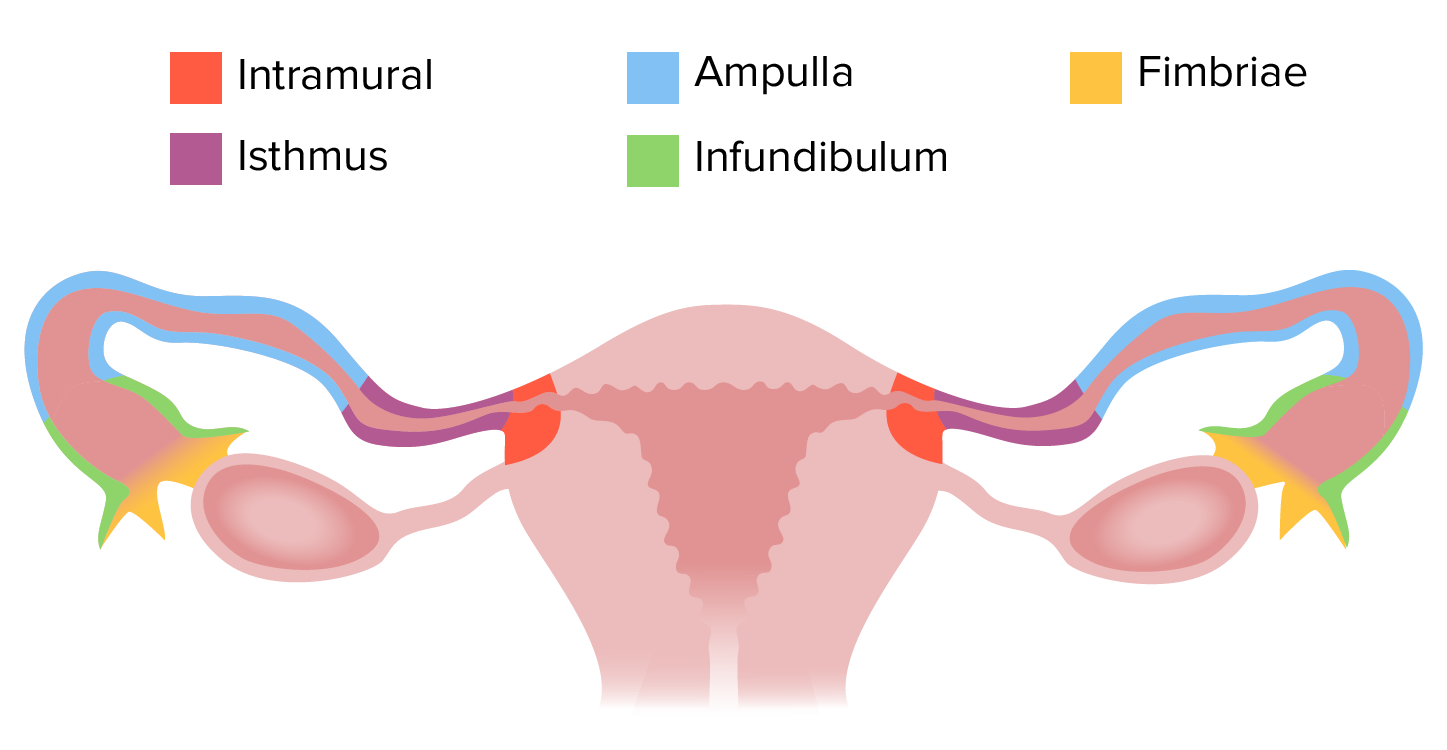
Structure of the fallopian tubes
Fallopian tubes are also known as uterine tubes. These paired, thin muscular tubes are attached to the uterus and are approximately 10 cm in total length. There are 4 parts, as follows, in order from lateral to medial.
Infundibulum:
Lateral-most part of the tubes, in close association with the ovariesOvariesOvaries are the paired gonads of the female reproductive system that contain haploid gametes known as oocytes. The ovaries are located intraperitoneally in the pelvis, just posterior to the broad ligament, and are connected to the pelvic sidewall and to the uterus by ligaments. These organs function to secrete hormones (estrogen and progesterone) and to produce the female germ cells (oocytes).Ovaries: Anatomy
Funnel-shaped with finger-like projections called fimbriaeFimbriaeThin, hairlike appendages, 1 to 20 microns in length and often occurring in large numbers, present on the cells of gram-negative bacteria, particularly enterobacteriaceae and Neisseria. Unlike flagella, they do not possess motility, but being protein (pilin) in nature, they possess antigenic and hemagglutinating properties. They are of medical importance because some fimbriae mediate the attachment of bacteria to cells via adhesins. Bacterial fimbriae refer to common pili, to be distinguished from the preferred use of ‘pili’.Escherichia coli
Opens into peritoneal cavityPeritoneal CavityThe space enclosed by the peritoneum. It is divided into two portions, the greater sac and the lesser sac or omental bursa, which lies behind the stomach. The two sacs are connected by the foramen of winslow, or epiploic foramen.Peritoneum: Anatomy
Ampulla:
The widest and longest portion of the tube
Usual site of fertilizationFertilizationTo undergo fertilization, the sperm enters the uterus, travels towards the ampulla of the fallopian tube, and encounters the oocyte. The zona pellucida (the outer layer of the oocyte) deteriorates along with the zygote, which travels towards the uterus and eventually forms a blastocyst, allowing for implantation to occur. Fertilization and First Week
Approximately 7–8 cm long
Isthmus:
Narrowing portion that approaches the uterus (at the uterine horns)
Thicker walls
Approximately 4 cm long
Uterine portion (also called the intramural or interstitial part):
Located within the uterine wall
Opens into uterine cavity through uterine ostium
Shortest segment (< 1 cm)
Schematic depiction of the 4 parts of the fallopian tubes
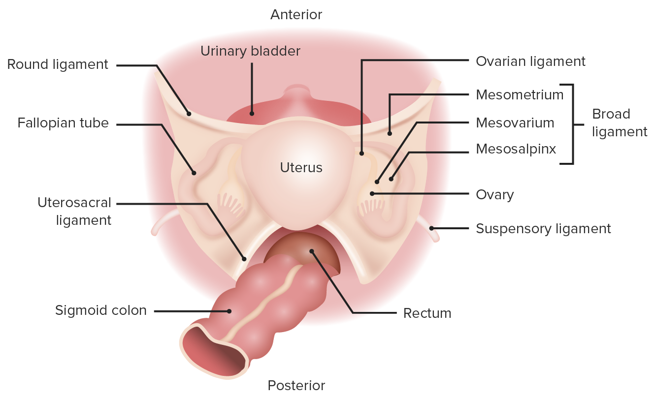
A thin sheet of peritoneumPeritoneumThe peritoneum is a serous membrane lining the abdominopelvic cavity. This lining is formed by connective tissue and originates from the mesoderm. The membrane lines both the abdominal walls (as parietal peritoneum) and all of the visceral organs (as visceral peritoneum).Peritoneum: Anatomy that drapes over the uterus and fallopian tubes like a sheet hanging over a clothesline
Connects the pelvic organs to the lateral pelvic wall
Divided into 3 parts: mesosalpinx, mesovarium, and mesometrium
Mesosalpinx:
Area adjacent to the fallopian tubes
Contains the tubal branches of the ovarian and ascending uterine arteriesArteriesArteries are tubular collections of cells that transport oxygenated blood and nutrients from the heart to the tissues of the body. The blood passes through the arteries in order of decreasing luminal diameter, starting in the largest artery (the aorta) and ending in the small arterioles. Arteries are classified into 3 types: large elastic arteries, medium muscular arteries, and small arteries and arterioles. Arteries: Histology
Mesovarium:
Area adjacent to the ovariesOvariesOvaries are the paired gonads of the female reproductive system that contain haploid gametes known as oocytes. The ovaries are located intraperitoneally in the pelvis, just posterior to the broad ligament, and are connected to the pelvic sidewall and to the uterus by ligaments. These organs function to secrete hormones (estrogen and progesterone) and to produce the female germ cells (oocytes).Ovaries: Anatomy
Contains the ovarian branches of the ovarian and ascending uterine arteriesArteriesArteries are tubular collections of cells that transport oxygenated blood and nutrients from the heart to the tissues of the body. The blood passes through the arteries in order of decreasing luminal diameter, starting in the largest artery (the aorta) and ending in the small arterioles. Arteries are classified into 3 types: large elastic arteries, medium muscular arteries, and small arteries and arterioles. Arteries: Histology
Inferiorly, contains the cardinal ligament and uretersUretersOne of a pair of thick-walled tubes that transports urine from the kidney pelvis to the urinary bladder.Urinary Tract: Anatomy
Found at the base of the broad ligament in the mesometrium
Contains the uterine vessels
Round ligament (ligamentum teresLigamentum teresA cord-like remnant structure formed from the closed left fetal umbilical vein. It is located along the lower edge of the falciform ligament.Liver: Anatomy):
A thickening of the broad ligament off the anterior surface of the uterus
Connects the anterior uterine horns to the anterior abdominal wallAbdominal wallThe outer margins of the abdomen, extending from the osteocartilaginous thoracic cage to the pelvis. Though its major part is muscular, the abdominal wall consists of at least seven layers: the skin, subcutaneous fat, deep fascia; abdominal muscles, transversalis fascia, extraperitoneal fat, and the parietal peritoneum.Surgical Anatomy of the Abdomen before passing through the inguinal canalInguinal canalThe tunnel in the lower anterior abdominal wall through which the spermatic cord, in the male; round ligament, in the female; nerves; and vessels pass. Its internal end is at the deep inguinal ring and its external end is at the superficial inguinal ring.Inguinal Canal: Anatomy and Hernias and terminating in the labia majoraLabia majoraVagina, Vulva, and Pelvic Floor: Anatomy
A thickened portion of the broad ligament within the mesovarium
Uterosacral ligament:
Connects the posteroinferior portion of the uterus (at the level of the cervix) to the sacrumSacrumFive fused vertebrae forming a triangle-shaped structure at the back of the pelvis. It articulates superiorly with the lumbar vertebrae, inferiorly with the coccyx, and anteriorly with the ilium of the pelvis. The sacrum strengthens and stabilizes the pelvis.Vertebral Column: Anatomy
A thicker, stronger ligament, providing important structural support (i.e., preventing uterine prolapseUterine prolapseDownward displacement of the uterus. It is classified in various degrees: in the first degree the uterine cervix is within the vaginal orifice; in the second degree the cervix is outside the orifice; in the third degree the entire uterus is outside the orifice.Pelvic Organ Prolapse into the vaginaVaginaThe vagina is the female genital canal, extending from the vulva externally to the cervix uteri internally. The structures have sexual, reproductive, and urinary functions and a rich blood supply, mainly arising from the internal iliac artery.Vagina, Vulva, and Pelvic Floor: Anatomy)
Posterosuperior view of the female pelvic anatomy depicting the broad ligament and the round ligament
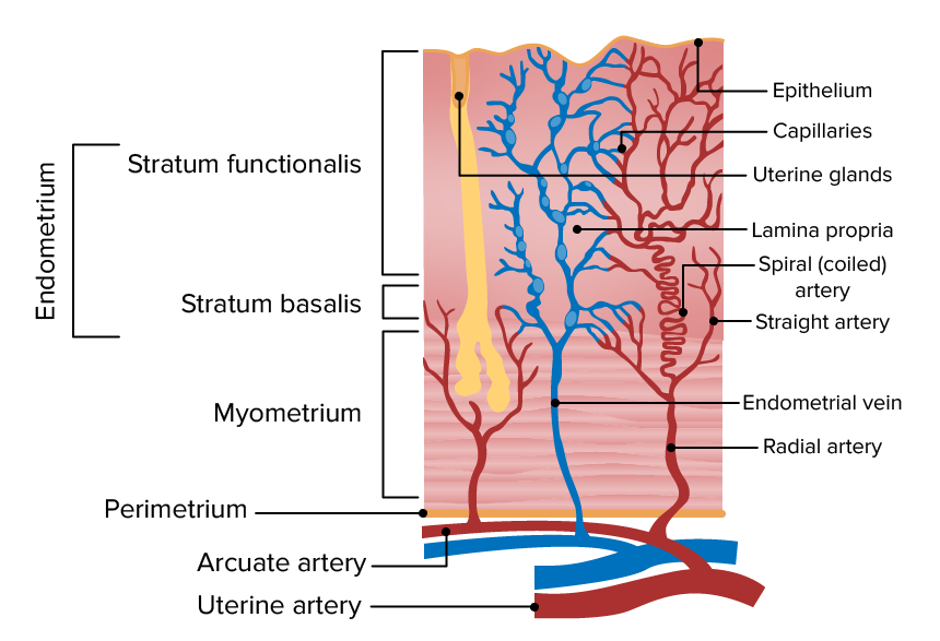
Histologically, the uterus is composed of 3 layers:
EndometriumEndometriumThe mucous membrane lining of the uterine cavity that is hormonally responsive during the menstrual cycle and pregnancy. The endometrium undergoes cyclic changes that characterize menstruation. After successful fertilization, it serves to sustain the developing embryo.Embryoblast and Trophoblast Development:
Lamina propriaLamina propriaWhipple’s Disease (also called stroma): made up of connective tissueConnective tissueConnective tissues originate from embryonic mesenchyme and are present throughout the body except inside the brain and spinal cord. The main function of connective tissues is to provide structural support to organs. Connective tissues consist of cells and an extracellular matrix.Connective Tissue: Histology
Divided into 2 layers:
Stratum functionalis: superficial layer that proliferates and sheds each month during the menstrual cycleMenstrual cycleThe menstrual cycle is the cyclic pattern of hormonal and tissular activity that prepares a suitable uterine environment for the fertilization and implantation of an ovum. The menstrual cycle involves both an endometrial and ovarian cycle that are dependent on one another for proper functioning. There are 2 phases of the ovarian cycle and 3 phases of the endometrial cycle.Menstrual Cycle
Stratum basalis: deeper layer; does not shed, and regenerates the functionalis each cycle
Supplied by small spiral arteriesSpiral arteriesPlacenta, Umbilical Cord, and Amniotic Cavity that constrict, rupture, and bleed during menstruationMenstruationThe periodic shedding of the endometrium and associated menstrual bleeding in the menstrual cycle of humans and primates. Menstruation is due to the decline in circulating progesterone, and occurs at the late luteal phase when luteolysis of the corpus luteum takes place.Menstrual Cycle
Contains the larger branches of the neurovasculature
Perimetrium:
Also known as the serosa
Outer layer, which is also the visceral layer of the peritoneumPeritoneumThe peritoneum is a serous membrane lining the abdominopelvic cavity. This lining is formed by connective tissue and originates from the mesoderm. The membrane lines both the abdominal walls (as parietal peritoneum) and all of the visceral organs (as visceral peritoneum).Peritoneum: Anatomy
Becomes the broad ligament laterally
Thin layer of connective tissueConnective tissueConnective tissues originate from embryonic mesenchyme and are present throughout the body except inside the brain and spinal cord. The main function of connective tissues is to provide structural support to organs. Connective tissues consist of cells and an extracellular matrix.Connective Tissue: Histology
Schematic representation of the multiple uterine layers
Image by Lecturio.
Microscopic anatomy of the cervix
Epithelial lining:
Ectocervix:
Lines the intravaginal part of the cervix; visible on speculum exam
The location where squamous and columnar epitheliumEpitheliumThe epithelium is a complex of specialized cellular organizations arranged into sheets and lining cavities and covering the surfaces of the body. The cells exhibit polarity, having an apical and a basal pole. Structures important for the epithelial integrity and function involve the basement membrane, the semipermeable sheet on which the cells rest, and interdigitations, as well as cellular junctions. Surface Epithelium: Histology overlap
Squamocolumnar junctionSquamocolumnar junctionEsophagus: Anatomy: transition point at which the epitheliumEpitheliumThe epithelium is a complex of specialized cellular organizations arranged into sheets and lining cavities and covering the surfaces of the body. The cells exhibit polarity, having an apical and a basal pole. Structures important for the epithelial integrity and function involve the basement membrane, the semipermeable sheet on which the cells rest, and interdigitations, as well as cellular junctions. Surface Epithelium: Histology becomes entirely stratified squamous (i.e., ectocervix).
Site of infection with human papillomaPapillomaA circumscribed benign epithelial tumor projecting from the surrounding surface; more precisely, a benign epithelial neoplasm consisting of villous or arborescent outgrowths of fibrovascular stroma covered by neoplastic cells.Cowden SyndromevirusVirusViruses are infectious, obligate intracellular parasites composed of a nucleic acid core surrounded by a protein capsid. Viruses can be either naked (non-enveloped) or enveloped. The classification of viruses is complex and based on many factors, including type and structure of the nucleoid and capsid, the presence of an envelope, the replication cycle, and the host range. Virology (HPVHPVHuman papillomavirus (HPV) is a nonenveloped, circular, double-stranded DNA virus belonging to the Papillomaviridae family. Humans are the only reservoir, and transmission occurs through close skin-to-skin or sexual contact. Human papillomaviruses infect basal epithelial cells and can affect cell-regulatory proteins to result in cell proliferation. Papillomavirus (HPV)), metaplasiaMetaplasiaA condition in which there is a change of one adult cell type to another similar adult cell type.Cellular Adaptation, and most cervical cancers
Stroma:
Made up primarily of fibroelastic connective tissueConnective tissueConnective tissues originate from embryonic mesenchyme and are present throughout the body except inside the brain and spinal cord. The main function of connective tissues is to provide structural support to organs. Connective tissues consist of cells and an extracellular matrix.Connective Tissue: Histology
< 10% smooth muscle
Necessary for the stretching capacity during childbirth
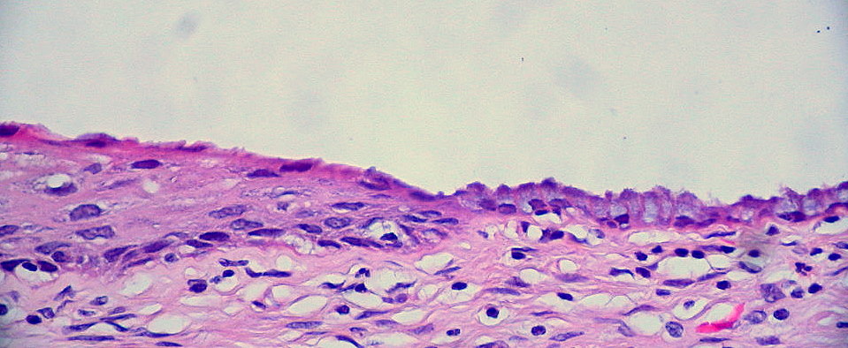
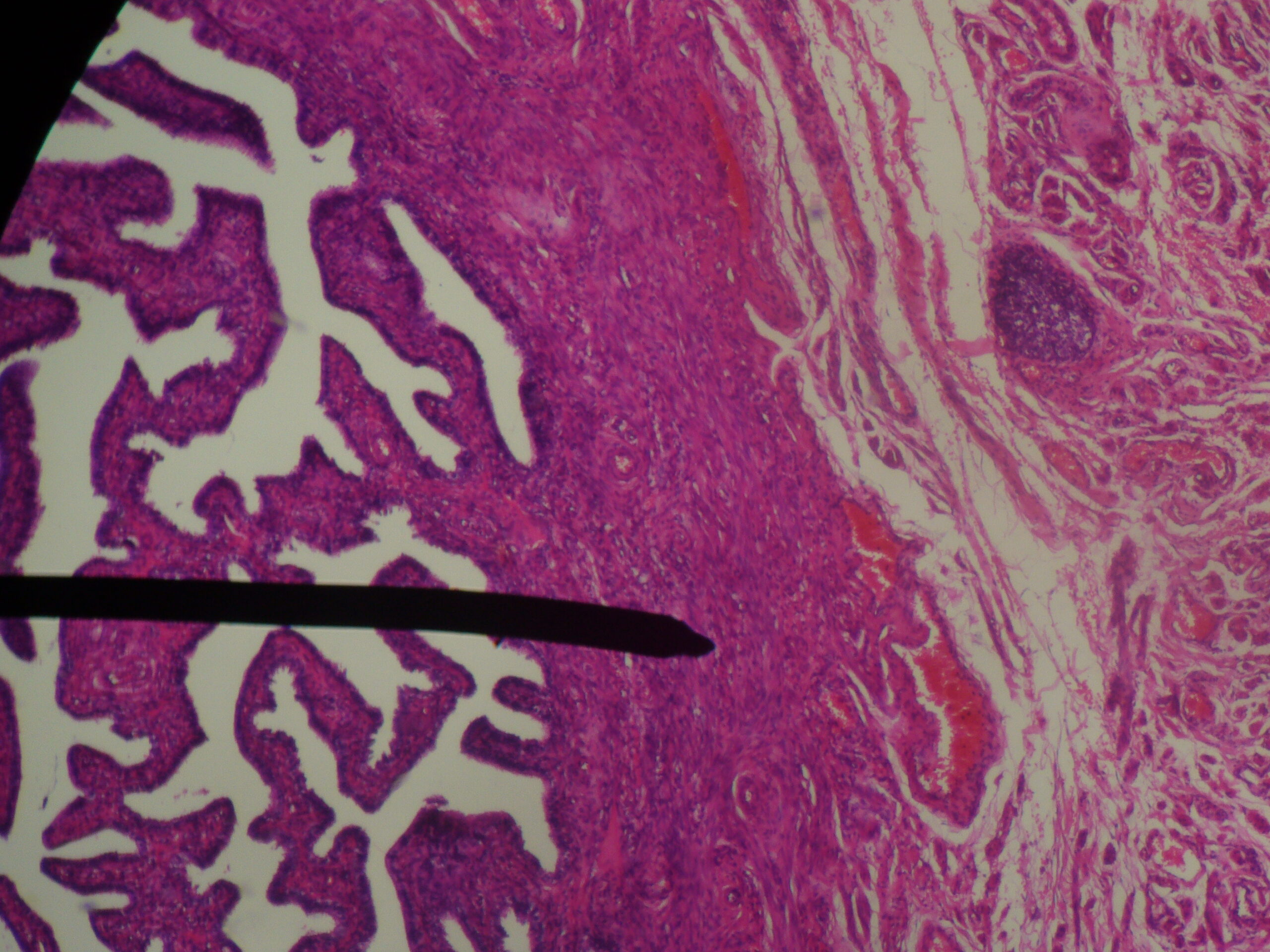
H&E-stained histologic slide depicting an unusually “clean” squamocolumnar junction of the cervix. This slide shows the boundary between the ectocervix, made up of stratified squamous epithelium on the left, and the endocervix, made up of columnar epithelium on the right.
Image: “Cervix: Normal Squamocolumnar Junction” by Ed Uthman. License: CC BY 2.0
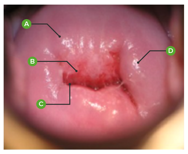
Normal parous (meaning the individual has had a vaginal birth) cervix, as viewed on colposcopy:
A. Exocervical mucosa
B. Transformation zone between exocervix and endocervix
C. Endocervical mucosa appearing at external cervical os
D. Nabothian cyst (mucus-filled cyst)
Image: “Images of VIA negative, VIA positive-cryotherapy eligible and VIA positive-cryotherapy ineligible lesions.” by Parham GP, et al. License: CC BY 4.0, edited by Lecturio.
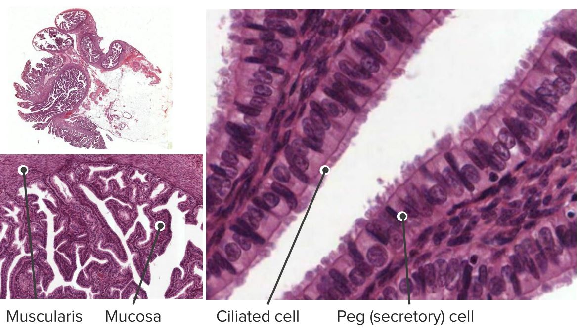
Assist in movement of sperm, oocytesOocytesFemale germ cells derived from oogonia and termed oocytes when they enter meiosis. The primary oocytes begin meiosis but are arrested at the diplotene state until ovulation at puberty to give rise to haploid secondary oocytes or ova (ovum).Ovaries: Anatomy, and embryos
Cilia beat more strongly in the presence of estrogenEstrogenCompounds that interact with estrogen receptors in target tissues to bring about the effects similar to those of estradiol. Estrogens stimulate the female reproductive organs, and the development of secondary female sex characteristics. Estrogenic chemicals include natural, synthetic, steroidal, or non-steroidal compounds.Ovaries: Anatomy
Peg cells (< 10%):
Nonciliated, secretory cells
Secretions are primarily under the influence of estrogenEstrogenCompounds that interact with estrogen receptors in target tissues to bring about the effects similar to those of estradiol. Estrogens stimulate the female reproductive organs, and the development of secondary female sex characteristics. Estrogenic chemicals include natural, synthetic, steroidal, or non-steroidal compounds.Ovaries: Anatomy
Secrete nutritious fluid to support fertilizationFertilizationTo undergo fertilization, the sperm enters the uterus, travels towards the ampulla of the fallopian tube, and encounters the oocyte. The zona pellucida (the outer layer of the oocyte) deteriorates along with the zygote, which travels towards the uterus and eventually forms a blastocyst, allowing for implantation to occur. Fertilization and First Week
Muscular layer:
Smooth muscle
↑ Concentrations of estrogenEstrogenCompounds that interact with estrogen receptors in target tissues to bring about the effects similar to those of estradiol. Estrogens stimulate the female reproductive organs, and the development of secondary female sex characteristics. Estrogenic chemicals include natural, synthetic, steroidal, or non-steroidal compounds.Ovaries: Anatomy present around ovulationOvulationThe discharge of an ovum from a rupturing follicle in the ovary.Menstrual Cycle stimulate peristaltic waves of contraction → enhances movement of sperm, oocytesOocytesFemale germ cells derived from oogonia and termed oocytes when they enter meiosis. The primary oocytes begin meiosis but are arrested at the diplotene state until ovulation at puberty to give rise to haploid secondary oocytes or ova (ovum).Ovaries: Anatomy, and embryos
Contains an inner circular and outer longitudinal layer
Serosa:
Thin outer layer made up of connective tissueConnective tissueConnective tissues originate from embryonic mesenchyme and are present throughout the body except inside the brain and spinal cord. The main function of connective tissues is to provide structural support to organs. Connective tissues consist of cells and an extracellular matrix.Connective Tissue: Histology
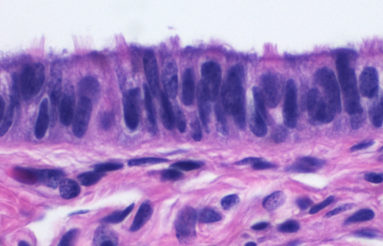
Histologic specimen showing the mucosa and muscularis layer in low power. The inset is a high-power magnification of the mucosal layer showing the ciliated epithelium with peg cells.
Image by Lecturio.
Human fallopian tube wall.
Image: “Fallopian tube (lamina propoia)” by Jpogi at English Wikipedia. License: Public Domain
Histology of ciliated columnar epithelium of the fallopian tube.
Image: “Histology of ciliated columnar epithelium of the fallopian tube” by Mikael Häggström. License: CC0 1.0
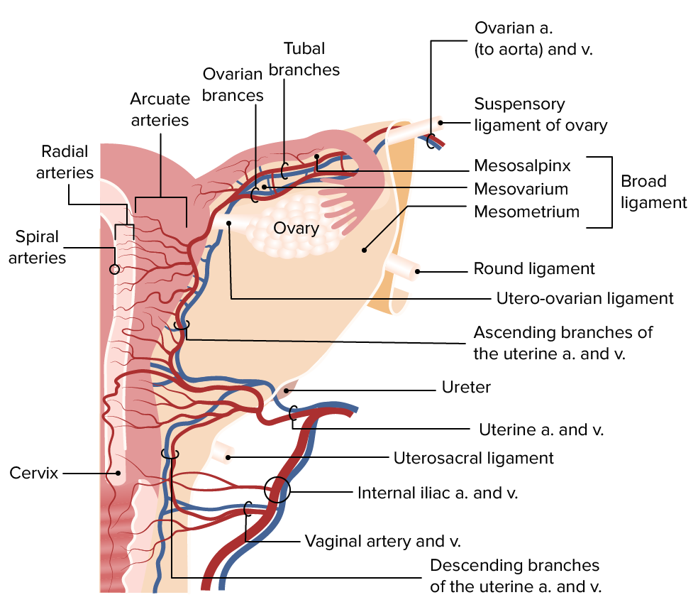
The primary blood supply to the uterus is via the uterine artery. The fallopian tubes are supplied by the anastomosis between the uterine and ovarian arteriesArteriesArteries are tubular collections of cells that transport oxygenated blood and nutrients from the heart to the tissues of the body. The blood passes through the arteries in order of decreasing luminal diameter, starting in the largest artery (the aorta) and ending in the small arterioles. Arteries are classified into 3 types: large elastic arteries, medium muscular arteries, and small arteries and arterioles. Arteries: Histology.
Uterine artery:
A branch of the anterior division of the internal iliac artery
Located within the broad ligament near the level of the cervix
Approaches the uterus at a 90-degree angle at the level of the internal cervical os → splits into ascending and descending branches
These branches run longitudinally along the lateral edges of the uterine body and cervix.
Ascending branches: anastomose with the ovarian artery
Runs along the pelvic wall → reaches the lateral side of the ovary
Splits into ovarian and tubal branches:
Both branches anastomose with the ascending branch of the uterine artery.
The tubal branch is often referred to as the utero-ovarian artery.
Supplying the myometrium and endometriumEndometriumThe mucous membrane lining of the uterine cavity that is hormonally responsive during the menstrual cycle and pregnancy. The endometrium undergoes cyclic changes that characterize menstruation. After successful fertilization, it serves to sustain the developing embryo.Embryoblast and Trophoblast Development:
Arcuate arteriesArteriesArteries are tubular collections of cells that transport oxygenated blood and nutrients from the heart to the tissues of the body. The blood passes through the arteries in order of decreasing luminal diameter, starting in the largest artery (the aorta) and ending in the small arterioles. Arteries are classified into 3 types: large elastic arteries, medium muscular arteries, and small arteries and arterioles. Arteries: Histology:
Branch off the ascending and descending uterine arteriesArteriesArteries are tubular collections of cells that transport oxygenated blood and nutrients from the heart to the tissues of the body. The blood passes through the arteries in order of decreasing luminal diameter, starting in the largest artery (the aorta) and ending in the small arterioles. Arteries are classified into 3 types: large elastic arteries, medium muscular arteries, and small arteries and arterioles. Arteries: Histology at an approximately 90-degree angle
Anastomose with arcuate arteriesArteriesArteries are tubular collections of cells that transport oxygenated blood and nutrients from the heart to the tissues of the body. The blood passes through the arteries in order of decreasing luminal diameter, starting in the largest artery (the aorta) and ending in the small arterioles. Arteries are classified into 3 types: large elastic arteries, medium muscular arteries, and small arteries and arterioles. Arteries: Histology coming from the oppose side
Tortuous vessels that supply the endometriumEndometriumThe mucous membrane lining of the uterine cavity that is hormonally responsive during the menstrual cycle and pregnancy. The endometrium undergoes cyclic changes that characterize menstruation. After successful fertilization, it serves to sustain the developing embryo.Embryoblast and Trophoblast Development between the glands
Rupture and constrict during mensesMensesThe periodic shedding of the endometrium and associated menstrual bleeding in the menstrual cycle of humans and primates. Menstruation is due to the decline in circulating progesterone, and occurs at the late luteal phase when luteolysis of the corpus luteum takes place.Menstrual Cycle → menstrual bleeding, ischemiaIschemiaA hypoperfusion of the blood through an organ or tissue caused by a pathologic constriction or obstruction of its blood vessels, or an absence of blood circulation.Ischemic Cell Damage, and endometrial shedding
Posterior view of the uterus showing the blood supply and venous drainage to the uterus, fallopian tubes, and ovary. The main blood supply to the uterus is via the uterine artery, a branch of the internal iliac. The ovarian artery also provides the uterus with arterial blood via an anastomosis with the ascending branch of the uterine artery. Arcuate arteries branch off of the uterine artery, supplying the myometrium.
Image by Lecturio.
Venous drainage
Uterine body:
Via the uterine venous plexus, which runs down the lateral side of the uterus
Located in the broad ligament near the level of the cervix
Drains into the uterine vein → into the internal iliac vein
Fallopian tubes: drain via tubal veinsVeinsVeins are tubular collections of cells, which transport deoxygenated blood and waste from the capillary beds back to the heart. Veins are classified into 3 types: small veins/venules, medium veins, and large veins. Each type contains 3 primary layers: tunica intima, tunica media, and tunica adventitia. Veins: Histology → ovarian veinsVeinsVeins are tubular collections of cells, which transport deoxygenated blood and waste from the capillary beds back to the heart. Veins are classified into 3 types: small veins/venules, medium veins, and large veins. Each type contains 3 primary layers: tunica intima, tunica media, and tunica adventitia. Veins: Histology
The primary lymphatic drainage for each section of the uterus and fallopian tubes is through:
Fallopian tubes:
Para-aortic nodes (follows drainage of the ovariesOvariesOvaries are the paired gonads of the female reproductive system that contain haploid gametes known as oocytes. The ovaries are located intraperitoneally in the pelvis, just posterior to the broad ligament, and are connected to the pelvic sidewall and to the uterus by ligaments. These organs function to secrete hormones (estrogen and progesterone) and to produce the female germ cells (oocytes).Ovaries: Anatomy)
Internal iliac nodes
Inguinal nodes (via the broad ligament)
FundusFundusThe superior portion of the body of the stomach above the level of the cardiac notch.Stomach: Anatomy:
Para-aortic nodes
Superficial inguinal nodes (areas near the round ligaments)
The uterus and tubes are innervated by the autonomic nervous systemAutonomic nervous systemThe ANS is a component of the peripheral nervous system that uses both afferent (sensory) and efferent (effector) neurons, which control the functioning of the internal organs and involuntary processes via connections with the CNS. The ANS consists of the sympathetic and parasympathetic nervous systems. Autonomic Nervous System: Anatomy (ANSANSThe ans is a component of the peripheral nervous system that uses both afferent (sensory) and efferent (effector) neurons, which control the functioning of the internal organs and involuntary processes via connections with the CNS. The ans consists of the sympathetic and parasympathetic nervous systems.Autonomic Nervous System: Anatomy). Nerve fibersNerve FibersSlender processes of neurons, including the axons and their glial envelopes (myelin sheath). Nerve fibers conduct nerve impulses to and from the central nervous system.Nervous System: Histology of the ANSANSThe ans is a component of the peripheral nervous system that uses both afferent (sensory) and efferent (effector) neurons, which control the functioning of the internal organs and involuntary processes via connections with the CNS. The ans consists of the sympathetic and parasympathetic nervous systems.Autonomic Nervous System: Anatomy pass via splanchnic nerves → inferior hypogastric plexus → uterovaginal plexus
Follow the sympathetic innervation in a retrograde direction
Visceral uterine painPainAn unpleasant sensation induced by noxious stimuli which are detected by nerve endings of nociceptive neurons.Pain: Types and Pathways from labor is felt in the T10–L2 dermatomesDermatomesSpinal Cord: Anatomy
Subperitoneal structures (lower uterus/cervix):
Follow the parasympathetic innervation in a retrograde direction
Visceral afferentAfferentNeurons which conduct nerve impulses to the central nervous system.Nervous System: Histology fibers unrelated to painPainAn unpleasant sensation induced by noxious stimuli which are detected by nerve endings of nociceptive neurons.Pain: Types and Pathways: follow the parasympathetic innervation
Clinical Relevance
Related anatomical structures
PelvisPelvisThe pelvis consists of the bony pelvic girdle, the muscular and ligamentous pelvic floor, and the pelvic cavity, which contains viscera, vessels, and multiple nerves and muscles. The pelvic girdle, composed of 2 “hip” bones and the sacrum, is a ring-like bony structure of the axial skeleton that links the vertebral column with the lower extremities.Pelvis: Anatomy: consists of the pelvic girdle, pelvic cavity, pelvic floorPelvic floorSoft tissue formed mainly by the pelvic diaphragm, which is composed of the two levator ani and two coccygeus muscles. The pelvic diaphragm lies just below the pelvic aperture (outlet) and separates the pelvic cavity from the perineum. It extends between the pubic bone anteriorly and the coccyx posteriorly.Vagina, Vulva, and Pelvic Floor: Anatomy, and all the viscera, vessels, and muscles contained within. The pelvic cavity houses various GI and urogenital structures.
OvariesOvariesOvaries are the paired gonads of the female reproductive system that contain haploid gametes known as oocytes. The ovaries are located intraperitoneally in the pelvis, just posterior to the broad ligament, and are connected to the pelvic sidewall and to the uterus by ligaments. These organs function to secrete hormones (estrogen and progesterone) and to produce the female germ cells (oocytes).Ovaries: Anatomy: paired gonadsGonadsThe gamete-producing glands, ovary or testis.Hormones: Overview and Types of the female reproductive system. The ovariesOvariesOvaries are the paired gonads of the female reproductive system that contain haploid gametes known as oocytes. The ovaries are located intraperitoneally in the pelvis, just posterior to the broad ligament, and are connected to the pelvic sidewall and to the uterus by ligaments. These organs function to secrete hormones (estrogen and progesterone) and to produce the female germ cells (oocytes).Ovaries: Anatomy are located intraperitoneally in the lesser pelvisLesser pelvisThe part of the pelvis, inferior to the pelvic brim, that comprises both the pelvic cavity and the part of the perineum lying inferior to the pelvic diaphragm.Pelvis: Anatomy, just posterior to the broad ligament.
VaginaVaginaThe vagina is the female genital canal, extending from the vulva externally to the cervix uteri internally. The structures have sexual, reproductive, and urinary functions and a rich blood supply, mainly arising from the internal iliac artery.Vagina, Vulva, and Pelvic Floor: Anatomy and vulvaVulvaThe vulva is the external genitalia of the female and includes the mons pubis, labia majora, labia minora, clitoris, vestibule, vestibular bulb, and greater vestibular glands. Vagina, Vulva, and Pelvic Floor: Anatomy: The vulvaVulvaThe vulva is the external genitalia of the female and includes the mons pubis, labia majora, labia minora, clitoris, vestibule, vestibular bulb, and greater vestibular glands. Vagina, Vulva, and Pelvic Floor: Anatomy is the external female genitalia and includes the mons pubisMons pubisVagina, Vulva, and Pelvic Floor: Anatomy, labia majoraLabia majoraVagina, Vulva, and Pelvic Floor: Anatomy, labia minoraLabia minoraVagina, Vulva, and Pelvic Floor: Anatomy, clitorisClitorisAn erectile structure homologous with the penis, situated beneath the anterior labial commissure, partially hidden between the anterior ends of the labia minora.Vagina, Vulva, and Pelvic Floor: Anatomy, vestibuleVestibuleAn oval, bony chamber of the inner ear, part of the bony labyrinth. It is continuous with bony cochlea anteriorly, and semicircular canals posteriorly. The vestibule contains two communicating sacs (utricle and saccule) of the balancing apparatus. The oval window on its lateral wall is occupied by the base of the stapes of the middle ear.Ear: Anatomy, vestibular bulb, and the greater vestibular glandsGreater vestibular glandsMucus-secreting glands situated on the posterior and lateral aspect of the vestibule of the vagina.Vagina, Vulva, and Pelvic Floor: Anatomy. The vaginaVaginaThe vagina is the female genital canal, extending from the vulva externally to the cervix uteri internally. The structures have sexual, reproductive, and urinary functions and a rich blood supply, mainly arising from the internal iliac artery.Vagina, Vulva, and Pelvic Floor: Anatomy is the genital canal in the female, extending from the vulvaVulvaThe vulva is the external genitalia of the female and includes the mons pubis, labia majora, labia minora, clitoris, vestibule, vestibular bulb, and greater vestibular glands. Vagina, Vulva, and Pelvic Floor: Anatomy to the cervix uteri.
Physiology
Menstrual cycleMenstrual cycleThe menstrual cycle is the cyclic pattern of hormonal and tissular activity that prepares a suitable uterine environment for the fertilization and implantation of an ovum. The menstrual cycle involves both an endometrial and ovarian cycle that are dependent on one another for proper functioning. There are 2 phases of the ovarian cycle and 3 phases of the endometrial cycle.Menstrual Cycle: cyclic pattern of hormonal and tissular activity responsible for the preparation of a suitable uterine environment for the implantationImplantationEndometrial implantation of embryo, mammalian at the blastocyst stage.Fertilization and First Week and development of a fertilized embryoEmbryoThe entity of a developing mammal, generally from the cleavage of a zygote to the end of embryonic differentiation of basic structures. For the human embryo, this represents the first two months of intrauterine development preceding the stages of the fetus.Fertilization and First Week
PregnancyPregnancyThe status during which female mammals carry their developing young (embryos or fetuses) in utero before birth, beginning from fertilization to birth.Pregnancy: Diagnosis, Physiology, and Care:fertilizationFertilizationTo undergo fertilization, the sperm enters the uterus, travels towards the ampulla of the fallopian tube, and encounters the oocyte. The zona pellucida (the outer layer of the oocyte) deteriorates along with the zygote, which travels towards the uterus and eventually forms a blastocyst, allowing for implantation to occur. Fertilization and First Week of the ovum and implantationImplantationEndometrial implantation of embryo, mammalian at the blastocyst stage.Fertilization and First Week into the uterine wall. PregnancyPregnancyThe status during which female mammals carry their developing young (embryos or fetuses) in utero before birth, beginning from fertilization to birth.Pregnancy: Diagnosis, Physiology, and Care usually lasts 40 weeks from the 1st day of the last menstrual periodLast menstrual periodThe 1st day of a woman’s last menstrual period. By convention, this date is usually used to date pregnancies.Pregnancy: Diagnosis, Physiology, and Care. Numerous physiologic changes to the uterus and many other organs occur during pregnancyPregnancyThe status during which female mammals carry their developing young (embryos or fetuses) in utero before birth, beginning from fertilization to birth.Pregnancy: Diagnosis, Physiology, and Care.
Clinical evaluation
Diagnostic procedures in gynecologyDiagnostic procedures in gynecologyDiagnostic procedures in gynecology are useful in identifying the presence of disease, determining the progression of disease, and monitoring the response of the organs to treatment. The major diagnostic procedures include speculum examinations, sonography (ultrasound), colposcopy, and cervical biopsy.Diagnostic Procedures in Gynecology: includes, among others, Pap smears and colposcopyColposcopyThe examination, therapy or surgery of the cervix and vagina by means of a specially designed endoscope introduced vaginally.Cervical Cancer Screening for the screeningScreeningPreoperative Care and diagnosis of cervical cancerCervical cancerCervical cancer, or invasive cervical carcinoma (ICC), is the 3rd most common cancer in women in the world, with > 50% of the cases being fatal. In the United States, ICC is the 13th most common cancer and the cause of < 3% of all cancer deaths due to the slow progression of precursor lesions and, more importantly, effective cancer screening. Cervical Cancer, invasive uterine tests for the diagnosis of pelvic conditions such as uterine biopsies, and mammographyMammographyRadiographic examination of the breast.Breast Cancer Screening for the diagnosis of breast conditions.
Imaging of the uterus and ovariesOvariesOvaries are the paired gonads of the female reproductive system that contain haploid gametes known as oocytes. The ovaries are located intraperitoneally in the pelvis, just posterior to the broad ligament, and are connected to the pelvic sidewall and to the uterus by ligaments. These organs function to secrete hormones (estrogen and progesterone) and to produce the female germ cells (oocytes).Ovaries: Anatomy: to assess abnormal uterine bleedingAbnormal Uterine BleedingAbnormal uterine bleeding is the medical term for abnormalities in the frequency, volume, duration, and regularity of the menstrual cycle. Abnormal uterine bleeding is classified using the acronym PALM-COEIN, with PALM representing the structural causes and COEIN indicating the non-structural causes. Abnormal Uterine Bleeding, pelvic painPainAn unpleasant sensation induced by noxious stimuli which are detected by nerve endings of nociceptive neurons.Pain: Types and Pathways, and other suspected pelvic pathologies. The 1st-line imaging method of choice for the uterus and ovariesOvariesOvaries are the paired gonads of the female reproductive system that contain haploid gametes known as oocytes. The ovaries are located intraperitoneally in the pelvis, just posterior to the broad ligament, and are connected to the pelvic sidewall and to the uterus by ligaments. These organs function to secrete hormones (estrogen and progesterone) and to produce the female germ cells (oocytes).Ovaries: Anatomy is almost always ultrasonography.
Uterine disorders
Congenital uterine abnormalities (müllerian anomalies): usually due to abnormal fusion of the paramesonephric ductsParamesonephric ductsA pair of ducts near the wolffian ducts in a developing embryo. In the male embryo, they degenerate with the appearance of testicular anti-mullerian hormone. In the absence of anti-mullerian hormone, mullerian ducts give rise to the female reproductive tract, including the oviducts; uterus; cervix; and vagina.Development of the Urogenital System, incomplete regressionRegressionCorneal Abrasions, Erosion, and Ulcers of the longitudinal septum, or complete agenesisAgenesisTeratogenic Birth Defects of part of the structures. Individuals may present with recurrent pregnancyPregnancyThe status during which female mammals carry their developing young (embryos or fetuses) in utero before birth, beginning from fertilization to birth.Pregnancy: Diagnosis, Physiology, and Care loss, infertilityInfertilityInfertility is the inability to conceive in the context of regular intercourse. The most common causes of infertility in women are related to ovulatory dysfunction or tubal obstruction, whereas, in men, abnormal sperm is a common cause. Infertility, preterm delivery, breech presentationBreech presentationA malpresentation of the fetus at near term or during obstetric labor with the fetal cephalic pole in the fundus of the uterus. There are three types of breech: the complete breech with flexed hips and knees; the incomplete breech with one or both hips partially or fully extended; the frank breech with flexed hips and extended knees.Fetal Malpresentation and Malposition, or placental abruptionPlacental AbruptionPremature separation of the normally implanted placenta from the uterus. Signs of varying degree of severity include uterine bleeding, uterine muscle hypertonia, and fetal distress or fetal death.Antepartum Hemorrhage. Surgical repair is possible in certain cases.
Endometrial hyperplasiaHyperplasiaAn increase in the number of cells in a tissue or organ without tumor formation. It differs from hypertrophy, which is an increase in bulk without an increase in the number of cells.Cellular Adaptation (EH) and endometrialcancer (EC): Endometrial hyperplasiaHyperplasiaAn increase in the number of cells in a tissue or organ without tumor formation. It differs from hypertrophy, which is an increase in bulk without an increase in the number of cells.Cellular Adaptation is abnormal growth of the uterine endometriumEndometriumThe mucous membrane lining of the uterine cavity that is hormonally responsive during the menstrual cycle and pregnancy. The endometrium undergoes cyclic changes that characterize menstruation. After successful fertilization, it serves to sustain the developing embryo.Embryoblast and Trophoblast Development, which is usually due to abnormal estrogenEstrogenCompounds that interact with estrogen receptors in target tissues to bring about the effects similar to those of estradiol. Estrogens stimulate the female reproductive organs, and the development of secondary female sex characteristics. Estrogenic chemicals include natural, synthetic, steroidal, or non-steroidal compounds.Ovaries: Anatomy stimulation or genetic mutationsGenetic MutationsCarcinogenesis leading to uncontrolled proliferation. Endometrial cancerEndometrial CancerEndometrial carcinoma (EC) is the most common gynecologic malignancy in the developed world, and it has several histologic types. Endometrioid carcinoma (known as type 1 EC) typically develops from atypical endometrial hyperplasia, is hormonally responsive, and carries a favorable prognosis.Endometrial Hyperplasia and Endometrial Cancer is the most common gynecologic malignancyMalignancyHemothorax in the developed world. The diagnosis is histologic, and management most often involves surgery, hormonal therapy, and adjuvantAdjuvantSubstances that augment, stimulate, activate, potentiate, or modulate the immune response at either the cellular or humoral level. The classical agents (freund’s adjuvant, bcg, corynebacterium parvum, et al.) contain bacterial antigens. Some are endogenous (e.g., histamine, interferon, transfer factor, tuftsin, interleukin-1). Their mode of action is either non-specific, resulting in increased immune responsiveness to a wide variety of antigens, or antigen-specific, i.e., affecting a restricted type of immune response to a narrow group of antigens. The therapeutic efficacy of many biological response modifiers is related to their antigen-specific immunoadjuvanticity.VaccinationradiationRadiationEmission or propagation of acoustic waves (sound), electromagnetic energy waves (such as light; radio waves; gamma rays; or x-rays), or a stream of subatomic particles (such as electrons; neutrons; protons; or alpha particles).Osteosarcoma therapy (for advanced disease).
Menstrual cycleMenstrual cycleThe menstrual cycle is the cyclic pattern of hormonal and tissular activity that prepares a suitable uterine environment for the fertilization and implantation of an ovum. The menstrual cycle involves both an endometrial and ovarian cycle that are dependent on one another for proper functioning. There are 2 phases of the ovarian cycle and 3 phases of the endometrial cycle.Menstrual Cycle abnormalities: alterations in frequency, volume, and/or duration of the menstrual cycleMenstrual cycleThe menstrual cycle is the cyclic pattern of hormonal and tissular activity that prepares a suitable uterine environment for the fertilization and implantation of an ovum. The menstrual cycle involves both an endometrial and ovarian cycle that are dependent on one another for proper functioning. There are 2 phases of the ovarian cycle and 3 phases of the endometrial cycle.Menstrual Cycle that are usually associated with the term abnormal uterine bleedingAbnormal Uterine BleedingAbnormal uterine bleeding is the medical term for abnormalities in the frequency, volume, duration, and regularity of the menstrual cycle. Abnormal uterine bleeding is classified using the acronym PALM-COEIN, with PALM representing the structural causes and COEIN indicating the non-structural causes. Abnormal Uterine Bleeding (AUBAUBAbnormal uterine bleeding is the medical term for abnormalities in the frequency, volume, duration, and regularity of the menstrual cycle. Abnormal uterine bleeding is classified using the acronym palm-coein, with palm representing the structural causes and coein indicating the non-structural causes.Abnormal Uterine Bleeding).
Endometrial polypsEndometrial polypsEndometrial polyps are pedunculated or sessile projections of the endometrium that result from overgrowth of endometrial glands and stroma around a central vascular stalk. Endometrial polyps are a few millimeters to a few centimeters in size, can occur anywhere within the uterine cavity, and, while usually benign, can be malignant, particularly in postmenopausal women. Endometrial Polyps: pedunculated or sessile projections of the endometriumEndometriumThe mucous membrane lining of the uterine cavity that is hormonally responsive during the menstrual cycle and pregnancy. The endometrium undergoes cyclic changes that characterize menstruation. After successful fertilization, it serves to sustain the developing embryo.Embryoblast and Trophoblast Development that result from overgrowth of endometrial glands and stroma around a central vascular stalk. Endometrial polypsEndometrial polypsEndometrial polyps are pedunculated or sessile projections of the endometrium that result from overgrowth of endometrial glands and stroma around a central vascular stalk. Endometrial polyps are a few millimeters to a few centimeters in size, can occur anywhere within the uterine cavity, and, while usually benign, can be malignant, particularly in postmenopausal women. Endometrial Polyps present with AUBAUBAbnormal uterine bleeding is the medical term for abnormalities in the frequency, volume, duration, and regularity of the menstrual cycle. Abnormal uterine bleeding is classified using the acronym palm-coein, with palm representing the structural causes and coein indicating the non-structural causes.Abnormal Uterine Bleeding or postmenopausal bleeding, though many are asymptomatic and discovered incidentally. These polyps are best diagnosed with saline infusion sonographySonographyThe visualization of deep structures of the body by recording the reflections or echoes of ultrasonic pulses directed into the tissues. Use of ultrasound for imaging or diagnostic purposes employs frequencies ranging from 1. 6 to 10 megahertz.Diagnostic Procedures in Gynecology (SISSISInfertility) and are usually treated with hysteroscopic resection.
Uterine leiomyomas: also known fibroidsFibroidsA benign tumor derived from smooth muscle tissue, also known as a fibroid tumor. They rarely occur outside of the uterus and the gastrointestinal tract but can occur in the skin and subcutaneous tissue, probably arising from the smooth muscle of small blood vessels in these tissues.Infertility. Uterine leiomyomas are common, benignBenignFibroadenoma, myometrial neoplasmsNeoplasmsNew abnormal growth of tissue. Malignant neoplasms show a greater degree of anaplasia and have the properties of invasion and metastasis, compared to benign neoplasms.Benign Bone Tumors that typically present with AUBAUBAbnormal uterine bleeding is the medical term for abnormalities in the frequency, volume, duration, and regularity of the menstrual cycle. Abnormal uterine bleeding is classified using the acronym palm-coein, with palm representing the structural causes and coein indicating the non-structural causes.Abnormal Uterine Bleeding, dysmenorrhea, and/or pelvic pressure/bulk symptoms. Uterine leiomyomas are usually diagnosed on pelvic ultrasonography and are best treated surgically if symptomatic.
EndometriosisEndometriosisEndometriosis is a common disease in which patients have endometrial tissue implanted outside of the uterus. Endometrial implants can occur anywhere in the pelvis, including the ovaries, the broad and uterosacral ligaments, the pelvic peritoneum, and the urinary and gastrointestinal tracts.Endometriosis: common disease in which ectopic normal endometrial tissueEndometrial tissueThe mucous membrane lining of the uterine cavity that is hormonally responsive during the menstrual cycle and pregnancy. The endometrium undergoes cyclic changes that characterize menstruation. After successful fertilization, it serves to sustain the developing embryo.Endometriosis is implanted outside the uterus. Individuals present with severe dysmenorrhea and/or other pelvic painPainAn unpleasant sensation induced by noxious stimuli which are detected by nerve endings of nociceptive neurons.Pain: Types and Pathways symptoms, such as dyspareuniaDyspareuniaRecurrent genital pain occurring during, before, or after sexual intercourse in either the male or the female.Primary Ovarian Insufficiency. Bleeding patterns are frequently normal.
Cervical disorders
Cervical cancerCervical cancerCervical cancer, or invasive cervical carcinoma (ICC), is the 3rd most common cancer in women in the world, with > 50% of the cases being fatal. In the United States, ICC is the 13th most common cancer and the cause of < 3% of all cancer deaths due to the slow progression of precursor lesions and, more importantly, effective cancer screening. Cervical Cancer: typically arises from the transformation zoneTransformation zoneDiagnostic Procedures in Gynecology out of premalignant lesions due to infection with high-risk HPVHPVHuman papillomavirus (HPV) is a nonenveloped, circular, double-stranded DNA virus belonging to the Papillomaviridae family. Humans are the only reservoir, and transmission occurs through close skin-to-skin or sexual contact. Human papillomaviruses infect basal epithelial cells and can affect cell-regulatory proteins to result in cell proliferation. Papillomavirus (HPV) strains. Early cervical neoplasia is asymptomatic, though it may present with contact bleeding (e.g., bleeding with intercourse). Diagnosis is often made by routine screeningScreeningPreoperative Care with a cervical Pap smearPap smearCytological preparation of cells collected from a mucosal surface and stained with Papanicolaou stain.Cervical Cancer Screening with cytology and high-risk human papillomavirusHuman papillomavirusHuman papillomavirus (HPV) is a nonenveloped, circular, double-stranded DNA virus belonging to the Papillomaviridae family. Humans are the only reservoir, and transmission occurs through close skin-to-skin or sexual contact. Human papillomaviruses infect basal epithelial cells and can affect cell-regulatory proteins to result in cell proliferation. Papillomavirus (HPV) (hrHPV) testing, and biopsyBiopsyRemoval and pathologic examination of specimens from the living body.Ewing Sarcoma.
Fallopian tube disorders
Ectopic pregnancyEctopic pregnancyEctopic pregnancy refers to the implantation of a fertilized egg (embryo) outside the uterine cavity. The main cause is disruption of the normal anatomy of the fallopian tube. Ectopic Pregnancy:implantationImplantationEndometrial implantation of embryo, mammalian at the blastocyst stage.Fertilization and First Week of the blastocystBlastocystA post-morula preimplantation mammalian embryo that develops from a 32-cell stage into a fluid-filled hollow ball of over a hundred cells. A blastocyst has two distinctive tissues. The outer layer of trophoblasts gives rise to extra-embryonic tissues. The inner cell mass gives rise to the embryonic disc and eventual embryo proper.Fertilization and First Week outside the uterine cavity. Individuals usually present with severe pelvic painPainAn unpleasant sensation induced by noxious stimuli which are detected by nerve endings of nociceptive neurons.Pain: Types and Pathways (may be unilateral), vaginal bleeding, and a positive pregnancyPregnancyThe status during which female mammals carry their developing young (embryos or fetuses) in utero before birth, beginning from fertilization to birth.Pregnancy: Diagnosis, Physiology, and Care test. If the tube ruptures with the growing pregnancyPregnancyThe status during which female mammals carry their developing young (embryos or fetuses) in utero before birth, beginning from fertilization to birth.Pregnancy: Diagnosis, Physiology, and Care, life-threatening hemorrhage can result. Diagnosis is made with ultrasonography and trending hCG levels. Management may be medical, with methotrexateMethotrexateAn antineoplastic antimetabolite with immunosuppressant properties. It is an inhibitor of tetrahydrofolate dehydrogenase and prevents the formation of tetrahydrofolate, necessary for synthesis of thymidylate, an essential component of DNA.Antimetabolite Chemotherapy, or surgical, with resection.
Pelvic inflammatory diseasePelvic inflammatory diseasePelvic inflammatory disease (PID) is defined as a polymicrobial infection of the upper female reproductive system. The disease can affect the uterus, fallopian tubes, ovaries, and adjacent structures. Pelvic inflammatory disease is closely linked with sexually transmitted diseases, most commonly caused by Chlamydia trachomatis, Neisseria gonorrhoeae, and Gardnerella vaginalis. Pelvic Inflammatory Disease (PIDPIDPelvic inflammatory disease (PID) is defined as a polymicrobial infection of the upper female reproductive system. The disease can affect the uterus, fallopian tubes, ovaries, and adjacent structures. Pelvic inflammatory disease is closely linked with sexually transmitted diseases, most commonly caused by Chlamydia trachomatis, Neisseria gonorrhoeae, and gardnerella vaginalis.Pelvic Inflammatory Disease): defined as a polymicrobial infection of the upper female reproductive system. This disease can affect the uterus, fallopian tubes, ovariesOvariesOvaries are the paired gonads of the female reproductive system that contain haploid gametes known as oocytes. The ovaries are located intraperitoneally in the pelvis, just posterior to the broad ligament, and are connected to the pelvic sidewall and to the uterus by ligaments. These organs function to secrete hormones (estrogen and progesterone) and to produce the female germ cells (oocytes).Ovaries: Anatomy, and adjacent structures and is often (though not always) caused by ascending cervicovaginal infectionsInfectionsInvasion of the host organism by microorganisms or their toxins or by parasites that can cause pathological conditions or diseases.Chronic Granulomatous Disease, most commonly from Chlamydia trachomatisChlamydia trachomatisType species of Chlamydia causing a variety of ocular and urogenital diseases.Chlamydia, Neisseria gonorrhoeaeNeisseria gonorrhoeaeA species of gram-negative, aerobic bacteria primarily found in purulent venereal discharges. It is the causative agent of gonorrhea.Neisseria, and organisms associated with bacterial vaginosisBacterial vaginosisPolymicrobial, nonspecific vaginitis associated with positive cultures of gardnerella vaginalis and other anaerobic organisms and a decrease in lactobacilli. It remains unclear whether the initial pathogenic event is caused by the growth of anaerobes or a primary decrease in lactobacilli.Vulvovaginitis, such as Gardnerella vaginalisGardnerella vaginalisPolymicrobial, nonspecific vaginitis associated with positive cultures of gardnerella vaginalis and other anaerobic organisms and a decrease in lactobacilli. It remains unclear whether the initial pathogenic event is caused by the growth of anaerobes or a primary decrease in lactobacilli.Vulvovaginitis. Management is with antibiotics.
References
Drake, R. (2019). Pelvis. In: Gray’s Anatomy for Students, 4th ed., Elsevier, pp. 469–478.
Gartner, L. P. (2018). Female reproductive system. In: BRS histology, 8th ed., Wolters Kluwer, pp. 346–349.
Saladin, K. S., Miller, L. (2004). Anatomy and Physiology, 3rd ed., McGraw-Hill Education, pp. 1050–1055.
Moore, K. L., Dalley, A. F. (2006). Clinically Oriented Anatomy, 5th ed., Lippincott Williams and Wilkins, pp. 415–427.
Create your free account or log in to continue reading!