Ischemic cell damage is the injury of a cell arising from reduced blood flowBlood flowBlood flow refers to the movement of a certain volume of blood through the vasculature over a given unit of time (e.g., mL per minute).Vascular Resistance, Flow, and Mean Arterial Pressure. The process involves hypoxia from interrupted blood supply, lack of nutrients, and accumulation of toxic metabolites. Damage to the cell can be reversible (function returns when blood flowBlood flowBlood flow refers to the movement of a certain volume of blood through the vasculature over a given unit of time (e.g., mL per minute).Vascular Resistance, Flow, and Mean Arterial Pressure resumes) or irreversible (the reversibility thresholdThresholdMinimum voltage necessary to generate an action potential (an all-or-none response)Skeletal Muscle Contraction has passed). While blood flowBlood flowBlood flow refers to the movement of a certain volume of blood through the vasculature over a given unit of time (e.g., mL per minute).Vascular Resistance, Flow, and Mean Arterial Pressure can be restored and allow cell recovery, reperfusion injury is possible in previously ischemic tissues. By producing calciumCalciumA basic element found in nearly all tissues. It is a member of the alkaline earth family of metals with the atomic symbol ca, atomic number 20, and atomic weight 40. Calcium is the most abundant mineral in the body and combines with phosphorus to form calcium phosphate in the bones and teeth. It is essential for the normal functioning of nerves and muscles and plays a role in blood coagulation (as factor IV) and in many enzymatic processes.Electrolytes overload, oxidative stressOxidative stressA disturbance in the prooxidant-antioxidant balance in favor of the former, leading to potential damage. Indicators of oxidative stress include damaged DNA bases, protein oxidation products, and lipid peroxidation products.Cell Injury and Death and inflammatory mechanisms involving immune cells, cytokinesCytokinesNon-antibody proteins secreted by inflammatory leukocytes and some non-leukocytic cells, that act as intercellular mediators. They differ from classical hormones in that they are produced by a number of tissue or cell types rather than by specialized glands. They generally act locally in a paracrine or autocrine rather than endocrine manner.Adaptive Immune Response, and the complement systemComplement systemSerum glycoproteins participating in the host defense mechanism of complement activation that creates the complement membrane attack complex. Included are glycoproteins in the various pathways of complement activation (classical complement pathway; alternative complement pathway; and lectin complement pathway).Innate Immunity: Barriers, Complement, and Cytokines, reperfusion can also lead to cell deathCell deathInjurious stimuli trigger the process of cellular adaptation, whereby cells respond to withstand the harmful changes in their environment. Overwhelmed adaptive mechanisms lead to cell injury. Mild stimuli produce reversible injury. If the stimulus is severe or persistent, injury becomes irreversible. Apoptosis is programmed cell death, a mechanism with both physiologic and pathologic effects.Cell Injury and Death (often by necrosis). Susceptibility to ischemia is affected by different factors, which include high metabolic activity, the presence of collateral circulationCirculationThe movement of the blood as it is pumped through the cardiovascular system.ABCDE Assessment, watershed areas, and the magnitude of ischemia. The organ most susceptible to ischemia is the brainBrainThe part of central nervous system that is contained within the skull (cranium). Arising from the neural tube, the embryonic brain is comprised of three major parts including prosencephalon (the forebrain); mesencephalon (the midbrain); and rhombencephalon (the hindbrain). The developed brain consists of cerebrum; cerebellum; and other structures in the brain stem.Nervous System: Anatomy, Structure, and Classification. Other susceptible organs include the heart, kidneysKidneysThe kidneys are a pair of bean-shaped organs located retroperitoneally against the posterior wall of the abdomen on either side of the spine. As part of the urinary tract, the kidneys are responsible for blood filtration and excretion of water-soluble waste in the urine.Kidneys: Anatomy, liverLiverThe liver is the largest gland in the human body. The liver is found in the superior right quadrant of the abdomen and weighs approximately 1.5 kilograms. Its main functions are detoxification, metabolism, nutrient storage (e.g., iron and vitamins), synthesis of coagulation factors, formation of bile, filtration, and storage of blood. Liver: Anatomy, and the large intestineLarge intestineThe large intestines constitute the last portion of the digestive system. The large intestine consists of the cecum, appendix, colon (with ascending, transverse, descending, and sigmoid segments), rectum, and anal canal. The primary function of the colon is to remove water and compact the stool prior to expulsion from the body via the rectum and anal canal. Colon, Cecum, and Appendix: Anatomy.
Ischemic cell injuryCell injuryThe cell undergoes a variety of changes in response to injury, which may or may not lead to cell death. Injurious stimuli trigger the process of cellular adaptation, whereby cells respond to withstand the harmful changes in their environment. Overwhelmed adaptive mechanisms lead to cell injury. Mild stimuli produce reversible injury. If the stimulus is severe or persistent, injury becomes irreversible. Cell Injury and Death is damage arising from a decrease in blood flowBlood flowBlood flow refers to the movement of a certain volume of blood through the vasculature over a given unit of time (e.g., mL per minute).Vascular Resistance, Flow, and Mean Arterial Pressure, which leads to hypoxia, lack of nutrients, and accumulation of toxic metabolites.
Hypoxia: decreased oxygen supply (blood flowBlood flowBlood flow refers to the movement of a certain volume of blood through the vasculature over a given unit of time (e.g., mL per minute).Vascular Resistance, Flow, and Mean Arterial Pressure often maintained)
Reperfusion injury: tissue damage from restoration of blood supply after an ischemic event
Cell injuryCell injuryThe cell undergoes a variety of changes in response to injury, which may or may not lead to cell death. Injurious stimuli trigger the process of cellular adaptation, whereby cells respond to withstand the harmful changes in their environment. Overwhelmed adaptive mechanisms lead to cell injury. Mild stimuli produce reversible injury. If the stimulus is severe or persistent, injury becomes irreversible. Cell Injury and Death
In cell injuryCell injuryThe cell undergoes a variety of changes in response to injury, which may or may not lead to cell death. Injurious stimuli trigger the process of cellular adaptation, whereby cells respond to withstand the harmful changes in their environment. Overwhelmed adaptive mechanisms lead to cell injury. Mild stimuli produce reversible injury. If the stimulus is severe or persistent, injury becomes irreversible. Cell Injury and Death, either the cells either cannot adapt or the maximum adaptive response to physiologic or pathologic stimuli is exceeded.
Ischemia and reperfusion injury are 2 causes of stimuli leading to cell injuryCell injuryThe cell undergoes a variety of changes in response to injury, which may or may not lead to cell death. Injurious stimuli trigger the process of cellular adaptation, whereby cells respond to withstand the harmful changes in their environment. Overwhelmed adaptive mechanisms lead to cell injury. Mild stimuli produce reversible injury. If the stimulus is severe or persistent, injury becomes irreversible. Cell Injury and Death and death.
Other injurious stimuliInjurious StimuliCell Injury and Death include physical causes such as trauma or radiationRadiationEmission or propagation of acoustic waves (sound), electromagnetic energy waves (such as light; radio waves; gamma rays; or x-rays), or a stream of subatomic particles (such as electrons; neutrons; protons; or alpha particles).Osteosarcoma, chemicals, loss of critical nutrients, and mutations.
Stages of cell injuryCell injuryThe cell undergoes a variety of changes in response to injury, which may or may not lead to cell death. Injurious stimuli trigger the process of cellular adaptation, whereby cells respond to withstand the harmful changes in their environment. Overwhelmed adaptive mechanisms lead to cell injury. Mild stimuli produce reversible injury. If the stimulus is severe or persistent, injury becomes irreversible. Cell Injury and Death and death:
The reversibility thresholdThresholdMinimum voltage necessary to generate an action potential (an all-or-none response)Skeletal Muscle Contraction for the cell has passed and cellular function cannot be restored.
The cell is committed to cell deathCell deathInjurious stimuli trigger the process of cellular adaptation, whereby cells respond to withstand the harmful changes in their environment. Overwhelmed adaptive mechanisms lead to cell injury. Mild stimuli produce reversible injury. If the stimulus is severe or persistent, injury becomes irreversible. Apoptosis is programmed cell death, a mechanism with both physiologic and pathologic effects.Cell Injury and Death.
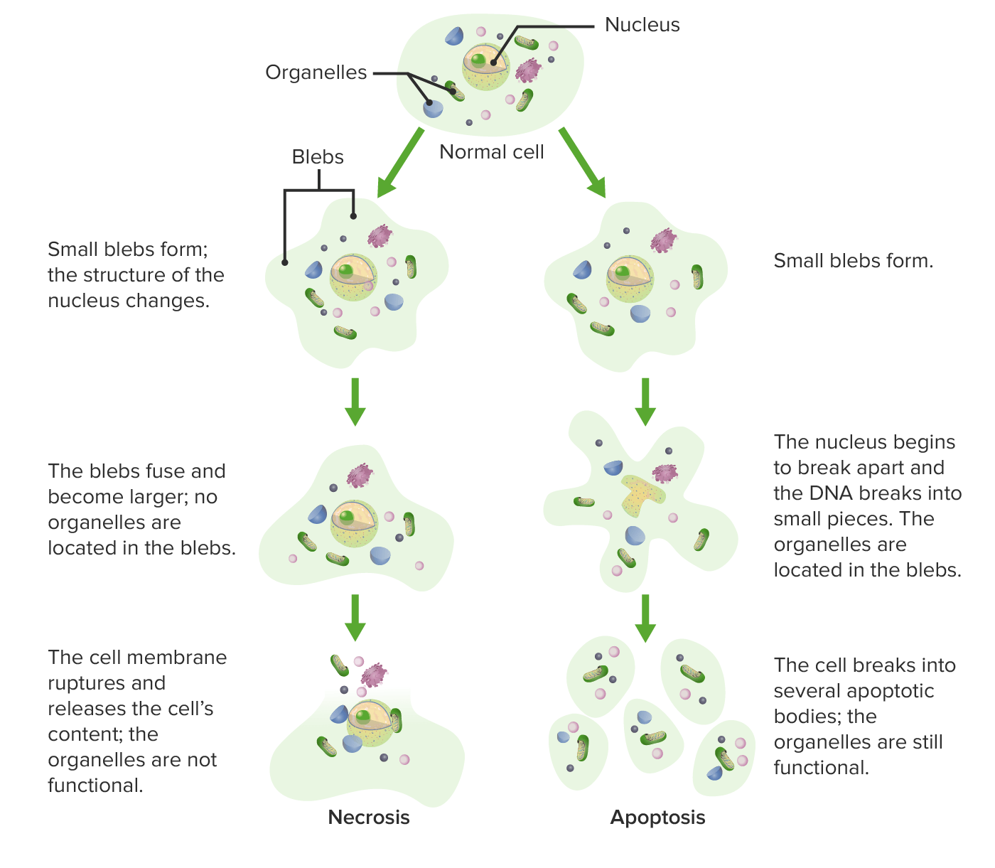
Cell deathCell deathInjurious stimuli trigger the process of cellular adaptation, whereby cells respond to withstand the harmful changes in their environment. Overwhelmed adaptive mechanisms lead to cell injury. Mild stimuli produce reversible injury. If the stimulus is severe or persistent, injury becomes irreversible. Apoptosis is programmed cell death, a mechanism with both physiologic and pathologic effects.Cell Injury and Death (via processes such as necrosis and apoptosis)
Cell deathCell deathInjurious stimuli trigger the process of cellular adaptation, whereby cells respond to withstand the harmful changes in their environment. Overwhelmed adaptive mechanisms lead to cell injury. Mild stimuli produce reversible injury. If the stimulus is severe or persistent, injury becomes irreversible. Apoptosis is programmed cell death, a mechanism with both physiologic and pathologic effects.Cell Injury and Death
Necrosis (most common cause):
Nonphysiological
Uncontrolled cell deathCell deathInjurious stimuli trigger the process of cellular adaptation, whereby cells respond to withstand the harmful changes in their environment. Overwhelmed adaptive mechanisms lead to cell injury. Mild stimuli produce reversible injury. If the stimulus is severe or persistent, injury becomes irreversible. Apoptosis is programmed cell death, a mechanism with both physiologic and pathologic effects.Cell Injury and Death after irreversible injury
Membrane damageMembrane DamageCell Injury and Death causes an influx of calciumCalciumA basic element found in nearly all tissues. It is a member of the alkaline earth family of metals with the atomic symbol ca, atomic number 20, and atomic weight 40. Calcium is the most abundant mineral in the body and combines with phosphorus to form calcium phosphate in the bones and teeth. It is essential for the normal functioning of nerves and muscles and plays a role in blood coagulation (as factor IV) and in many enzymatic processes.Electrolytes → organelle swellingSwellingInflammation → digestive enzyme release, which leads to:
Programmed cell deathCell deathInjurious stimuli trigger the process of cellular adaptation, whereby cells respond to withstand the harmful changes in their environment. Overwhelmed adaptive mechanisms lead to cell injury. Mild stimuli produce reversible injury. If the stimulus is severe or persistent, injury becomes irreversible. Apoptosis is programmed cell death, a mechanism with both physiologic and pathologic effects.Cell Injury and Death
Activated by the release of proapoptotic molecules from the mitochondriaMitochondriaSemiautonomous, self-reproducing organelles that occur in the cytoplasm of all cells of most, but not all, eukaryotes. Each mitochondrion is surrounded by a double limiting membrane. The inner membrane is highly invaginated, and its projections are called cristae. Mitochondria are the sites of the reactions of oxidative phosphorylation, which result in the formation of ATP. They contain distinctive ribosomes, transfer RNAs; amino Acyl tRNA synthetases; and elongation and termination factors. Mitochondria depend upon genes within the nucleus of the cells in which they reside for many essential messenger RNAs. Mitochondria are believed to have arisen from aerobic bacteria that established a symbiotic relationship with primitive protoeukaryotes.The Cell: Organelles (physiologic response)
Extrinsic pathways:
Fas (CD95) → Fas ligandFas ligandA transmembrane protein belonging to the tumor necrosis factor superfamily that was originally discovered on cells of the lymphoid-myeloid lineage, including activated T-lymphocytes and natural killer cells. It plays an important role in immune homeostasis and cell-mediated toxicity by binding to the fas receptor and triggering apoptosis.Tumor Necrosis Factor (TNF) (FasL)
TumorTumorInflammation necrosis factor (TNFTNFTumor necrosis factor (TNF) is a major cytokine, released primarily by macrophages in response to stimuli. The presence of microbial products and dead cells and injury are among the stimulating factors. This protein belongs to the TNF superfamily, a group of ligands and receptors performing functions in inflammatory response, morphogenesis, and cell proliferation. Tumor Necrosis Factor (TNF))-α → TNFTNFTumor necrosis factor (TNF) is a major cytokine, released primarily by macrophages in response to stimuli. The presence of microbial products and dead cells and injury are among the stimulating factors. This protein belongs to the TNF superfamily, a group of ligands and receptors performing functions in inflammatory response, morphogenesis, and cell proliferation. Tumor Necrosis Factor (TNF)receptorReceptorReceptors are proteins located either on the surface of or within a cell that can bind to signaling molecules known as ligands (e.g., hormones) and cause some type of response within the cell.Receptors 1 (TNR1)
Intrinsic pathwayIntrinsic pathwayThe intrinsic pathway is mainly responsible for the amplification of factor X activationHemostasis (mitochondrial pathway):
DNA damageDNA DamageInjuries to DNA that introduce deviations from its normal, intact structure and which may, if left unrepaired, result in a mutation or a block of DNA replication. These deviations may be caused by physical or chemical agents and occur by natural or unnatural, introduced circumstances. They include the introduction of illegitimate bases during replication or by deamination or other modification of bases; the loss of a base from the DNA backbone leaving an abasic site; single-strand breaks; double strand breaks; and intrastrand (pyrimidine dimers) or interstrand crosslinking. Damage can often be repaired (DNA repair). If the damage is extensive, it can induce apoptosis.DNA Repair Mechanisms → p53 activated → cell cycleCell cycleThe phases of the cell cycle include interphase (G1, S, and G2) and mitosis (prophase, metaphase, anaphase, and telophase). The cell’s progression through these phases is punctuated by checkpoints regulated by cyclins, cyclin-dependent kinases, tumor suppressors, and their antagonists.Cell Cycle arrest → p53 activates apoptosis
↑ Proapoptotic proteinsProteinsLinear polypeptides that are synthesized on ribosomes and may be further modified, crosslinked, cleaved, or assembled into complex proteins with several subunits. The specific sequence of amino acids determines the shape the polypeptide will take, during protein folding, and the function of the protein.Energy Homeostasis (e.g., BAK and BAX), ↓ antiapoptotic proteinsProteinsLinear polypeptides that are synthesized on ribosomes and may be further modified, crosslinked, cleaved, or assembled into complex proteins with several subunits. The specific sequence of amino acids determines the shape the polypeptide will take, during protein folding, and the function of the protein.Energy Homeostasis (e.g., Bcl-2BCL-2The B-cell leukemia/lymphoma-2 genes, responsible for blocking apoptosis in normal cells, and associated with follicular lymphoma when overexpressed. Overexpression results from the t(14; 18) translocation. The human c-bcl-2 gene is located at 18q24 on the long arm of chromosome 18.Non-Hodgkin Lymphomas) → mitochondriaMitochondriaSemiautonomous, self-reproducing organelles that occur in the cytoplasm of all cells of most, but not all, eukaryotes. Each mitochondrion is surrounded by a double limiting membrane. The inner membrane is highly invaginated, and its projections are called cristae. Mitochondria are the sites of the reactions of oxidative phosphorylation, which result in the formation of ATP. They contain distinctive ribosomes, transfer RNAs; amino Acyl tRNA synthetases; and elongation and termination factors. Mitochondria depend upon genes within the nucleus of the cells in which they reside for many essential messenger RNAs. Mitochondria are believed to have arisen from aerobic bacteria that established a symbiotic relationship with primitive protoeukaryotes.The Cell: Organelles release cytochrome c
Cytochrome c binds apoptosis protease-activating factor (APAF)-1 → activation of caspase and endonuclease
PerforinPerforinA calcium-dependent pore-forming protein synthesized in cytolytic lymphocytes and sequestered in secretory granules. Upon immunological reaction between a cytolytic lymphocyte and a target cell, perforin is released at the plasma membrane and polymerizes into transmembrane tubules (forming pores) which lead to death of a target cell.Lymphocytes: Histology/granzyme pathwayGranzyme pathwayA family of serine endopeptidases found in the secretory granules of leukocytes such as cytotoxic T-lymphocytes and natural killer cells. When secreted into the intercellular space granzymes act to eliminate transformed and virus-infected host cells.Innate Immunity: Phagocytes and Antigen Presentation:
Utilized by cytotoxicCytotoxicParvovirus B19T cellsT cellsLymphocytes responsible for cell-mediated immunity. Two types have been identified – cytotoxic (t-lymphocytes, cytotoxic) and helper T-lymphocytes (t-lymphocytes, helper-inducer). They are formed when lymphocytes circulate through the thymus gland and differentiate to thymocytes. When exposed to an antigen, they divide rapidly and produce large numbers of new T cells sensitized to that antigen.T cells: Types and Functions and natural killer cellsNatural killer cellsA specialized subset of T-lymphocytes that exhibit features of innate immunity similar to that of natural killer cells. They are reactive to glycolipids presented in the context of the major histocompatibility complex (MHC) class I-like molecule, CD1D antigen.Lymphocytes: Histology
PerforinPerforinA calcium-dependent pore-forming protein synthesized in cytolytic lymphocytes and sequestered in secretory granules. Upon immunological reaction between a cytolytic lymphocyte and a target cell, perforin is released at the plasma membrane and polymerizes into transmembrane tubules (forming pores) which lead to death of a target cell.Lymphocytes: Histology pores are created in target cells, which allow entry of caspase-like granzyme.
Dead cells are replaced by phospholipidsPhospholipidsLipids containing one or more phosphate groups, particularly those derived from either glycerol (phosphoglycerides) or sphingosine (sphingolipids). They are polar lipids that are of great importance for the structure and function of cell membranes and are the most abundant of membrane lipids, although not stored in large amounts in the system.Lipid Metabolism and myelin figuresMyelin figuresLarge whorled phospholipid precipitates (from the damaged membrane), which are phagocytosed or degraded to fatty acidsCell Injury and Death, resulting in clarification or phagocytosisPhagocytosisThe engulfing and degradation of microorganisms; other cells that are dead, dying, or pathogenic; and foreign particles by phagocytic cells (phagocytes).Innate Immunity: Phagocytes and Antigen Presentation by macrophagesMacrophagesThe relatively long-lived phagocytic cell of mammalian tissues that are derived from blood monocytes. Main types are peritoneal macrophages; alveolar macrophages; histiocytes; kupffer cells of the liver; and osteoclasts. They may further differentiate within chronic inflammatory lesions to epithelioid cells or may fuse to form foreign body giant cells or langhans giant cells.Innate Immunity: Phagocytes and Antigen Presentation.
Differences between apoptosis and necrosis at structural level
↓ Supply of blood: mechanical arterial obstruction (most common):
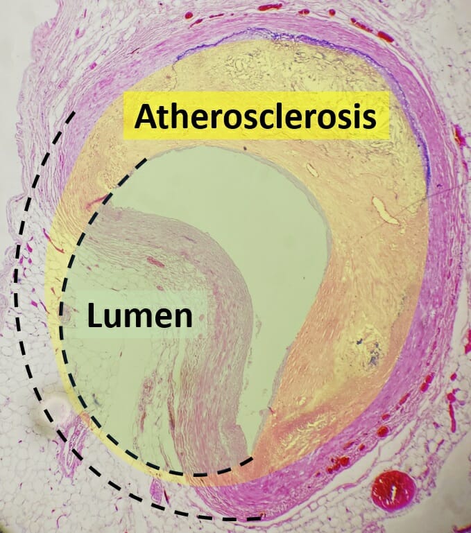
AtherosclerosisAtherosclerosisAtherosclerosis is a common form of arterial disease in which lipid deposition forms a plaque in the blood vessel walls. Atherosclerosis is an incurable disease, for which there are clearly defined risk factors that often can be reduced through a change in lifestyle and behavior of the patient. Atherosclerosis: plaquePlaquePrimary Skin Lesions building up in arterial walls
ThromboembolismThromboembolismObstruction of a blood vessel (embolism) by a blood clot (thrombus) in the blood stream.Systemic Lupus Erythematosus: blockage of a blood vessel by a clot dislodged from another source in the body
↓ Venous drainage of blood: venous flowFlowBlood flows through the heart, arteries, capillaries, and veins in a closed, continuous circuit. Flow is the movement of volume per unit of time. Flow is affected by the pressure gradient and the resistance fluid encounters between 2 points. Vascular resistance is the opposition to flow, which is caused primarily by blood friction against vessel walls.Vascular Resistance, Flow, and Mean Arterial Pressure stops:
Deep vein thrombosisThrombosisFormation and development of a thrombus or blood clot in the blood vessel.Epidemic Typhus: a blood clot in the deep veinsVeinsVeins are tubular collections of cells, which transport deoxygenated blood and waste from the capillary beds back to the heart. Veins are classified into 3 types: small veins/venules, medium veins, and large veins. Each type contains 3 primary layers: tunica intima, tunica media, and tunica adventitia. Veins: Histology
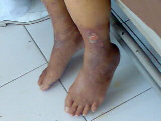
Peripheral venous disease: progressive stenosisStenosisHypoplastic Left Heart Syndrome (HLHS) of veinsVeinsVeins are tubular collections of cells, which transport deoxygenated blood and waste from the capillary beds back to the heart. Veins are classified into 3 types: small veins/venules, medium veins, and large veins. Each type contains 3 primary layers: tunica intima, tunica media, and tunica adventitia. Veins: Histology
ShockShockShock is a life-threatening condition associated with impaired circulation that results in tissue hypoxia. The different types of shock are based on the underlying cause: distributive (↑ cardiac output (CO), ↓ systemic vascular resistance (SVR)), cardiogenic (↓ CO, ↑ SVR), hypovolemic (↓ CO, ↑ SVR), obstructive (↓ CO), and mixed. Types of Shock:
Cardiogenic: ↓ left ventricular function (e.g., congestive heart failureHeart FailureA heterogeneous condition in which the heart is unable to pump out sufficient blood to meet the metabolic need of the body. Heart failure can be caused by structural defects, functional abnormalities (ventricular dysfunction), or a sudden overload beyond its capacity. Chronic heart failure is more common than acute heart failure which results from sudden insult to cardiac function, such as myocardial infarction.Total Anomalous Pulmonary Venous Return (TAPVR))
Distributive: septic (from sepsisSepsisSystemic inflammatory response syndrome with a proven or suspected infectious etiology. When sepsis is associated with organ dysfunction distant from the site of infection, it is called severe sepsis. When sepsis is accompanied by hypotension despite adequate fluid infusion, it is called septic shock.Sepsis and Septic Shock), neurogenic (e.g., spinal cordSpinal cordThe spinal cord is the major conduction pathway connecting the brain to the body; it is part of the CNS. In cross section, the spinal cord is divided into an H-shaped area of gray matter (consisting of synapsing neuronal cell bodies) and a surrounding area of white matter (consisting of ascending and descending tracts of myelinated axons). Spinal Cord: Anatomy injury), or anaphylactic
Obstructive: lack of cardiac outflow (e.g., tension pneumothoraxTension PneumothoraxPneumothorax/tamponadeTamponadePericardial effusion, usually of rapid onset, exceeding ventricular filling pressures and causing collapse of the heart with a markedly reduced cardiac output.Pericarditis or pulmonary embolismPulmonary EmbolismPulmonary embolism (PE) is a potentially fatal condition that occurs as a result of intraluminal obstruction of the main pulmonary artery or its branches. The causative factors include thrombi, air, amniotic fluid, and fat. In PE, gas exchange is impaired due to the decreased return of deoxygenated blood to the lungs. Pulmonary Embolism)
Histopathology of a collapsed coronary artery due to atherosclerosis
Image: “Histopathology of coronary artery atherosclerosis, annotated” by Mikael Häggström. License: CC0 1.0
Ulcers on the extremities caused by peripheral venous disease
Image: “Peripheral Vascular Disease” by Wfnicdao. License: Public Domain
Mechanism of injury
↓ Oxygen availability (aerobic metabolism is interrupted) → reduced ATP production → failure of energy-dependent systems:
Plasma membranePlasma membraneA cell membrane (also known as the plasma membrane or plasmalemma) is a biological membrane that separates the cell contents from the outside environment. A cell membrane is composed of a phospholipid bilayer and proteins that function to protect cellular DNA and mediate the exchange of ions and molecules.The Cell: Cell Membrane +Na+-K+pumpPumpACES and RUSH: Resuscitation Ultrasound Protocols (Na⁺, K⁺-ATPase) fails → sodiumSodiumA member of the alkali group of metals. It has the atomic symbol na, atomic number 11, and atomic weight 23.Hyponatremia enters the cell → cell swells
Anaerobic metabolism compensates for the ATP loss → depleted glycogen → ↑ lactic acid → ↓ intracellular pHpHThe quantitative measurement of the acidity or basicity of a solution.Acid-Base Balance → impaired enzymesEnzymesEnzymes are complex protein biocatalysts that accelerate chemical reactions without being consumed by them. Due to the body’s constant metabolic needs, the absence of enzymes would make life unsustainable, as reactions would occur too slowly without these molecules. Basics of Enzymes
Impaired enzymesEnzymesEnzymes are complex protein biocatalysts that accelerate chemical reactions without being consumed by them. Due to the body’s constant metabolic needs, the absence of enzymes would make life unsustainable, as reactions would occur too slowly without these molecules. Basics of Enzymes lead to reduced protein synthesisSynthesisPolymerase Chain Reaction (PCR) → detachment of ribosomesRibosomesMulticomponent ribonucleoprotein structures found in the cytoplasm of all cells, and in mitochondria, and plastids. They function in protein biosynthesis via genetic translation.The Cell: Organelles
Microscopic changes:
Loss of microvilli and formation of “blebs” in the cytoplasm and on the cell membraneCell MembraneA cell membrane (also known as the plasma membrane or plasmalemma) is a biological membrane that separates the cell contents from the outside environment. A cell membrane is composed of a phospholipid bilayer and proteins that function to protect cellular DNA and mediate the exchange of ions and molecules. The Cell: Cell Membrane
Cell and organellesOrganellesA cell is a complex unit that performs several complex functions. An organelle is a specialized subunit within a cell that fulfills a specific role or function. Organelles are enclosed within their own lipid bilayers or are unbound by membranes. The Cell: Organelles swell
Cells start to lose functionality.
↑ Intracellular concentrations of water, sodiumSodiumA member of the alkali group of metals. It has the atomic symbol na, atomic number 11, and atomic weight 23.Hyponatremia, and chlorideChlorideInorganic compounds derived from hydrochloric acid that contain the Cl- ion.Electrolytes, but ↓ potassiumPotassiumAn element in the alkali group of metals with an atomic symbol k, atomic number 19, and atomic weight 39. 10. It is the chief cation in the intracellular fluid of muscle and other cells. Potassium ion is a strong electrolyte that plays a significant role in the regulation of fluid volume and maintenance of the water-electrolyte balance.Hyperkalemia
All changes are reversible if perfusion and oxygenation are restored.
If ischemia persists, the tissue succumbs to irreversible injury and death.
Recovery can be achieved (especially with reversible injury).
Can paradoxically worsen the injury and lead to cell deathCell deathInjurious stimuli trigger the process of cellular adaptation, whereby cells respond to withstand the harmful changes in their environment. Overwhelmed adaptive mechanisms lead to cell injury. Mild stimuli produce reversible injury. If the stimulus is severe or persistent, injury becomes irreversible. Apoptosis is programmed cell death, a mechanism with both physiologic and pathologic effects.Cell Injury and Death → ischemia-reperfusion injury:
Reperfusion can exacerbate the damage and injure distant organs when mediators are released into the bloodstream.
A clinically significant consideration in the treatment of myocardial infarctionMyocardial infarctionMI is ischemia and death of an area of myocardial tissue due to insufficient blood flow and oxygenation, usually from thrombus formation on a ruptured atherosclerotic plaque in the epicardial arteries. Clinical presentation is most commonly with chest pain, but women and patients with diabetes may have atypical symptoms.Myocardial Infarction and stroke
Mechanism of injury
Perfusion is restored, which brings damaging pathways:
Oxidative stressOxidative stressA disturbance in the prooxidant-antioxidant balance in favor of the former, leading to potential damage. Indicators of oxidative stress include damaged DNA bases, protein oxidation products, and lipid peroxidation products.Cell Injury and Death: ↑ production of reactive oxygen speciesReactive oxygen speciesMolecules or ions formed by the incomplete one-electron reduction of oxygen. These reactive oxygen intermediates include singlet oxygen; superoxides; peroxides; hydroxyl radical; and hypochlorous acid. They contribute to the microbicidal activity of phagocytes, regulation of signal transduction and gene expression, and the oxidative damage to nucleic acids; proteins; and lipids.Metabolic Dysfunction-associated Steatotic Liver Disease (MASLD) (ROS) or free radicals (molecules with an unpaired electron in the outer orbit):
Produced from leukocytesLeukocytesWhite blood cells. These include granular leukocytes (basophils; eosinophils; and neutrophils) as well as non-granular leukocytes (lymphocytes and monocytes).White Myeloid Cells: Histology and damaged cells
Overload of intracellular calciumCalciumA basic element found in nearly all tissues. It is a member of the alkaline earth family of metals with the atomic symbol ca, atomic number 20, and atomic weight 40. Calcium is the most abundant mineral in the body and combines with phosphorus to form calcium phosphate in the bones and teeth. It is essential for the normal functioning of nerves and muscles and plays a role in blood coagulation (as factor IV) and in many enzymatic processes.Electrolytes:
↑ CalciumCalciumA basic element found in nearly all tissues. It is a member of the alkaline earth family of metals with the atomic symbol ca, atomic number 20, and atomic weight 40. Calcium is the most abundant mineral in the body and combines with phosphorus to form calcium phosphate in the bones and teeth. It is essential for the normal functioning of nerves and muscles and plays a role in blood coagulation (as factor IV) and in many enzymatic processes.Electrolytes → opening of mitochondrial permeability transition pore (mPTPMPTPParkinson’s Disease) → ATP depletionATP depletionCell Injury and Death
↑ CalciumCalciumA basic element found in nearly all tissues. It is a member of the alkaline earth family of metals with the atomic symbol ca, atomic number 20, and atomic weight 40. Calcium is the most abundant mineral in the body and combines with phosphorus to form calcium phosphate in the bones and teeth. It is essential for the normal functioning of nerves and muscles and plays a role in blood coagulation (as factor IV) and in many enzymatic processes.Electrolytes → cellular enzymesEnzymesEnzymes are complex protein biocatalysts that accelerate chemical reactions without being consumed by them. Due to the body’s constant metabolic needs, the absence of enzymes would make life unsustainable, as reactions would occur too slowly without these molecules. Basics of Enzymes (e.g., proteaseProteaseEnzyme of the human immunodeficiency virus that is required for post-translational cleavage of gag and gag-pol precursor polyproteins into functional products needed for viral assembly. HIV protease is an aspartic protease encoded by the amino terminus of the pol gene.HIV Infection and AIDS, phospholipase, ATPase, endonuclease) → membrane and nuclear damage
LeukocytesLeukocytesWhite blood cells. These include granular leukocytes (basophils; eosinophils; and neutrophils) as well as non-granular leukocytes (lymphocytes and monocytes).White Myeloid Cells: Histology and cytokinesCytokinesNon-antibody proteins secreted by inflammatory leukocytes and some non-leukocytic cells, that act as intercellular mediators. They differ from classical hormones in that they are produced by a number of tissue or cell types rather than by specialized glands. They generally act locally in a paracrine or autocrine rather than endocrine manner.Adaptive Immune Response → recruit more immune cells → ↑ inflammationInflammationInflammation is a complex set of responses to infection and injury involving leukocytes as the principal cellular mediators in the body’s defense against pathogenic organisms. Inflammation is also seen as a response to tissue injury in the process of wound healing. The 5 cardinal signs of inflammation are pain, heat, redness, swelling, and loss of function. Inflammation (“sterileSterileBasic ProceduresinflammationInflammationInflammation is a complex set of responses to infection and injury involving leukocytes as the principal cellular mediators in the body’s defense against pathogenic organisms. Inflammation is also seen as a response to tissue injury in the process of wound healing. The 5 cardinal signs of inflammation are pain, heat, redness, swelling, and loss of function. Inflammation”)
Complement systemComplement systemSerum glycoproteins participating in the host defense mechanism of complement activation that creates the complement membrane attack complex. Included are glycoproteins in the various pathways of complement activation (classical complement pathway; alternative complement pathway; and lectin complement pathway).Innate Immunity: Barriers, Complement, and Cytokines activation → complement proteinsProteinsLinear polypeptides that are synthesized on ribosomes and may be further modified, crosslinked, cleaved, or assembled into complex proteins with several subunits. The specific sequence of amino acids determines the shape the polypeptide will take, during protein folding, and the function of the protein.Energy HomeostasisbindBINDHyperbilirubinemia of the Newborn ischemic tissues of antibodiesAntibodiesImmunoglobulins (Igs), also known as antibodies, are glycoprotein molecules produced by plasma cells that act in immune responses by recognizing and binding particular antigens. The various Ig classes are IgG (the most abundant), IgM, IgE, IgD, and IgA, which differ in their biologic features, structure, target specificity, and distribution.Immunoglobulins: Types and Functions → ↑ inflammationInflammationInflammation is a complex set of responses to infection and injury involving leukocytes as the principal cellular mediators in the body’s defense against pathogenic organisms. Inflammation is also seen as a response to tissue injury in the process of wound healing. The 5 cardinal signs of inflammation are pain, heat, redness, swelling, and loss of function. Inflammation
The combination of the mechanisms induce:
Damage to DNADNAA deoxyribonucleotide polymer that is the primary genetic material of all cells. Eukaryotic and prokaryotic organisms normally contain DNA in a double-stranded state, yet several important biological processes transiently involve single-stranded regions. DNA, which consists of a polysugar-phosphate backbone possessing projections of purines (adenine and guanine) and pyrimidines (thymine and cytosine), forms a double helix that is held together by hydrogen bonds between these purines and pyrimidines (adenine to thymine and guanine to cytosine).DNA Types and Structure, structural proteinsStructural proteinsProteins and Peptides, and lipidsLipidsLipids are a diverse group of hydrophobic organic molecules, which include fats, oils, sterols, and waxes.Fatty Acids and Lipids
Further activation of proinflammatory and prothrombotic cascades
Cellular architecture is lost and cell deathCell deathInjurious stimuli trigger the process of cellular adaptation, whereby cells respond to withstand the harmful changes in their environment. Overwhelmed adaptive mechanisms lead to cell injury. Mild stimuli produce reversible injury. If the stimulus is severe or persistent, injury becomes irreversible. Apoptosis is programmed cell death, a mechanism with both physiologic and pathologic effects.Cell Injury and Death follows.
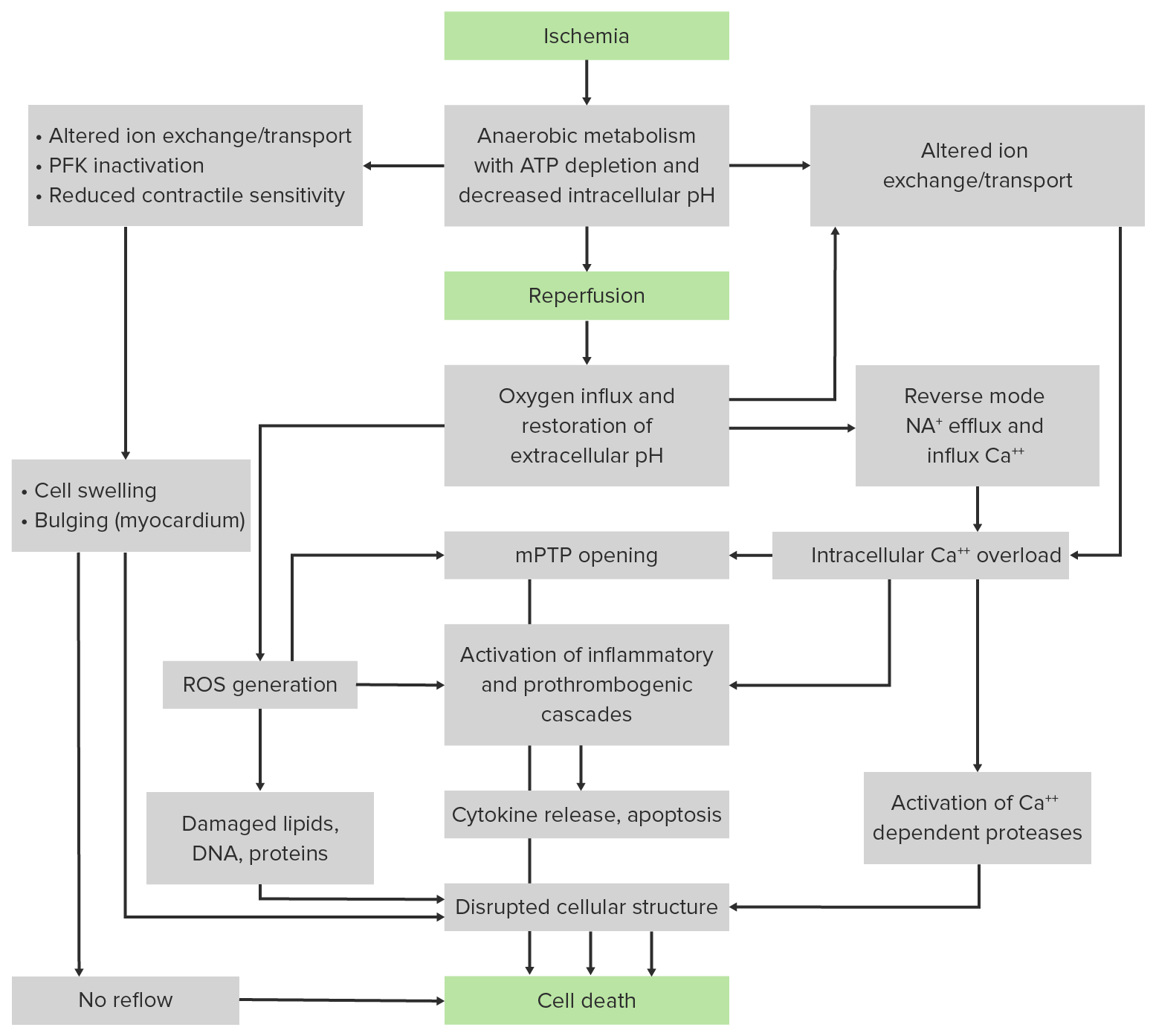
Flowchart summarizing the major pathologic events contributing to ischemic (upper panel) and reperfusion (middle panel) components of tissue injury: In prolonged ischemia, hypoxia leads to depletion of ATP and reduced intracellular pH (from lactate accumulation). ATP-dependent ion transport mechanisms become deranged, causing cellular calcium overload, swelling/rupture, and death. When oxygen levels are restored (reperfusion), reactive oxygen species (ROS) are generated. Proinflammatory changes also occur: Neutrophils infiltrate ischemic tissues and worsen the ischemic injury. The pathologic events lead to the opening of the mitochondrial permeability transition pore (mPTP) in the inner mitochondrial membrane, which allow passage of molecules into the mitochondria and further impair ATP production. PFK: phosphofructokinase
Infarct: area of necrotic cells in an organ, arising mainly from hypoxia and ischemia:
Red or “hemorrhagic” infarct:
Affects organs with multiple blood supplies or loose parenchyma allowing blood to leak into tissue (e.g., lungsLungsLungs are the main organs of the respiratory system. Lungs are paired viscera located in the thoracic cavity and are composed of spongy tissue. The primary function of the lungs is to oxygenate blood and eliminate CO2. Lungs: Anatomy)
From venous infarcts: The vein is blocked, but the artery delivers blood.
Reperfusion injury: The restoration of blood flowBlood flowBlood flow refers to the movement of a certain volume of blood through the vasculature over a given unit of time (e.g., mL per minute).Vascular Resistance, Flow, and Mean Arterial Pressure causes blood to leak through damaged vessels.
Pale/white or “anemic” infarct: injury to organs with a single arterial supply or solid parenchyma (e.g., kidney, heart)
BrainBrainThe part of central nervous system that is contained within the skull (cranium). Arising from the neural tube, the embryonic brain is comprised of three major parts including prosencephalon (the forebrain); mesencephalon (the midbrain); and rhombencephalon (the hindbrain). The developed brain consists of cerebrum; cerebellum; and other structures in the brain stem.Nervous System: Anatomy, Structure, and Classification:
The most susceptible organ to ischemia
The least amount of time before irreversibility occurs
Most susceptible organs to reduced blood supply after the brainBrainThe part of central nervous system that is contained within the skull (cranium). Arising from the neural tube, the embryonic brain is comprised of three major parts including prosencephalon (the forebrain); mesencephalon (the midbrain); and rhombencephalon (the hindbrain). The developed brain consists of cerebrum; cerebellum; and other structures in the brain stem.Nervous System: Anatomy, Structure, and Classification: The heart/myocardiumMyocardiumThe muscle tissue of the heart. It is composed of striated, involuntary muscle cells connected to form the contractile pump to generate blood flow.Heart: Anatomy is the 2nd and the kidneysKidneysThe kidneys are a pair of bean-shaped organs located retroperitoneally against the posterior wall of the abdomen on either side of the spine. As part of the urinary tract, the kidneys are responsible for blood filtration and excretion of water-soluble waste in the urine.Kidneys: Anatomy are the 3rd.
Both skinSkinThe skin, also referred to as the integumentary system, is the largest organ of the body. The skin is primarily composed of the epidermis (outer layer) and dermis (deep layer). The epidermis is primarily composed of keratinocytes that undergo rapid turnover, while the dermis contains dense layers of connective tissue.Skin: Structure and Functions and skeletal muscle tolerate longer periods of ischemia:
Often seen in the emergency application of tourniquets (sometimes for hours) with little injury to the tissues
Release (after the 1st 2 hours) followed by reapplication of compressionCompressionBlunt Chest Trauma produces minimal injuries.
Anatomically, some organs have watershed areas (border zone):
The regions have dual blood supply, but are located at the most distal reaches of the arteriesArteriesArteries are tubular collections of cells that transport oxygenated blood and nutrients from the heart to the tissues of the body. The blood passes through the arteries in order of decreasing luminal diameter, starting in the largest artery (the aorta) and ending in the small arterioles. Arteries are classified into 3 types: large elastic arteries, medium muscular arteries, and small arteries and arterioles. Arteries: Histology.
Susceptible to ischemia
BrainBrainThe part of central nervous system that is contained within the skull (cranium). Arising from the neural tube, the embryonic brain is comprised of three major parts including prosencephalon (the forebrain); mesencephalon (the midbrain); and rhombencephalon (the hindbrain). The developed brain consists of cerebrum; cerebellum; and other structures in the brain stem.Nervous System: Anatomy, Structure, and Classification
With high metabolic activity and low carbohydrate stores, the brainBrainThe part of central nervous system that is contained within the skull (cranium). Arising from the neural tube, the embryonic brain is comprised of three major parts including prosencephalon (the forebrain); mesencephalon (the midbrain); and rhombencephalon (the hindbrain). The developed brain consists of cerebrum; cerebellum; and other structures in the brain stem.Nervous System: Anatomy, Structure, and Classification has the highest susceptibility to ischemia.
Ischemia occurs when an embolus or thrombus (ischemic strokeIschemic StrokeAn ischemic stroke (also known as cerebrovascular accident) is an acute neurologic injury that occurs as a result of brain ischemia; this condition may be due to cerebral blood vessel occlusion by thrombosis or embolism, or rarely due to systemic hypoperfusion. Ischemic Stroke) reduces the blood flowBlood flowBlood flow refers to the movement of a certain volume of blood through the vasculature over a given unit of time (e.g., mL per minute).Vascular Resistance, Flow, and Mean Arterial Pressure:
Survival of tissue depends on:
Collateral circulationCirculationThe movement of the blood as it is pumped through the cardiovascular system.ABCDE Assessment
NeuronsNeuronsThe basic cellular units of nervous tissue. Each neuron consists of a body, an axon, and dendrites. Their purpose is to receive, conduct, and transmit impulses in the nervous system.Nervous System: Histology die within 5 minutes in the case of complete blockage.
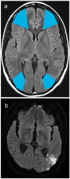
Watershed areas:
The border zones of the arterial territories
The area between the anterior and middle cerebral arteryMiddle cerebral arteryThe largest of the cerebral arteries. It trifurcates into temporal, frontal, and parietal branches supplying blood to most of the parenchyma of these lobes in the cerebral cortex. These are the areas involved in motor, sensory, and speech activities.Cerebrovascular System: Anatomy distribution is at highest risk.
Infarcts develop after significant hypotensionHypotensionHypotension is defined as low blood pressure, specifically < 90/60 mm Hg, and is most commonly a physiologic response. Hypotension may be mild, serious, or life threatening, depending on the cause. Hypotension.
Watershed areas and infarcts seen on MRI: a: Watershed areas between the anterior and middle cerebral arterial territories are seen in the anterior blue shade. Watershed areas between the middle and posterior arterial territories are seen in the posterior blue shade. b: Occipital watershed infarct is seen at the boundaries of the middle and the posterior arterial territories.
Image: “Watershed territories” by Clothilde Isabel et al. License: CC BY 4.0
Heart
In the setting severe ischemia:
Injury to the myocardiumMyocardiumThe muscle tissue of the heart. It is composed of striated, involuntary muscle cells connected to form the contractile pump to generate blood flow.Heart: Anatomy is potentially reversible within 30 minutes.
The most susceptible tissue in the heart is the subendocardial muscle of the left ventricle.
Damage from cardiac ischemia leads to:
Stable anginaStable anginaPersistent and reproducible chest discomfort usually precipitated by a physical exertion that dissipates upon cessation of such an activity. The symptoms are manifestations of myocardial ischemia.Stable and Unstable Angina:
Angina (chest painPainAn unpleasant sensation induced by noxious stimuli which are detected by nerve endings of nociceptive neurons.Pain: Types and Pathways) subsides within 15 minutes of rest or with administration of nitroglycerinNitroglycerinA volatile vasodilator which relieves angina pectoris by stimulating guanylate cyclase and lowering cytosolic calcium. It is also sometimes used for tocolysis and explosives.Nitrates.
Unstable or crescendo angina: Angina lasting > 20 minutes at rest or with minimal exertion (troponin levels are normal).
Non-ST-elevation myocardial infarctionMyocardial infarctionMI is ischemia and death of an area of myocardial tissue due to insufficient blood flow and oxygenation, usually from thrombus formation on a ruptured atherosclerotic plaque in the epicardial arteries. Clinical presentation is most commonly with chest pain, but women and patients with diabetes may have atypical symptoms.Myocardial Infarction (NSTEMI):
Myocardial infarctionMyocardial infarctionMI is ischemia and death of an area of myocardial tissue due to insufficient blood flow and oxygenation, usually from thrombus formation on a ruptured atherosclerotic plaque in the epicardial arteries. Clinical presentation is most commonly with chest pain, but women and patients with diabetes may have atypical symptoms.Myocardial Infarction with angina and increased troponin
Not associated with elevation of the ST segmentST segmentIsoelectric segment between the s wave and the initial deflection of the t wave.Electrocardiogram (ECG) on ECGECGAn electrocardiogram (ECG) is a graphic representation of the electrical activity of the heart plotted against time. Adhesive electrodes are affixed to the skin surface allowing measurement of cardiac impulses from many angles. The ECG provides 3-dimensional information about the conduction system of the heart, the myocardium, and other cardiac structures. Electrocardiogram (ECG)
STEMI: a myocardial infarctionMyocardial infarctionMI is ischemia and death of an area of myocardial tissue due to insufficient blood flow and oxygenation, usually from thrombus formation on a ruptured atherosclerotic plaque in the epicardial arteries. Clinical presentation is most commonly with chest pain, but women and patients with diabetes may have atypical symptoms.Myocardial Infarction with angina and elevation of the ST segmentST segmentIsoelectric segment between the s wave and the initial deflection of the t wave.Electrocardiogram (ECG) on ECGECGAn electrocardiogram (ECG) is a graphic representation of the electrical activity of the heart plotted against time. Adhesive electrodes are affixed to the skin surface allowing measurement of cardiac impulses from many angles. The ECG provides 3-dimensional information about the conduction system of the heart, the myocardium, and other cardiac structures. Electrocardiogram (ECG)
Kidney
Vulnerable due to:
A significant amount of cardiac outputCardiac outputThe volume of blood passing through the heart per unit of time. It is usually expressed as liters (volume) per minute so as not to be confused with stroke volume (volume per beat).Cardiac Mechanics (25%) moving to the kidneysKidneysThe kidneys are a pair of bean-shaped organs located retroperitoneally against the posterior wall of the abdomen on either side of the spine. As part of the urinary tract, the kidneys are responsible for blood filtration and excretion of water-soluble waste in the urine.Kidneys: Anatomy
Limited collateral blood supply from extrarenal sites
High metabolic activity
Shows pale/white infarct when ischemic damage occurs
Ischemia can occur in cases of:
HypotensionHypotensionHypotension is defined as low blood pressure, specifically < 90/60 mm Hg, and is most commonly a physiologic response. Hypotension may be mild, serious, or life threatening, depending on the cause. Hypotension
SepsisSepsisSystemic inflammatory response syndrome with a proven or suspected infectious etiology. When sepsis is associated with organ dysfunction distant from the site of infection, it is called severe sepsis. When sepsis is accompanied by hypotension despite adequate fluid infusion, it is called septic shock.Sepsis and Septic Shock
Surgery
Interruption to complete obstruction of blood supply noted in:
Cardioembolic disease (e.g., atrial fibrillationAtrial fibrillationAtrial fibrillation (AF or Afib) is a supraventricular tachyarrhythmia and the most common kind of arrhythmia. It is caused by rapid, uncontrolled atrial contractions and uncoordinated ventricular responses. Atrial Fibrillation)
Renal arteryRenal arteryA branch of the abdominal aorta which supplies the kidneys, adrenal glands and ureters.Glomerular Filtration injury
HypercoagulableHypercoagulableHypercoagulable states (also referred to as thrombophilias) are a group of hematologic diseases defined by an increased risk of clot formation (i.e., thrombosis) due to either an increase in procoagulants, a decrease in anticoagulants, or a decrease in fibrinolysis. Hypercoagulable States state
Areas most affected:
Proximal tubuleProximal tubuleThe renal tubule portion that extends from the bowman capsule in the kidney cortex into the kidney medulla. The proximal tubule consists of a convoluted proximal segment in the cortex, and a distal straight segment descending into the medulla where it forms the u-shaped loop of henle.Tubular System (S3S3Heart Sounds segment): minimal capacity to produce energy in anaerobic conditions
The medullary thick ascending limbThick ascending limbRenal Sodium and Water Regulation of the loop of HenleLoop of HenleThe U-shaped portion of the renal tubule in the kidney medulla, consisting of a descending limb and an ascending limb. It is situated between the proximal kidney tubule and the distal kidney tubule.Tubular System
LiverLiverThe liver is the largest gland in the human body. The liver is found in the superior right quadrant of the abdomen and weighs approximately 1.5 kilograms. Its main functions are detoxification, metabolism, nutrient storage (e.g., iron and vitamins), synthesis of coagulation factors, formation of bile, filtration, and storage of blood. Liver: Anatomy
With a complex vasculature and high metabolic activity, hepatic injury results from severe hypoperfusion.
Can occur with an interruption of blood supply to the liverLiverThe liver is the largest gland in the human body. The liver is found in the superior right quadrant of the abdomen and weighs approximately 1.5 kilograms. Its main functions are detoxification, metabolism, nutrient storage (e.g., iron and vitamins), synthesis of coagulation factors, formation of bile, filtration, and storage of blood. Liver: Anatomy:
Hepatic sickle cell crisis
Hepatic arteryHepatic arteryA branch of the celiac artery that distributes to the stomach, pancreas, duodenum, liver, gallbladder, and greater omentum.Liver: AnatomythrombosisThrombosisFormation and development of a thrombus or blood clot in the blood vessel.Epidemic Typhus
Other systemic conditions (e.g., shockShockShock is a life-threatening condition associated with impaired circulation that results in tissue hypoxia. The different types of shock are based on the underlying cause: distributive (↑ cardiac output (CO), ↓ systemic vascular resistance (SVR)), cardiogenic (↓ CO, ↑ SVR), hypovolemic (↓ CO, ↑ SVR), obstructive (↓ CO), and mixed. Types of Shock, respiratory failureRespiratory failureRespiratory failure is a syndrome that develops when the respiratory system is unable to maintain oxygenation and/or ventilation. Respiratory failure may be acute or chronic and is classified as hypoxemic, hypercapnic, or a combination of the two. Respiratory Failure)
Interruption of hepatic blood supply manifests with:
Elevation of transaminasesTransaminasesA subclass of enzymes of the transferase class that catalyze the transfer of an amino group from a donor (generally an amino acid) to an acceptor (generally a 2-keto acid). Most of these enzymes are pyridoxyl phosphate proteins.Autoimmune Hepatitis
Occasionally with GI symptoms (e.g., nauseaNauseaAn unpleasant sensation in the stomach usually accompanied by the urge to vomit. Common causes are early pregnancy, sea and motion sickness, emotional stress, intense pain, food poisoning, and various enteroviruses.Antiemetics, abdominal painAbdominal PainAcute Abdomen)
Often accompanied by other end-organ hypoperfusion (e.g., renal ischemia presenting as ↑ creatinine)
Area most affected: zone 3 (area closest to and around the central vein)
Intestine
Extensive collateral circulationCirculationThe movement of the blood as it is pumped through the cardiovascular system.ABCDE Assessment (protective against hypoperfusion)
Even if mesenteric blood flowBlood flowBlood flow refers to the movement of a certain volume of blood through the vasculature over a given unit of time (e.g., mL per minute).Vascular Resistance, Flow, and Mean Arterial Pressure decreases by 75% for up to 12 hours, injury is minimal due to collateral circulationCirculationThe movement of the blood as it is pumped through the cardiovascular system.ABCDE Assessment.
Sources of ischemia:
Mesenteric arterial occlusion (thrombosisThrombosisFormation and development of a thrombus or blood clot in the blood vessel.Epidemic Typhus or emboli)
Venous thrombosisVenous thrombosisThe formation or presence of a blood clot (thrombus) within a vein.Budd-Chiari Syndrome (↑ resistanceResistancePhysiologically, the opposition to flow of air caused by the forces of friction. As a part of pulmonary function testing, it is the ratio of driving pressure to the rate of air flow.Ventilation: Mechanics of Breathing from venous flowFlowBlood flows through the heart, arteries, capillaries, and veins in a closed, continuous circuit. Flow is the movement of volume per unit of time. Flow is affected by the pressure gradient and the resistance fluid encounters between 2 points. Vascular resistance is the opposition to flow, which is caused primarily by blood friction against vessel walls.Vascular Resistance, Flow, and Mean Arterial Pressure → bowel edemaEdemaEdema is a condition in which excess serous fluid accumulates in the body cavity or interstitial space of connective tissues. Edema is a symptom observed in several medical conditions. It can be categorized into 2 types, namely, peripheral (in the extremities) and internal (in an organ or body cavity). Edema and ischemia)
Nonocclusive mesenteric ischemiaMesenteric IschemiaMesenteric ischemia is a rare, life-threatening condition caused by inadequate blood flow through the mesenteric vessels, which results in ischemia and necrosis of the intestinal wall. Mesenteric ischemia can be either acute or chronic. Mesenteric Ischemia (splanchnic hypoperfusion)
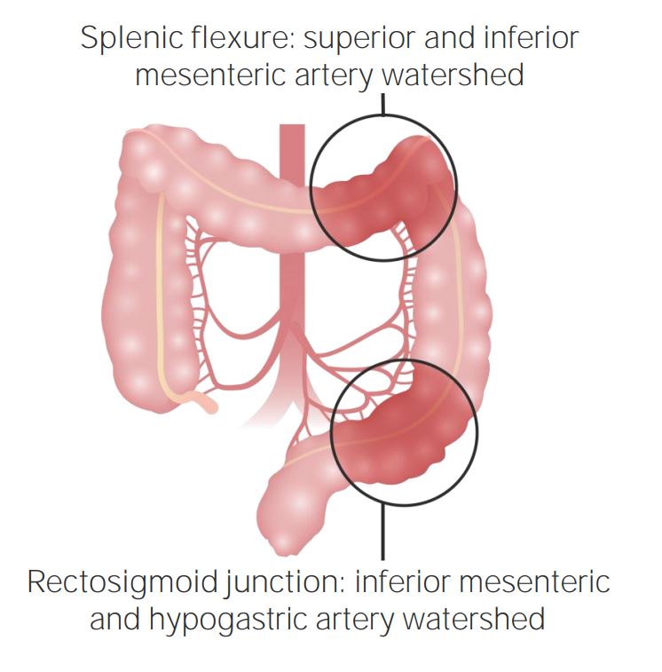
Watershed areas (large intestineLarge intestineThe large intestines constitute the last portion of the digestive system. The large intestine consists of the cecum, appendix, colon (with ascending, transverse, descending, and sigmoid segments), rectum, and anal canal. The primary function of the colon is to remove water and compact the stool prior to expulsion from the body via the rectum and anal canal. Colon, Cecum, and Appendix: Anatomy): splenic flexureSplenic flexureSmall Intestine: Anatomy:
Blood supply from the narrow terminal branches of the superior mesenteric arterySuperior mesenteric arteryA large vessel supplying the whole length of the small intestine except the superior part of the duodenum. It also supplies the cecum and the ascending part of the colon and about half the transverse part of the colon. It arises from the anterior surface of the aorta below the celiac artery at the level of the first lumbar vertebra.Small Intestine: Anatomy
Blood supply from the narrow terminal branches of the inferior mesenteric arteryInferior mesenteric arteryThe artery supplying nearly all the left half of the transverse colon, the whole of the descending colon, the sigmoid colon, and the greater part of the rectum. It is smaller than the superior mesenteric artery and arises from the aorta above its bifurcation into the common iliac arteries.Small Intestine: Anatomy
Sudeck’s point: area of weakness
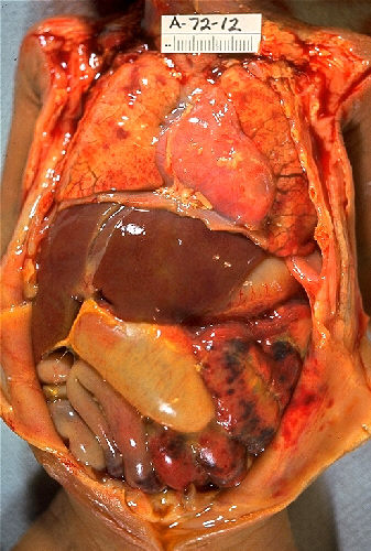
Necrotizing enterocolitis in an infant: Notice the patches of necrosis in the intestine.
Image: “Gross pathology of neonatal necrotizing enterocolitis” by Centers for Disease Control and Prevention. License: Public Domain
Watershed areas of the colon
Image by Lecturio.
References
El Sabbahy, M., Vaidya, V.S. (2011). Ischemic kidney injury and mechanisms of tissue repair. Wiley interdisciplinary reviews. Systems biology and medicine, 3(5), 606–618. https://doi.org/10.1002/wsbm.133
Lee, J.M., Grabb, M.C., Zipfel, G.J., Choi, D.W. (2000). Brain tissue responses to ischemia. The Journal of clinical investigation, 106(6), 723–731. https://doi.org/10.1172/JCI11003
Mitchell, R., Connolly, A. (2021). The Heart. In Kumar, V., Abbas, A., Aster, J., Robbins, S. (Eds.),Robbins and Cotran Pathologic Basis of Disease (10th ed., pp. 527–555). Elsevier, Inc.
Oakes, S. (2021). Cell injury, cell death and adaptations. In Kumar, V., Abbas, A., Aster, J., Robbins, S. (Eds.),Robbins and Cotran Pathologic Basis of Disease (10th ed., pp. 55–57). Elsevier, Inc.
Create your free account or log in to continue reading!