Cardiac mechanics refers to how the heart muscle pumps blood and the factors that affect Affect The feeling-tone accompaniment of an idea or mental representation. It is the most direct psychic derivative of instinct and the psychic representative of the various bodily changes by means of which instincts manifest themselves. Psychiatric Assessment the heart's pumping function. Stroke volume Stroke volume The amount of blood pumped out of the heart per beat, not to be confused with cardiac output (volume/time). It is calculated as the difference between the end-diastolic volume and the end-systolic volume. Cardiac Cycle (the volume of blood pumped out during each contraction) is affected by 3 key factors: preload, afterload, and inotropy (also known as contractility). Preload is how much the ventricle has stretched by the end of diastole Diastole Post-systolic relaxation of the heart, especially the heart ventricles. Cardiac Cycle (and thus how much blood has filled the ventricles). Afterload is the pressures in the aorta Aorta The main trunk of the systemic arteries. Mediastinum and Great Vessels: Anatomy that ventricular contraction must overcome in order to open the aortic valve Aortic valve The valve between the left ventricle and the ascending aorta which prevents backflow into the left ventricle. Heart: Anatomy and eject blood into the aorta Aorta The main trunk of the systemic arteries. Mediastinum and Great Vessels: Anatomy. Inotropy is the strength of the muscle contraction itself (independent of the preload), which is primarily related to how much intracellular Ca CA Condylomata acuminata are a clinical manifestation of genital HPV infection. Condylomata acuminata are described as raised, pearly, flesh-colored, papular, cauliflower-like lesions seen in the anogenital region that may cause itching, pain, or bleeding. Condylomata Acuminata (Genital Warts)2+ is present.
Last updated: 5 Aug, 2022
Overview:
Phases:
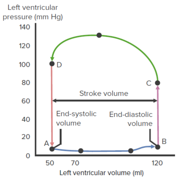
Left ventricular pressure–volume loop:
This diagram illustrates the
relationship
Relationship
A connection, association, or involvement between 2 or more parties.
Clinician–Patient Relationship between left intraventricular pressure and volume throughout the
cardiac cycle
Cardiac cycle
The cardiac cycle describes a complete contraction and relaxation of all 4 chambers of the heart during a standard heartbeat. The cardiac cycle includes 7 phases, which together describe the cycle of ventricular filling, isovolumetric contraction, ventricular ejection, and isovolumetric relaxation.
Cardiac Cycle. The segment from point A to point B represents
ventricular filling
Ventricular filling
Cardiac Cycle. The
mitral valve
Mitral valve
The valve between the left atrium and left ventricle of the heart.
Heart: Anatomy opens at A and closes at B. The segment from point B to point C represents
isovolumetric contraction
Isovolumetric contraction
Cardiac Cycle. The
aortic valve
Aortic valve
The valve between the left ventricle and the ascending aorta which prevents backflow into the left ventricle.
Heart: Anatomy opens at C. The curved line from point C to point D represents
ventricular ejection
Ventricular ejection
Cardiac Cycle. The
aortic valve
Aortic valve
The valve between the left ventricle and the ascending aorta which prevents backflow into the left ventricle.
Heart: Anatomy closes at D. The segment from point D to point A represents
isovolumetric relaxation
Isovolumetric relaxation
Cardiac Cycle.
Preload is a measure of how much the cardiomyocytes have stretched by the end of diastole Diastole Post-systolic relaxation of the heart, especially the heart ventricles. Cardiac Cycle. The Frank-Starling law is associated with preload effects on SV: ↑ preload = ↑ SV.
The length–tension relationship Relationship A connection, association, or involvement between 2 or more parties. Clinician–Patient Relationship explains how the Frank-Starling law works. This law applies to striated muscle Striated muscle One of two types of muscle in the body, characterized by the array of bands observed under microscope. Striated muscles can be divided into two subtypes: the cardiac muscle and the skeletal muscle. Muscle Tissue: Histology: skeletal and cardiac muscles.
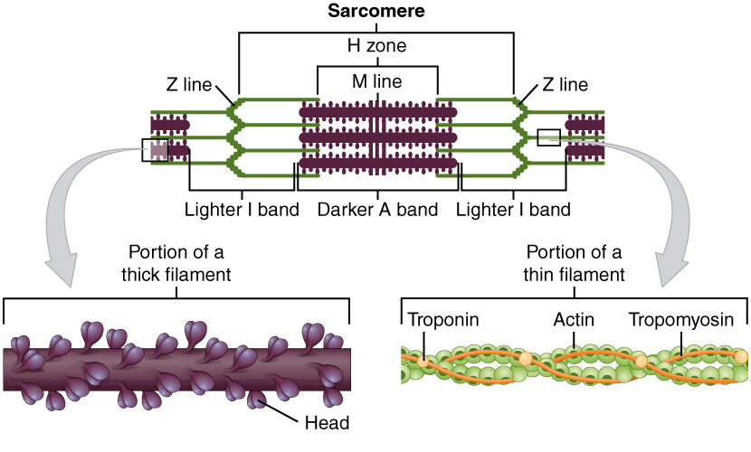
Diagram depicting the microscopic structure of sarcomeres, actin Actin Filamentous proteins that are the main constituent of the thin filaments of muscle fibers. The filaments (known also as filamentous or f-actin) can be dissociated into their globular subunits; each subunit is composed of a single polypeptide 375 amino acids long. This is known as globular or g-actin. In conjunction with myosins, actin is responsible for the contraction and relaxation of muscle. Skeletal Muscle Contraction, and myosin Myosin A diverse superfamily of proteins that function as translocating proteins. They share the common characteristics of being able to bind actins and hydrolyze mgATP. Myosins generally consist of heavy chains which are involved in locomotion, and light chains which are involved in regulation. Within the structure of myosin heavy chain are three domains: the head, the neck and the tail. The head region of the heavy chain contains the actin binding domain and mgATPase domain which provides energy for locomotion. The neck region is involved in binding the light-chains. The tail region provides the anchoring point that maintains the position of the heavy chain. The superfamily of myosins is organized into structural classes based upon the type and arrangement of the subunits they contain. Skeletal Muscle Contraction
Image: “The sarcomere Sarcomere The repeating contractile units of the myofibril, delimited by z bands along its length. Muscle Tissue: Histology, the region from one Z-line Z-line Esophagus: Anatomy to the next Z-line Z-line Esophagus: Anatomy, is the functional unit of a skeletal muscle fiber” by OpenStax College. License: CC BY 4.0
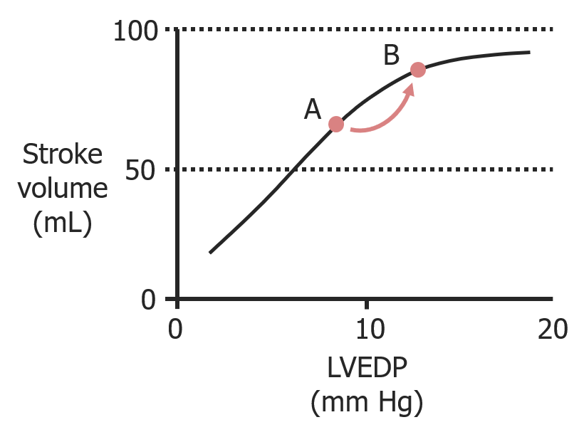
As left ventricular end-diastolic pressure (LVEDP) increases owing to increased filling, stroke volume Stroke volume The amount of blood pumped out of the heart per beat, not to be confused with cardiac output (volume/time). It is calculated as the difference between the end-diastolic volume and the end-systolic volume. Cardiac Cycle increases as well.
Image by Lecturio.
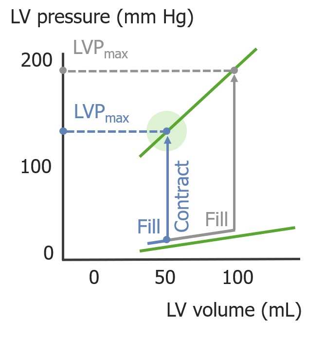
As the left ventricular (LV) volume increases during diastole Diastole Post-systolic relaxation of the heart, especially the heart ventricles. Cardiac Cycle, the LV pressure (LVP) generated during systole Systole Period of contraction of the heart, especially of the heart ventricles. Cardiac Cycle increases.
Image by Lecturio.
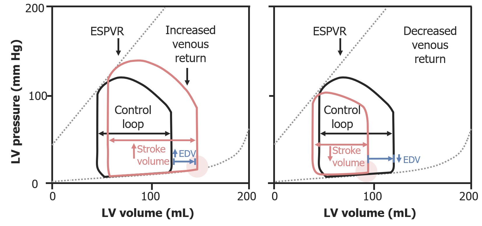
Pressure–volume loops illustrating the Frank-Starling law:
On the left, increased preload from increased venous return results in a greater
EDV
EDV
Cardiac Cycle, which increases
stroke volume
Stroke volume
The amount of blood pumped out of the heart per beat, not to be confused with cardiac output (volume/time). It is calculated as the difference between the end-diastolic volume and the end-systolic volume.
Cardiac Cycle. On the right, preload is reduced, and therefore,
stroke volume
Stroke volume
The amount of blood pumped out of the heart per beat, not to be confused with cardiac output (volume/time). It is calculated as the difference between the end-diastolic volume and the end-systolic volume.
Cardiac Cycle is reduced.
ESPVR: end-systolic pressure–volume
relationship
Relationship
A connection, association, or involvement between 2 or more parties.
Clinician–Patient Relationship
EDV
EDV
Cardiac Cycle:
end-diastolic volume
End-diastolic volume
Cardiac Cycle
Afterload is the resistance Resistance Physiologically, the opposition to flow of air caused by the forces of friction. As a part of pulmonary function testing, it is the ratio of driving pressure to the rate of air flow. Ventilation: Mechanics of Breathing in the aorta Aorta The main trunk of the systemic arteries. Mediastinum and Great Vessels: Anatomy that prevents blood from leaving the heart. Afterload represents the pressure the LV needs to overcome to eject blood into the aorta Aorta The main trunk of the systemic arteries. Mediastinum and Great Vessels: Anatomy.
↑ Afterload:
↓ Afterload:
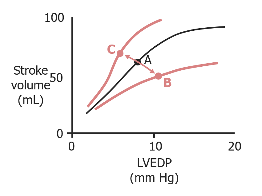
Frank-Starling curves and the effects of afterload on
stroke volume
Stroke volume
The amount of blood pumped out of the heart per beat, not to be confused with cardiac output (volume/time). It is calculated as the difference between the end-diastolic volume and the end-systolic volume.
Cardiac Cycle and left ventricular end-diastolic pressure (LVEDP):
When afterload increases (A to B), preload increases (↑ LVEDP) but
stroke volume
Stroke volume
The amount of blood pumped out of the heart per beat, not to be confused with cardiac output (volume/time). It is calculated as the difference between the end-diastolic volume and the end-systolic volume.
Cardiac Cycle decreases.
Pressure–volume loops illustrating the effects of afterload:
On the left, an increase in afterload (increased aortic pressure) requires higher pressures to be achieved during
isovolumetric contraction
Isovolumetric contraction
Cardiac Cycle; ultimately, this reduces
stroke volume
Stroke volume
The amount of blood pumped out of the heart per beat, not to be confused with cardiac output (volume/time). It is calculated as the difference between the end-diastolic volume and the end-systolic volume.
Cardiac Cycle. On the right, a decreased afterload lowers the required pressure to open the
aortic valve
Aortic valve
The valve between the left ventricle and the ascending aorta which prevents backflow into the left ventricle.
Heart: Anatomy; this results in an increased
stroke volume
Stroke volume
The amount of blood pumped out of the heart per beat, not to be confused with cardiac output (volume/time). It is calculated as the difference between the end-diastolic volume and the end-systolic volume.
Cardiac Cycle.
ESV
ESV
Cardiac Cycle:
end-systolic volume
End-systolic volume
Cardiac Cycle
EDV
EDV
Cardiac Cycle:
end-diastolic volume
End-diastolic volume
Cardiac Cycle
Inotropy is a measure of the force of contraction, independent of changes in preload.
Inotropy is increased as a result of:
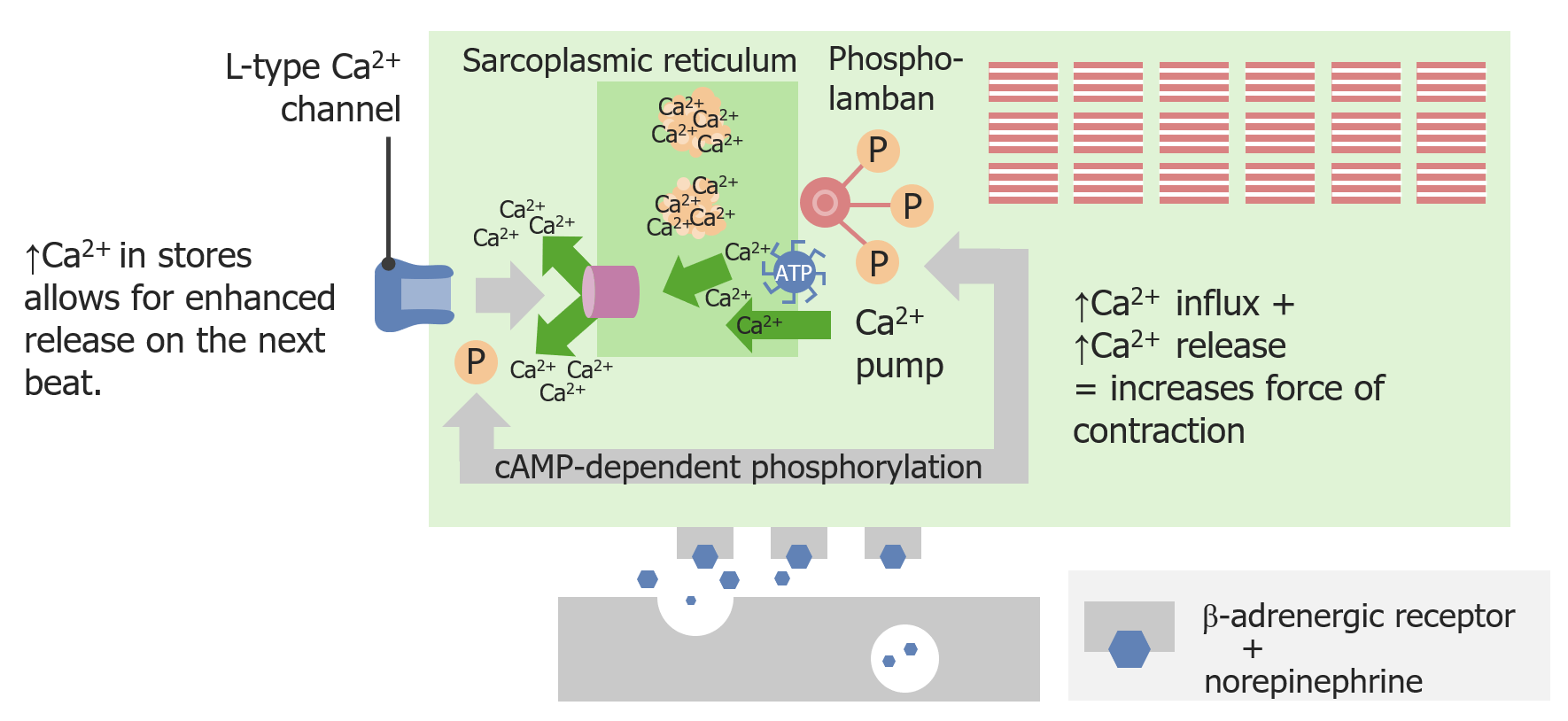
Sympathetic activation increases inotropy:
Norepinephrine
Norepinephrine
Precursor of epinephrine that is secreted by the adrenal medulla and is a widespread central and autonomic neurotransmitter. Norepinephrine is the principal transmitter of most postganglionic sympathetic fibers, and of the diffuse projection system in the brain that arises from the locus ceruleus.
Receptors and Neurotransmitters of the CNS binding to a β-adrenergic
receptor
Receptor
Receptors are proteins located either on the surface of or within a cell that can bind to signaling molecules known as ligands (e.g., hormones) and cause some type of response within the cell.
Receptors generates
cAMP
cAMP
An adenine nucleotide containing one phosphate group which is esterified to both the 3′- and 5′-positions of the sugar moiety. It is a second messenger and a key intracellular regulator, functioning as a mediator of activity for a number of hormones, including epinephrine, glucagon, and acth.
Phosphodiesterase Inhibitors, which causes the
release
Release
Release of a virus from the host cell following virus assembly and maturation. Egress can occur by host cell lysis, exocytosis, or budding through the plasma membrane.
Virology of
Ca
CA
Condylomata acuminata are a clinical manifestation of genital HPV infection. Condylomata acuminata are described as raised, pearly, flesh-colored, papular, cauliflower-like lesions seen in the anogenital region that may cause itching, pain, or bleeding.
Condylomata Acuminata (Genital Warts)2+ from the
sarcoplasmic reticulum
Sarcoplasmic Reticulum
A network of tubules and sacs in the cytoplasm of skeletal muscle fibers that assist with muscle contraction and relaxation by releasing and storing calcium ions.
Muscle Tissue: Histology (SR). This provides more intracellular
Ca
CA
Condylomata acuminata are a clinical manifestation of genital HPV infection. Condylomata acuminata are described as raised, pearly, flesh-colored, papular, cauliflower-like lesions seen in the anogenital region that may cause itching, pain, or bleeding.
Condylomata Acuminata (Genital Warts)2+ to
bind
BIND
Hyperbilirubinemia of the Newborn to more
myofilaments
Myofilaments
Refers to individual proteins that together cause muscle contraction.
Muscle Tissue: Histology, leading to a greater force of contraction.
↑ Inotropy:
↓ Inotropy:
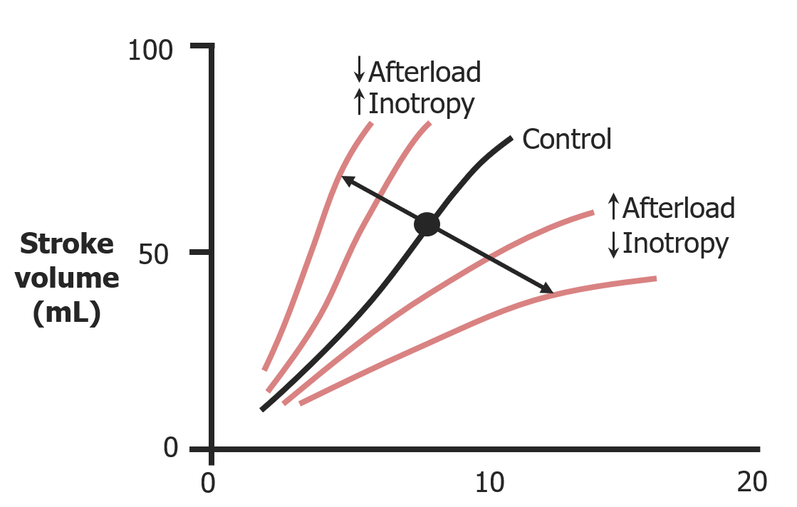
Effects of both inotropy and afterload on stroke volume Stroke volume The amount of blood pumped out of the heart per beat, not to be confused with cardiac output (volume/time). It is calculated as the difference between the end-diastolic volume and the end-systolic volume. Cardiac Cycle
Image by Lecturio.
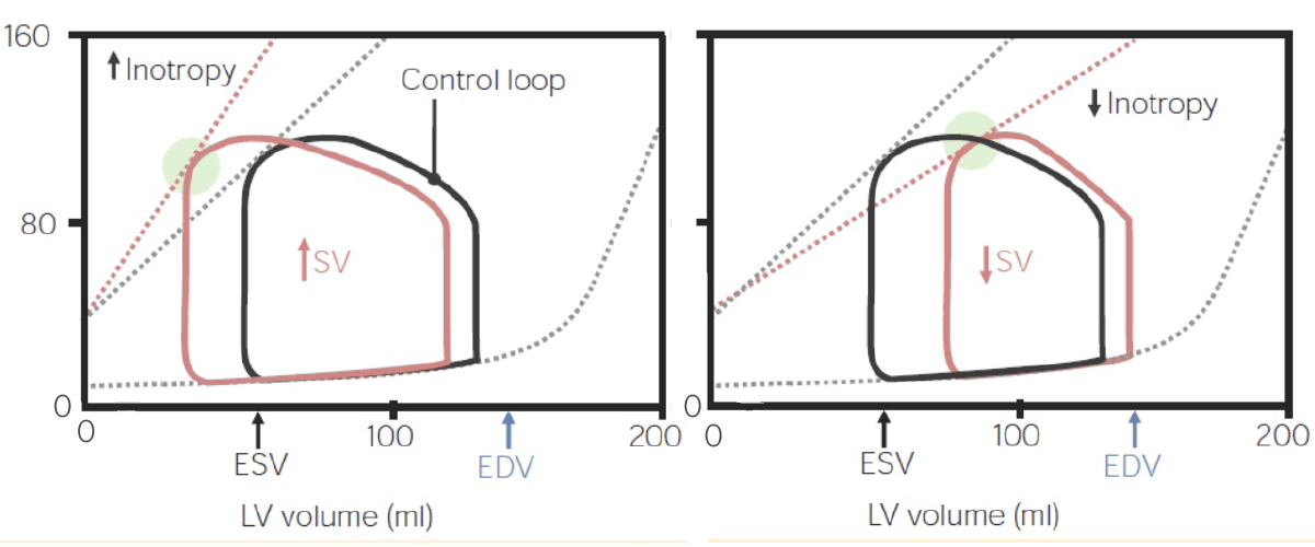
Pressure–volume loops illustrating the effects of inotropy:
On the left, as inotropy increases, so does the
stroke volume
Stroke volume
The amount of blood pumped out of the heart per beat, not to be confused with cardiac output (volume/time). It is calculated as the difference between the end-diastolic volume and the end-systolic volume.
Cardiac Cycle, leading to a smaller
end-systolic volume
End-systolic volume
Cardiac Cycle (
ESV
ESV
Cardiac Cycle). On the right, as inotropy decreases, so does the
stroke volume
Stroke volume
The amount of blood pumped out of the heart per beat, not to be confused with cardiac output (volume/time). It is calculated as the difference between the end-diastolic volume and the end-systolic volume.
Cardiac Cycle, so there is a larger volume of blood left at the end of
systole
Systole
Period of contraction of the heart, especially of the heart ventricles.
Cardiac Cycle.
The following are common conditions that affect Affect The feeling-tone accompaniment of an idea or mental representation. It is the most direct psychic derivative of instinct and the psychic representative of the various bodily changes by means of which instincts manifest themselves. Psychiatric Assessment cardiac mechanics.
Pressure–volume loop, illustrating the changes that occur during the fight-or-flight response:
The end-systolic pressure-volume
relationship
Relationship
A connection, association, or involvement between 2 or more parties.
Clinician–Patient Relationship (ESPVR) is such that, as
end-diastolic volume
End-diastolic volume
Cardiac Cycle increases (↑ preload) as result of sympathetic
nervous system
Nervous system
The nervous system is a small and complex system that consists of an intricate network of neural cells (or neurons) and even more glial cells (for support and insulation). It is divided according to its anatomical components as well as its functional characteristics. The brain and spinal cord are referred to as the central nervous system, and the branches of nerves from these structures are referred to as the peripheral nervous system.
Nervous System: Anatomy, Structure, and Classification (SNS) activation,
stroke volume
Stroke volume
The amount of blood pumped out of the heart per beat, not to be confused with cardiac output (volume/time). It is calculated as the difference between the end-diastolic volume and the end-systolic volume.
Cardiac Cycle increases owing to the Frank-Starling law. The SNS also increases inotropy, which also contributes to an increase in
stroke volume
Stroke volume
The amount of blood pumped out of the heart per beat, not to be confused with cardiac output (volume/time). It is calculated as the difference between the end-diastolic volume and the end-systolic volume.
Cardiac Cycle.