Condylomata acuminata are a clinical manifestation of genital HPV HPV Human papillomavirus (HPV) is a nonenveloped, circular, double-stranded DNA virus belonging to the Papillomaviridae family. Humans are the only reservoir, and transmission occurs through close skin-to-skin or sexual contact. Human papillomaviruses infect basal epithelial cells and can affect cell-regulatory proteins to result in cell proliferation. Papillomavirus (HPV) infection. Condylomata acuminata are described as raised, pearly, flesh-colored, papular, cauliflower-like lesions seen in the anogenital region that may cause itching, pain Pain An unpleasant sensation induced by noxious stimuli which are detected by nerve endings of nociceptive neurons. Pain: Types and Pathways, or bleeding. Sexual contact is a common route of spread for HPV HPV Human papillomavirus (HPV) is a nonenveloped, circular, double-stranded DNA virus belonging to the Papillomaviridae family. Humans are the only reservoir, and transmission occurs through close skin-to-skin or sexual contact. Human papillomaviruses infect basal epithelial cells and can affect cell-regulatory proteins to result in cell proliferation. Papillomavirus (HPV). While seen in all populations and ages, condylomata acuminata is most often seen in adolescence. HPV HPV Human papillomavirus (HPV) is a nonenveloped, circular, double-stranded DNA virus belonging to the Papillomaviridae family. Humans are the only reservoir, and transmission occurs through close skin-to-skin or sexual contact. Human papillomaviruses infect basal epithelial cells and can affect cell-regulatory proteins to result in cell proliferation. Papillomavirus (HPV) types 6 and 11 are responsible for 90% of warts Warts Benign epidermal proliferations or tumors; some are viral in origin. Female Genitourinary Examination and are considered low risk for malignancy Malignancy Hemothorax; however, other types of HPV HPV Human papillomavirus (HPV) is a nonenveloped, circular, double-stranded DNA virus belonging to the Papillomaviridae family. Humans are the only reservoir, and transmission occurs through close skin-to-skin or sexual contact. Human papillomaviruses infect basal epithelial cells and can affect cell-regulatory proteins to result in cell proliferation. Papillomavirus (HPV) may also be present. Lesions typically self-resolve over months to years; however, they can be removed via cryotherapy Cryotherapy A form of therapy consisting in the local or general use of cold. The selective destruction of tissue by extreme cold or freezing is cryosurgery. Chondrosarcoma or topical antimitotic agents. While there is currently no treatment for HPV HPV Human papillomavirus (HPV) is a nonenveloped, circular, double-stranded DNA virus belonging to the Papillomaviridae family. Humans are the only reservoir, and transmission occurs through close skin-to-skin or sexual contact. Human papillomaviruses infect basal epithelial cells and can affect cell-regulatory proteins to result in cell proliferation. Papillomavirus (HPV) infection, it can be prevented through vaccination Vaccination Vaccination is the administration of a substance to induce the immune system to develop protection against a disease. Unlike passive immunization, which involves the administration of pre-performed antibodies, active immunization constitutes the administration of a vaccine to stimulate the body to produce its own antibodies. Vaccination.
Last updated: Jan 20, 2026
Condylomata acuminata are specifically lesions created by HPV HPV Human papillomavirus (HPV) is a nonenveloped, circular, double-stranded DNA virus belonging to the Papillomaviridae family. Humans are the only reservoir, and transmission occurs through close skin-to-skin or sexual contact. Human papillomaviruses infect basal epithelial cells and can affect cell-regulatory proteins to result in cell proliferation. Papillomavirus (HPV).
Infection to resolution:
Patients Patients Individuals participating in the health care system for the purpose of receiving therapeutic, diagnostic, or preventive procedures. Clinician–Patient Relationship are often asymptomatic, presenting only for the appearance of lesions. The diagnosis of HPV HPV Human papillomavirus (HPV) is a nonenveloped, circular, double-stranded DNA virus belonging to the Papillomaviridae family. Humans are the only reservoir, and transmission occurs through close skin-to-skin or sexual contact. Human papillomaviruses infect basal epithelial cells and can affect cell-regulatory proteins to result in cell proliferation. Papillomavirus (HPV) may cause significant psychosocial distress, given the associated stigma.

Multiple enlarged warts present in the anal region of a patient
Image: “4151” by Dr. Wiesner. License: Public Domain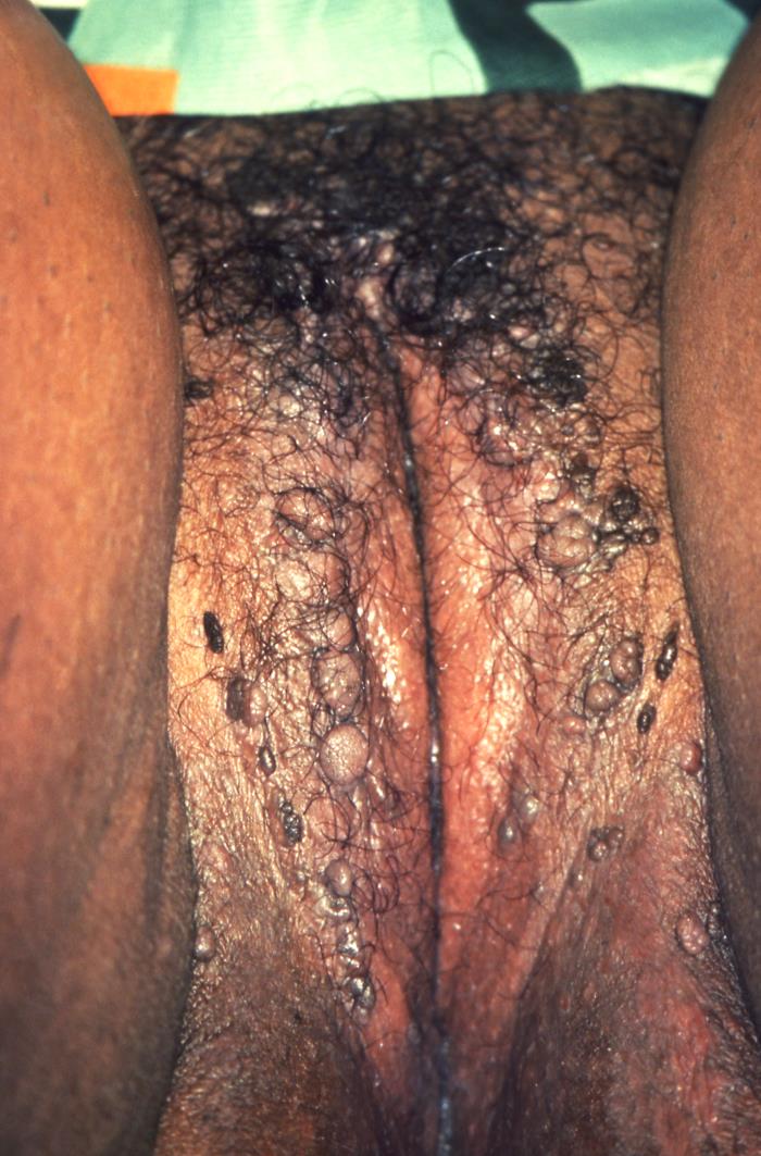
Numerous genital warts on the labia majora:
Condylomata acuminata appear as flesh-colored, raised, cauliflower-like lesions of the anogenital area. In women, they can be seen on the labia majora, vagina, cervix, and anus.

Genital warts present on the shaft of a penis in this patient:
Condylomata acuminata appear as flesh-colored, raised, cauliflower-like lesions of the anogenital area. In men, they can be seen on the glans penis, scrotum, urethra, and anus.
Spontaneous and complete resolution can take up to 24 months.