

Ewing sarcoma (ES) is a primary bone Bone Bone is a compact type of hardened connective tissue composed of bone cells, membranes, an extracellular mineralized matrix, and central bone marrow. The 2 primary types of bone are compact and spongy. Bones: Structure and Types malignancy Malignancy Hemothorax derived from primitive round cells affecting primarily children and teenagers. Ewing sarcoma commonly presents with a painful mass Mass Three-dimensional lesion that occupies a space within the breast Imaging of the Breast, swelling Swelling Inflammation, and pathologic bone fractures Bone fractures Breaks in bones. Bones: Remodeling and Healing. Diagnosis is established with imaging and biopsy. Treatment involves systemic chemotherapy Chemotherapy Osteosarcoma and local control of the tumor Tumor Inflammation with surgical resection or radiation Radiation Emission or propagation of acoustic waves (sound), electromagnetic energy waves (such as light; radio waves; gamma rays; or x-rays), or a stream of subatomic particles (such as electrons; neutrons; protons; or alpha particles). Osteosarcoma. With proper treatment, the overall 5-year survival is over 70%.
Last updated: Sep 29, 2026
Ewing sarcoma (ES) is a malignant sarcoma derived from undifferentiated primitive round cells that usually arises in bone Bone Bone is a compact type of hardened connective tissue composed of bone cells, membranes, an extracellular mineralized matrix, and central bone marrow. The 2 primary types of bone are compact and spongy. Bones: Structure and Types but may be extraosseous. It is biologically distinct from osteosarcoma Osteosarcoma Osteosarcoma is a primary malignant tumor of the bone characterized by the production of osteoid or immature bone by the tumor cells. The disease is most common in children and young adults and most frequently affects growth plates of the long bones, although it can involve any bone. Osteosarcoma because it does not produce osteoid Osteoid Bones: Development and Ossification.
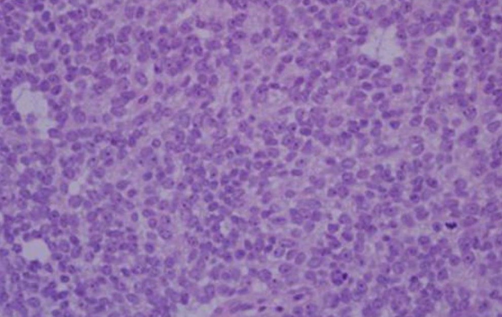
A hematoxylin and eosin (H&E) stained section from an Ewing sarcoma tumor magnified at 400x:
Sheets of small round cells with a high nuclear:cytoplasmic ratio
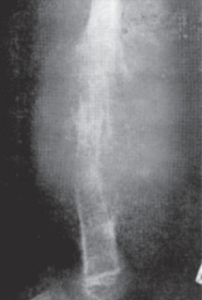
X-ray of Ewing sarcoma of the femur (large destructive lesion)
Image: “Ewing’s sarcoma of the femur” by Vyankat G Vohar. License: CC BY 2.0, cropped by Lecturio.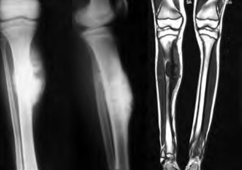
Ewing sarcoma:
Anteroposterior and lateral radiographs with T1-weighted coronal section of MRI showing involvement of proximal ⅓ diaphysis of the right tibia
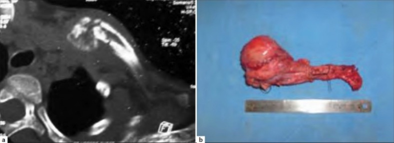
Ewing sarcoma:
a: Axial section of noncontrast CT scan showing Ewing sarcoma of the right clavicle
b: Photograph showing the resected right clavicle with tumor in situ