Bone is a compact type of hardened connective tissueConnective tissueConnective tissues originate from embryonic mesenchyme and are present throughout the body except inside the brain and spinal cord. The main function of connective tissues is to provide structural support to organs. Connective tissues consist of cells and an extracellular matrix.Connective Tissue: Histology composed of bone cells, membranes, an extracellular mineralized matrix, and central bone marrowBone marrowThe soft tissue filling the cavities of bones. Bone marrow exists in two types, yellow and red. Yellow marrow is found in the large cavities of large bones and consists mostly of fat cells and a few primitive blood cells. Red marrow is a hematopoietic tissue and is the site of production of erythrocytes and granular leukocytes. Bone marrow is made up of a framework of connective tissue containing branching fibers with the frame being filled with marrow cells.Bone Marrow: Composition and Hematopoiesis. The 2 primary types of bone are compact and spongy. Because the matrix is mineralized (rather than aqueous), nutrients and waste cannot diffuse through the matrix. Bone has developed a unique structure to allow the functions to occur. The structure of bone allows the bone to be hard, but not too brittle, and gives bone the strength to resist compressive and bending forces. As a result, bone is ideally suited for the functions of support, protection of vital organs, and movement. In addition, bone produces blood cells in the marrow and is the body's primary storage site for calciumCalciumA basic element found in nearly all tissues. It is a member of the alkaline earth family of metals with the atomic symbol ca, atomic number 20, and atomic weight 40. Calcium is the most abundant mineral in the body and combines with phosphorus to form calcium phosphate in the bones and teeth. It is essential for the normal functioning of nerves and muscles and plays a role in blood coagulation (as factor IV) and in many enzymatic processes.Electrolytes.
Osseous tissue: a type of connective tissueConnective tissueConnective tissues originate from embryonic mesenchyme and are present throughout the body except inside the brain and spinal cord. The main function of connective tissues is to provide structural support to organs. Connective tissues consist of cells and an extracellular matrix.Connective Tissue: Histology hardened by the deposition of mineralsMineralsElectrolytes (primarily calciumCalciumA basic element found in nearly all tissues. It is a member of the alkaline earth family of metals with the atomic symbol ca, atomic number 20, and atomic weight 40. Calcium is the most abundant mineral in the body and combines with phosphorus to form calcium phosphate in the bones and teeth. It is essential for the normal functioning of nerves and muscles and plays a role in blood coagulation (as factor IV) and in many enzymatic processes.Electrolytes and phosphatePhosphateInorganic salts of phosphoric acid.Electrolytes)
Blood
Bone marrowBone marrowThe soft tissue filling the cavities of bones. Bone marrow exists in two types, yellow and red. Yellow marrow is found in the large cavities of large bones and consists mostly of fat cells and a few primitive blood cells. Red marrow is a hematopoietic tissue and is the site of production of erythrocytes and granular leukocytes. Bone marrow is made up of a framework of connective tissue containing branching fibers with the frame being filled with marrow cells.Bone Marrow: Composition and Hematopoiesis
CartilageCartilageCartilage is a type of connective tissue derived from embryonic mesenchyme that is responsible for structural support, resilience, and the smoothness of physical actions. Perichondrium (connective tissue membrane surrounding cartilage) compensates for the absence of vasculature in cartilage by providing nutrition and support. Cartilage: Histology and fibrousFibrousFibrocystic Changeconnective tissueConnective tissueConnective tissues originate from embryonic mesenchyme and are present throughout the body except inside the brain and spinal cord. The main function of connective tissues is to provide structural support to organs. Connective tissues consist of cells and an extracellular matrix.Connective Tissue: Histology
Adipose tissueAdipose tissueAdipose tissue is a specialized type of connective tissue that has both structural and highly complex metabolic functions, including energy storage, glucose homeostasis, and a multitude of endocrine capabilities. There are three types of adipose tissue, white adipose tissue, brown adipose tissue, and beige or “brite” adipose tissue, which is a transitional form.Adipose Tissue: Histology
Nervous tissue
Bone can refer to:
The entire organ
The osseous tissue within the organ only
Function of bones
Protection (e.g., the skullSkullThe skull (cranium) is the skeletal structure of the head supporting the face and forming a protective cavity for the brain. The skull consists of 22 bones divided into the viscerocranium (facial skeleton) and the neurocranium.Skull: Anatomy protects the brainBrainThe part of central nervous system that is contained within the skull (cranium). Arising from the neural tube, the embryonic brain is comprised of three major parts including prosencephalon (the forebrain); mesencephalon (the midbrain); and rhombencephalon (the hindbrain). The developed brain consists of cerebrum; cerebellum; and other structures in the brain stem.Nervous System: Anatomy, Structure, and Classification, the ribsRibsA set of twelve curved bones which connect to the vertebral column posteriorly, and terminate anteriorly as costal cartilage. Together, they form a protective cage around the internal thoracic organs.Chest Wall: Anatomy protect the heart and lungsLungsLungs are the main organs of the respiratory system. Lungs are paired viscera located in the thoracic cavity and are composed of spongy tissue. The primary function of the lungs is to oxygenate blood and eliminate CO2. Lungs: Anatomy)
Support
Movement
Blood cell formationBlood cell formationThe development and formation of various types of blood cells. Hematopoiesis can take place in the bone marrow (medullary) or outside the bone marrow (extramedullary hematopoiesis).Bone Marrow: Composition and Hematopoiesis
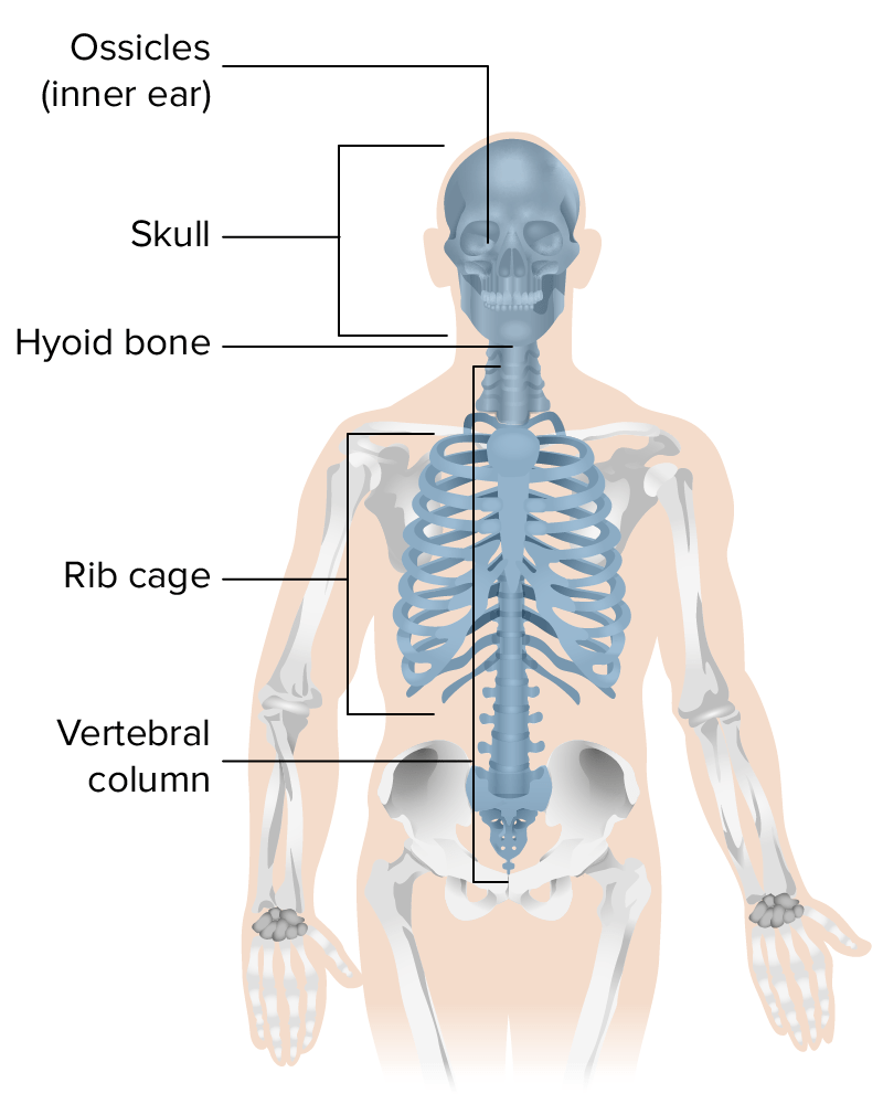
SkullSkullThe skull (cranium) is the skeletal structure of the head supporting the face and forming a protective cavity for the brain. The skull consists of 22 bones divided into the viscerocranium (facial skeleton) and the neurocranium.Skull: Anatomy
Vertebral columnVertebral columnThe human spine, or vertebral column, is the most important anatomical and functional axis of the human body. It consists of 7 cervical vertebrae, 12 thoracic vertebrae, and 5 lumbar vertebrae and is limited cranially by the skull and caudally by the sacrum. Vertebral Column: Anatomy and sacrumSacrumFive fused vertebrae forming a triangle-shaped structure at the back of the pelvis. It articulates superiorly with the lumbar vertebrae, inferiorly with the coccyx, and anteriorly with the ilium of the pelvis. The sacrum strengthens and stabilizes the pelvis.Vertebral Column: Anatomy
Rib cageRib cageThe bony thoracic enclosure consisting of the vertebral column; the ribs; the sternum; and the costal cartilage.Chest Wall: Anatomy: ribsRibsA set of twelve curved bones which connect to the vertebral column posteriorly, and terminate anteriorly as costal cartilage. Together, they form a protective cage around the internal thoracic organs.Chest Wall: Anatomy and sternumSternumA long, narrow, and flat bone commonly known as breastbone occurring in the midsection of the anterior thoracic segment or chest region, which stabilizes the rib cage and serves as the point of origin for several muscles that move the arms, head, and neck.Chest Wall: Anatomy
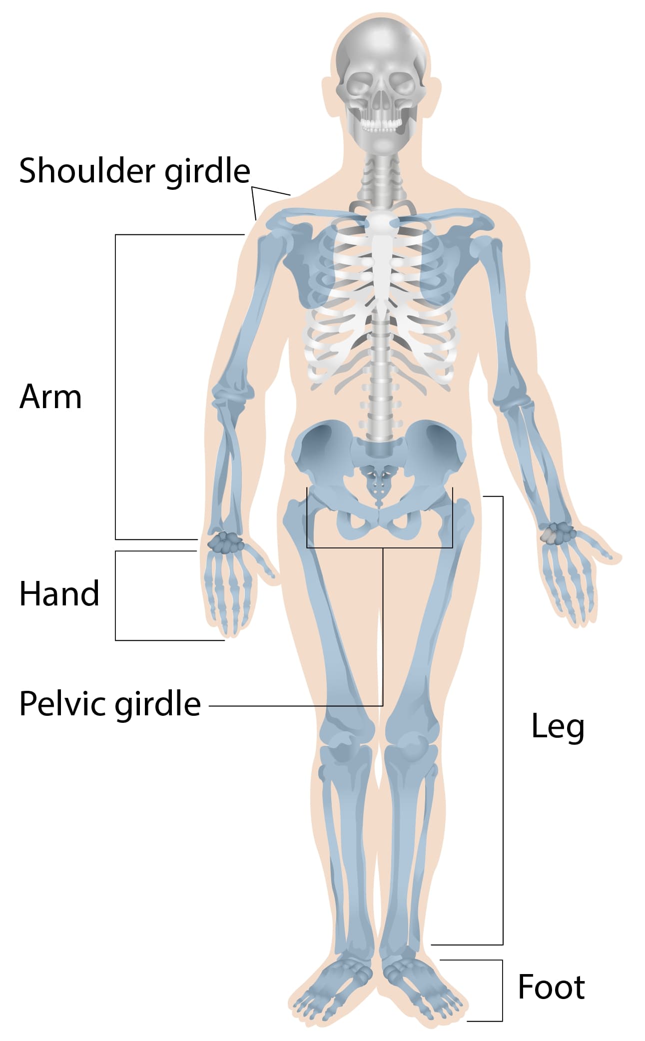
Appendicular bones:
ClavicleClavicleA bone on the ventral side of the shoulder girdle, which in humans is commonly called the collar bone.Clavicle Fracture
Scapula
Arms and hands
Pelvic girdle
Legs and feet
Illustration representing the bones that form the axial skeleton
Image by Lecturio.
Illustration representing the bones that form the appendicular skeleton
Image by Lecturio.
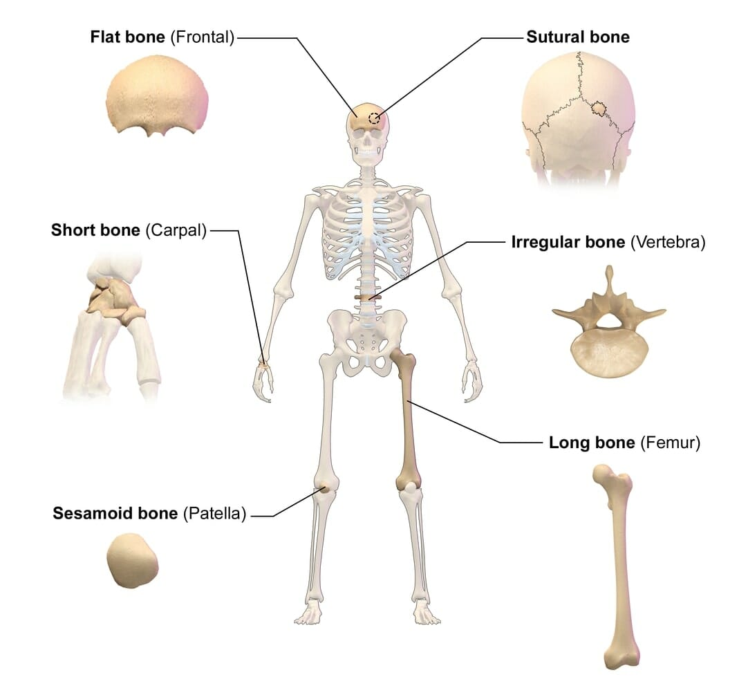
Classification of bones by shape
Long bones: length greater than width:
Upper extremity: humerusHumerusBone in humans and primates extending from the shoulder joint to the elbow joint.Arm: Anatomy, radiusRadiusThe outer shorter of the two bones of the forearm, lying parallel to the ulna and partially revolving around it.Forearm: Anatomy, ulnaUlnaThe inner and longer bone of the forearm.Forearm: Anatomy, metacarpalsMetacarpalsThe five cylindrical bones of the metacarpus, articulating with the carpal bones proximally and the phalanges of fingers distally.Wrist Joint: Anatomy, and phalangesPhalangesBones that make up the skeleton of the fingers, consisting of two for the thumb, and three for each of the other fingers.Hand: Anatomy
Lower extremity: femur, tibiaTibiaThe second longest bone of the skeleton. It is located on the medial side of the lower leg, articulating with the fibula laterally, the talus distally, and the femur proximally.Knee Joint: Anatomy, fibulaFibulaThe bone of the lower leg lateral to and smaller than the tibia. In proportion to its length, it is the most slender of the long bones.Leg: Anatomy, metatarsals, and phalangesPhalangesBones that make up the skeleton of the fingers, consisting of two for the thumb, and three for each of the other fingers.Hand: Anatomy
Short bones: length approximately equal to width:
Carpal bonesCarpal bonesThe eight bones of the wrist: scaphoid bone; lunate bone; triquetrum bone; pisiform bone; trapezium bone; trapezoid bone; capitate bone; and hamate bone.Carpal Tunnel Syndrome (wrist)
Tarsal bonesTarsal BonesThe seven bones which form the tarsus – namely, calcaneus; talus; cuboid, navicular, and the internal, middle, and external cuneiforms.Foot: Anatomy (ankle)
Flat bones: enclose and protect soft organs:
Most skullSkullThe skull (cranium) is the skeletal structure of the head supporting the face and forming a protective cavity for the brain. The skull consists of 22 bones divided into the viscerocranium (facial skeleton) and the neurocranium.Skull: Anatomy bones
RibsRibsA set of twelve curved bones which connect to the vertebral column posteriorly, and terminate anteriorly as costal cartilage. Together, they form a protective cage around the internal thoracic organs.Chest Wall: Anatomy
SternumSternumA long, narrow, and flat bone commonly known as breastbone occurring in the midsection of the anterior thoracic segment or chest region, which stabilizes the rib cage and serves as the point of origin for several muscles that move the arms, head, and neck.Chest Wall: Anatomy
Scapula
Irregular bones: bones not fitting into other categories:
Vertebrae
Some skullSkullThe skull (cranium) is the skeletal structure of the head supporting the face and forming a protective cavity for the brain. The skull consists of 22 bones divided into the viscerocranium (facial skeleton) and the neurocranium.Skull: Anatomy bones (e.g., sphenoid, facial bones)
Illustration representing the classification of bones by shape
Image: “Classification of Bones By Shape” by BruceBlaus. License: CC BY 3.0, cropped by Lecturio.
Function: strength to withstand compressive forces
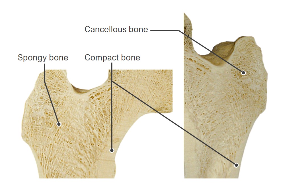
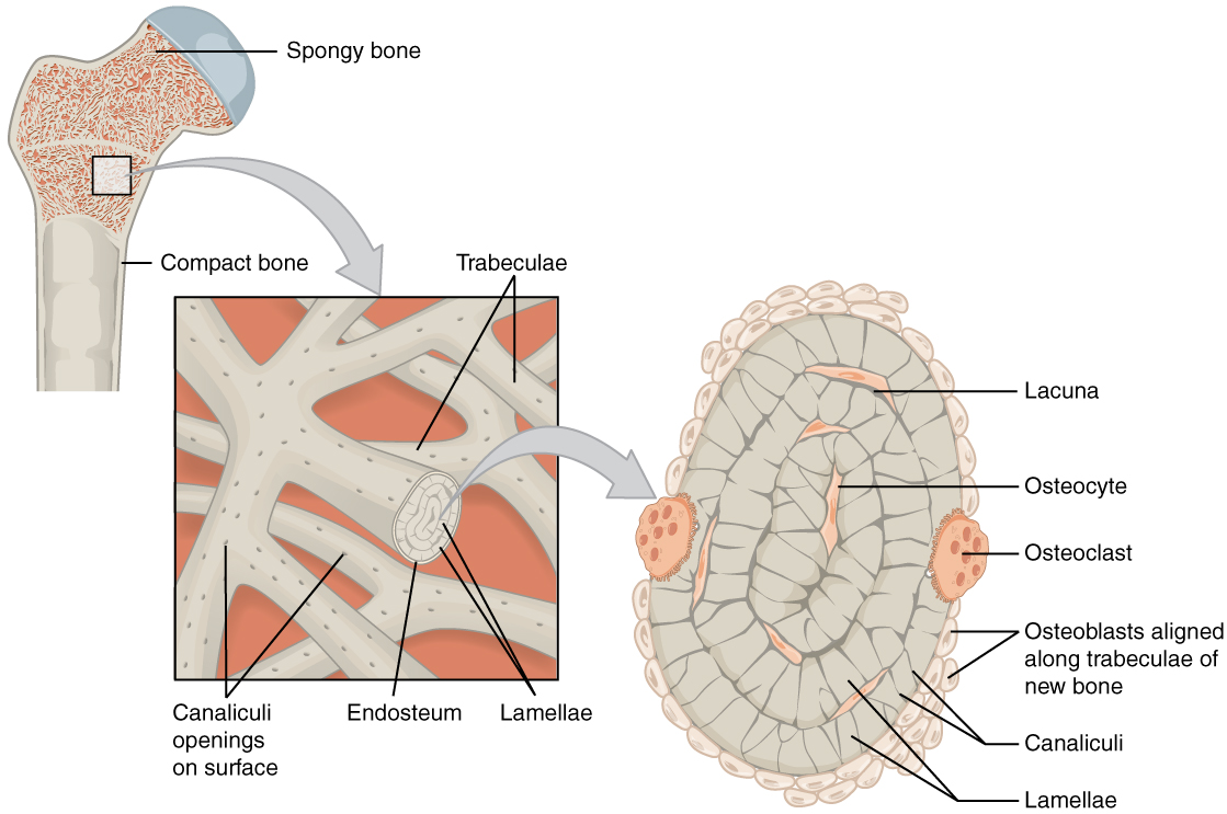
Spongy (cancellous) bone:
Loosely organized, inner-layer osseous tissue
Consists of a lattice of small, thin pieces of osseous tissue called trabeculae or bony spicules:
Transfers force on the bone to the outer compact bone
Constantly reforming to meet the body’s needs (e.g., exercise increases the trabeculae; prolonged weightlessness in space reduces the trabeculae)
Locations: internal to compact bone:
At the ends of long bones
In the middle of short, flat, and irregular bones
Image of the internal structure of a femur head: Note the compact bone along the outside and spongy/cancellous bone in the center.
Image by Lecturio.
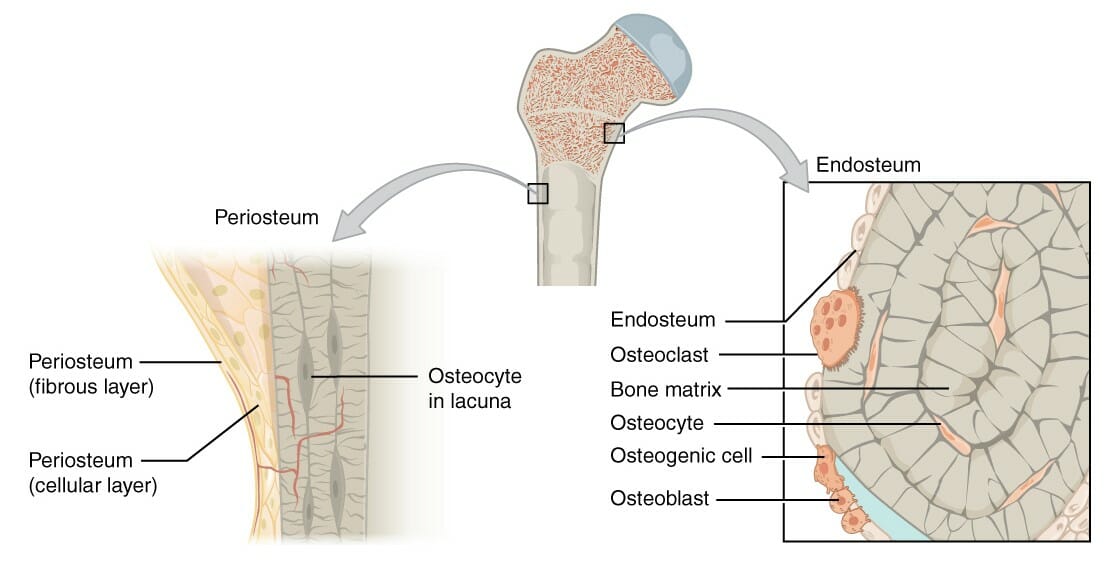
Bone membranes
Two primary membranes surround bone tissue: the periosteum (externally) and the endosteum (internally).
Periosteum:
Outer layer surrounding bone on the external surface (except at the joints, which are covered in articular cartilageCartilageCartilage is a type of connective tissue derived from embryonic mesenchyme that is responsible for structural support, resilience, and the smoothness of physical actions. Perichondrium (connective tissue membrane surrounding cartilage) compensates for the absence of vasculature in cartilage by providing nutrition and support. Cartilage: Histology)
Tough outer layer of collagenCollagenA polypeptide substance comprising about one third of the total protein in mammalian organisms. It is the main constituent of skin; connective tissue; and the organic substance of bones (bone and bones) and teeth (tooth).Connective Tissue: Histology
Sharpey fibers: collagenCollagenA polypeptide substance comprising about one third of the total protein in mammalian organisms. It is the main constituent of skin; connective tissue; and the organic substance of bones (bone and bones) and teeth (tooth).Connective Tissue: Histology fibers from the fibrousFibrousFibrocystic Change layer of the periosteum:
Continuous with muscle tendons on top of the bone
Penetrate deep into the bone matrix to secure the periosteum and overlying muscle to the bone
Osteogenic layer of periosteum:
Contains bone-forming cells:
OsteoblastsOsteoblastsBone-forming cells which secrete an extracellular matrix. Hydroxyapatite crystals are then deposited into the matrix to form bone.Bones: Development and Ossification
OsteoclastsOsteoclastsA large multinuclear cell associated with the bone resorption. An odontoclast, also called cementoclast, is cytomorphologically the same as an osteoclast and is involved in cementum resorption.Bones: Development and Ossification
Contains the same bone-forming cells as the osteogenic layer of the periosteum
Membranes of the bone, periosteum, and endosteum:
The periosteum lines the external surface of the bone and the endosteum lines the internal surface of the bone.
Image: “Figure 6.8 Periosteum and Endosteum” by OpenStax College. License: CC BY 4.0
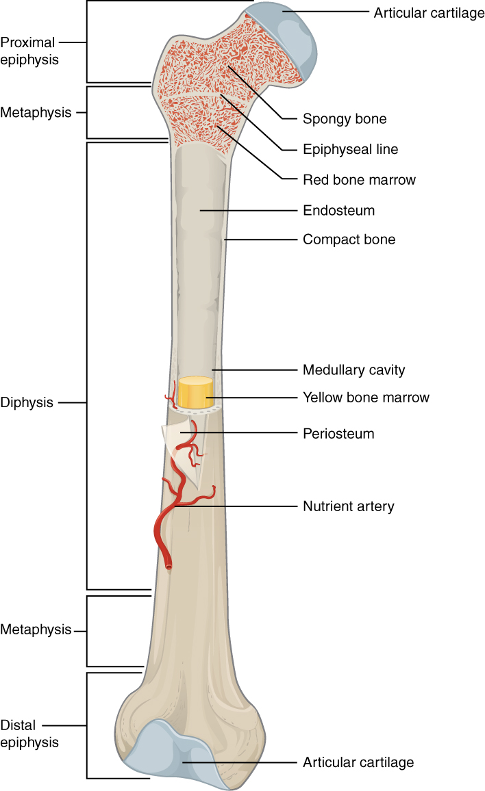
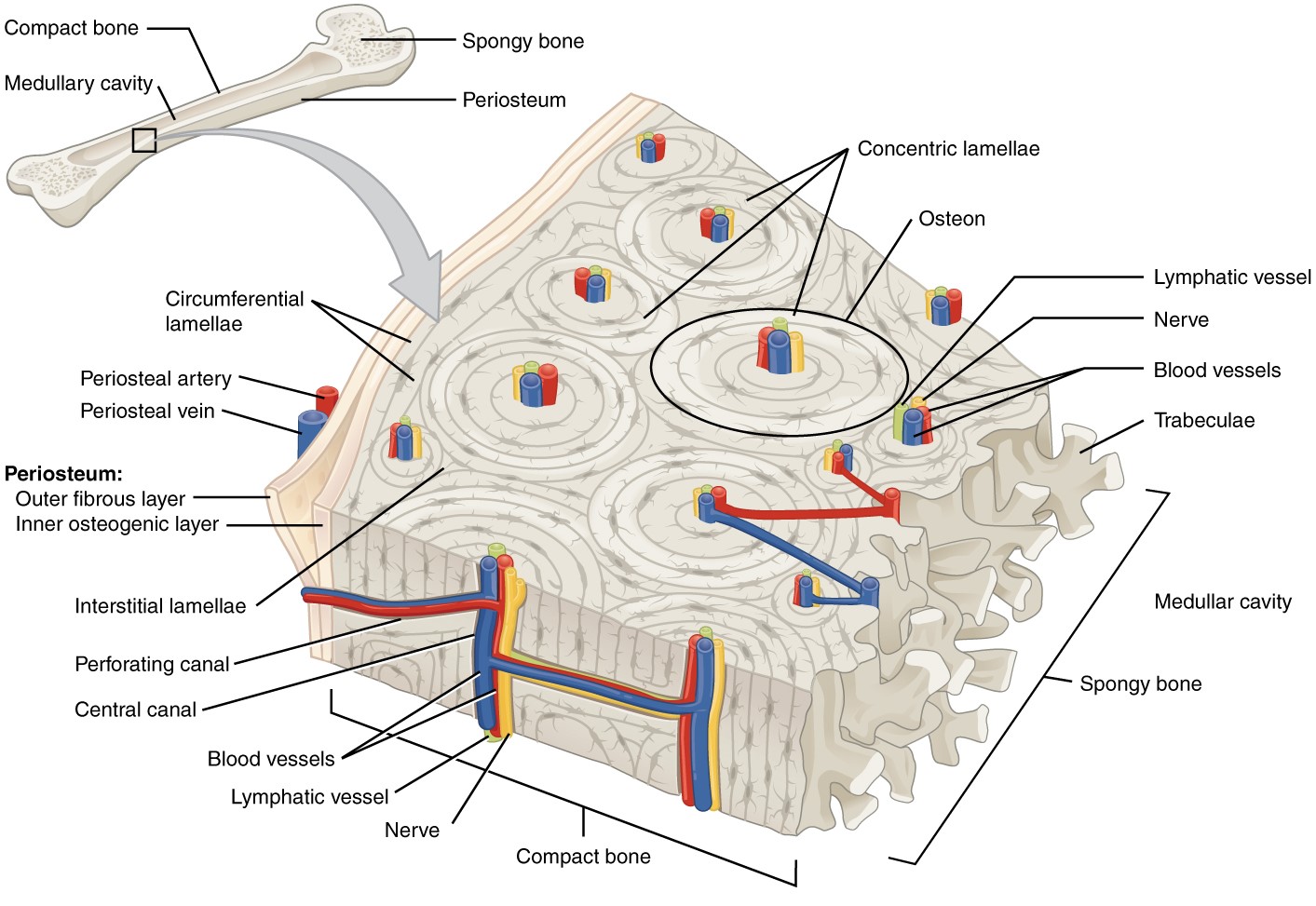
Structure of long bones
The 3 primary anatomic regions of long bones:
Diaphysis:
The shaft
Forms the long axis of long bones
Consists of a thick layer of compact bone, surrounding a central medullary cavityMedullary CavityEwing Sarcomacontaining bone marrowBone marrowThe soft tissue filling the cavities of bones. Bone marrow exists in two types, yellow and red. Yellow marrow is found in the large cavities of large bones and consists mostly of fat cells and a few primitive blood cells. Red marrow is a hematopoietic tissue and is the site of production of erythrocytes and granular leukocytes. Bone marrow is made up of a framework of connective tissue containing branching fibers with the frame being filled with marrow cells.Bone Marrow: Composition and Hematopoiesis
Epiphysis:
Ends of the bones (at joints)
Wider than the diaphysis:
Strengthens the joint
↑ Surface area for tendon and ligament attachment
Primarily composed of spongy bone
Outer layer of compact bone
Covered in articular cartilageCartilageCartilage is a type of connective tissue derived from embryonic mesenchyme that is responsible for structural support, resilience, and the smoothness of physical actions. Perichondrium (connective tissue membrane surrounding cartilage) compensates for the absence of vasculature in cartilage by providing nutrition and support. Cartilage: Histology:
A type of hyaline cartilageHyaline cartilageA type of cartilage characterized by a homogeneous amorphous matrix containing predominantly type II collagen and ground substance. Hyaline cartilage is found in articular cartilage; costal cartilage; laryngeal cartilages; and the nasal septum.Cartilage: Histology
Articular cartilageCartilageCartilage is a type of connective tissue derived from embryonic mesenchyme that is responsible for structural support, resilience, and the smoothness of physical actions. Perichondrium (connective tissue membrane surrounding cartilage) compensates for the absence of vasculature in cartilage by providing nutrition and support. Cartilage: Histology + lubricating fluid → ↓ friction → significantly easier joint movement
Acts as a shockShockShock is a life-threatening condition associated with impaired circulation that results in tissue hypoxia. The different types of shock are based on the underlying cause: distributive (↑ cardiac output (CO), ↓ systemic vascular resistance (SVR)), cardiogenic (↓ CO, ↑ SVR), hypovolemic (↓ CO, ↑ SVR), obstructive (↓ CO), and mixed. Types of Shock absorber
Metaphysis:
In between the epiphysis and diaphysis
Remnant of the epiphyseal plateEpiphyseal plateThe area between the epiphysis and the diaphysis within which bone growth occurs.Cartilage: Histology or line: the hyaline cartilageHyaline cartilageA type of cartilage characterized by a homogeneous amorphous matrix containing predominantly type II collagen and ground substance. Hyaline cartilage is found in articular cartilage; costal cartilage; laryngeal cartilages; and the nasal septum.Cartilage: Histology allowed for bone elongationElongationPolymerase Chain Reaction (PCR) in childhood
Bone marrow inside the femur
Image: “603 Anatomy of Long Bone” by OpenStax College. License: CC BY 4.0
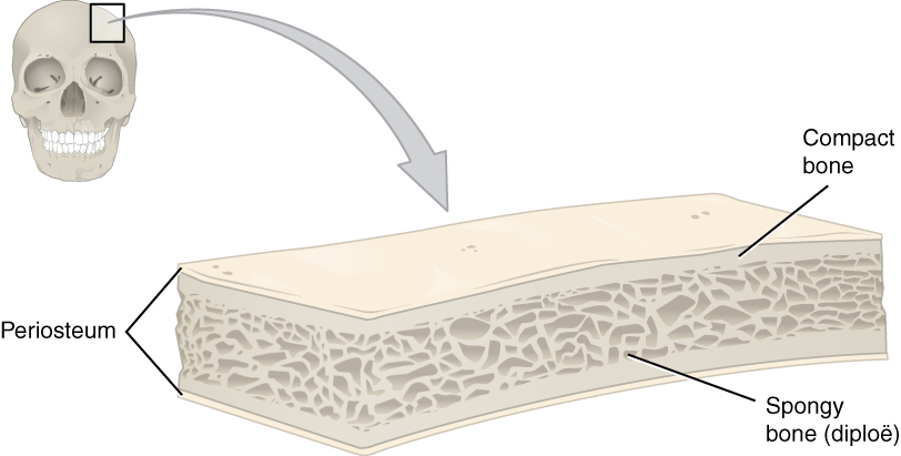
Structure of short, irregular, and flat bones
Outer layers: thin plates of periosteum-covered compact bone
Inner layer: endosteum-covered spongy bone
In flat bones, the inner spongy bone is:
Known as diploë
Sandwiched between 2 layers of compact bone
Structure of a flat bone
Image: “Cross-section of a flat bone showing the spongy bone (diploë) lined on either side by a layer of compact bone” by OpenStax College. License: CC BY 4.0
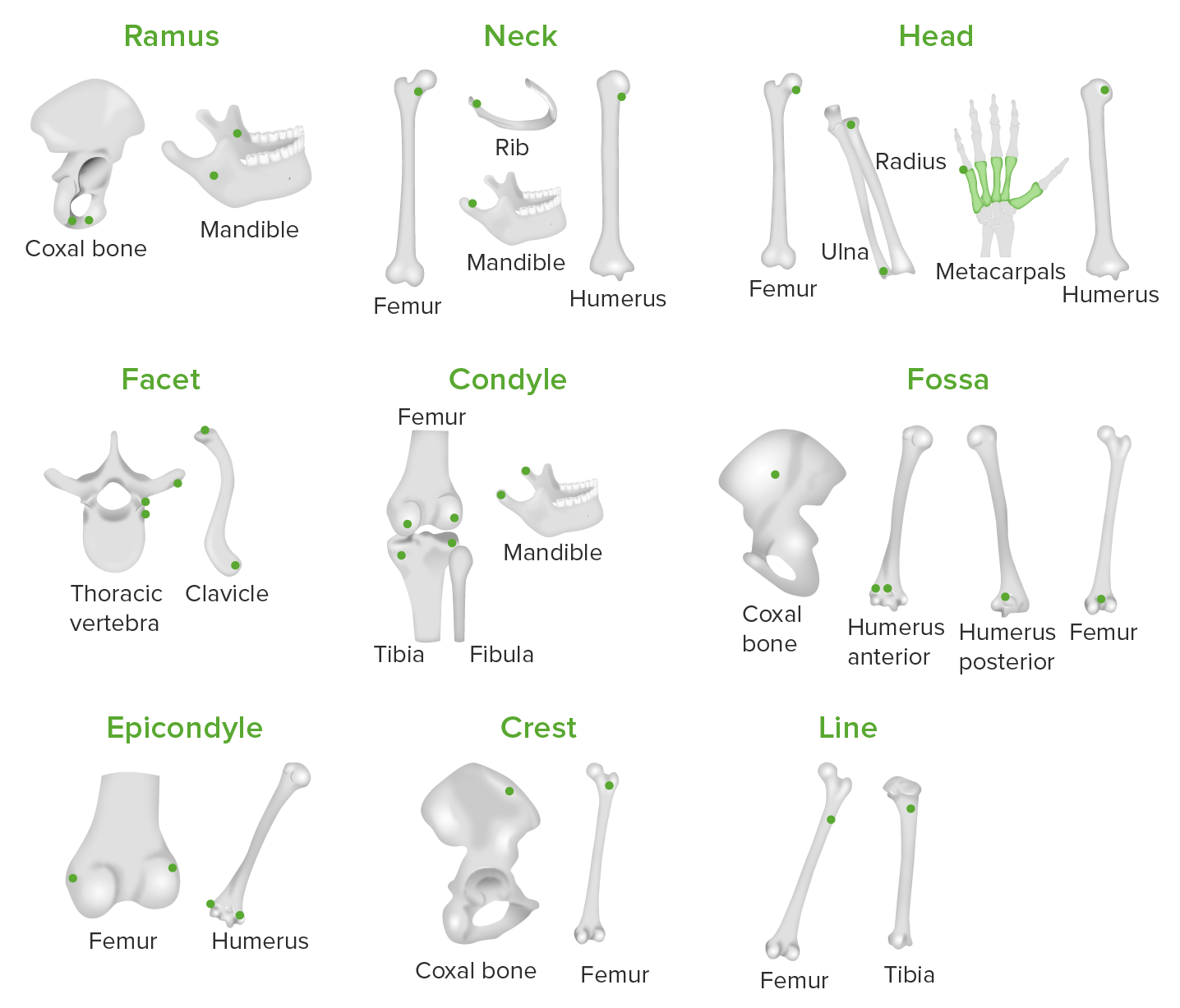
Bone markings
Bone markings are areas of bone where tendons, ligaments, and fasciaFasciaLayers of connective tissue of variable thickness. The superficial fascia is found immediately below the skin; the deep fascia invests muscles, nerves, and other organs.Cellulitis attach, including articulations, projections, and holes.
Articular markings:
Condyle: rounded surface at an articular area
Epicondyle: eminence superior to a condyle
Facet: flat surface where bones articulate
Projections:
Crest: ridge of a bone
Process: prominence feature
Protuberance: projection of bone
SpineSpineThe human spine, or vertebral column, is the most important anatomical and functional axis of the human body. It consists of 7 cervical vertebrae, 12 thoracic vertebrae, and 5 lumbar vertebrae and is limited cranially by the skull and caudally by the sacrum.Vertebral Column: Anatomy: a sharp process
Tubercle: smaller rounded process
Tuberosity: rough surface
Holes:
Canal: channel through bone
Foramen: passage through a bone
Fossa: hollow or depressed area
Groove or sulcus: elongated depression or furrow
Sinus: irregularly shaped cavernous area
Illustration representing various types of bone markings on different bones in the body where tendons, ligaments, and fascia attach to the bones: The green dots represent the bone markings present on the different bones in the body.
Image by Lecturio.
Illustration representing various types of bone markings on different bones in the body where tendons, ligaments, and fascia attach to the bones: The green dots represent the bone markings present on the different bones in the body.
Image by Lecturio.
Bone Cells and Matrix
The 2 primary components of bone are cells and matrix.
Bone cells
Bone contains a relatively small number of cells compared to the amount of matrix. In addition to other functions, the cells synthesize and break down the bone. Four major types of osseous cells exist:
Stem cells arising from embryonic fibroblastsFibroblastsConnective tissue cells which secrete an extracellular matrix rich in collagen and other macromolecules.Sarcoidosis
Can differentiate into osteoblastsOsteoblastsBone-forming cells which secrete an extracellular matrix. Hydroxyapatite crystals are then deposited into the matrix to form bone.Bones: Development and Ossification → stimulated by stress (e.g., from exercise) and fractures
Found in the endosteum and osteogenic periosteum
OsteoblastsOsteoblastsBone-forming cells which secrete an extracellular matrix. Hydroxyapatite crystals are then deposited into the matrix to form bone.Bones: Development and Ossification:
Synthesize the collagenCollagenA polypeptide substance comprising about one third of the total protein in mammalian organisms. It is the main constituent of skin; connective tissue; and the organic substance of bones (bone and bones) and teeth (tooth).Connective Tissue: Histology matrix (organic portion of bone)
Deposit calciumCalciumA basic element found in nearly all tissues. It is a member of the alkaline earth family of metals with the atomic symbol ca, atomic number 20, and atomic weight 40. Calcium is the most abundant mineral in the body and combines with phosphorus to form calcium phosphate in the bones and teeth. It is essential for the normal functioning of nerves and muscles and plays a role in blood coagulation (as factor IV) and in many enzymatic processes.Electrolytes salts on the matrix (mineralization)
OsteocytesOsteocytesMature osteoblasts that have become embedded in the bone matrix. They occupy a small cavity, called lacuna, in the matrix and are connected to adjacent osteocytes via protoplasmic projections called canaliculi.Bones: Development and Ossification:
OsteoblastsOsteoblastsBone-forming cells which secrete an extracellular matrix. Hydroxyapatite crystals are then deposited into the matrix to form bone.Bones: Development and Ossification trapped in the bone the osteoblastsOsteoblastsBone-forming cells which secrete an extracellular matrix. Hydroxyapatite crystals are then deposited into the matrix to form bone.Bones: Development and Ossification created
Primary role: sense strain and communicate the message to surface osteoblastsOsteoblastsBone-forming cells which secrete an extracellular matrix. Hydroxyapatite crystals are then deposited into the matrix to form bone.Bones: Development and Ossification
OsteoclastsOsteoclastsA large multinuclear cell associated with the bone resorption. An odontoclast, also called cementoclast, is cytomorphologically the same as an osteoclast and is involved in cementum resorption.Bones: Development and Ossification:
Release of stored calciumCalciumA basic element found in nearly all tissues. It is a member of the alkaline earth family of metals with the atomic symbol ca, atomic number 20, and atomic weight 40. Calcium is the most abundant mineral in the body and combines with phosphorus to form calcium phosphate in the bones and teeth. It is essential for the normal functioning of nerves and muscles and plays a role in blood coagulation (as factor IV) and in many enzymatic processes.Electrolytes (to maintain tightly regulated calciumCalciumA basic element found in nearly all tissues. It is a member of the alkaline earth family of metals with the atomic symbol ca, atomic number 20, and atomic weight 40. Calcium is the most abundant mineral in the body and combines with phosphorus to form calcium phosphate in the bones and teeth. It is essential for the normal functioning of nerves and muscles and plays a role in blood coagulation (as factor IV) and in many enzymatic processes.Electrolytes levels)
Develop from the fusion of monocytesMonocytesLarge, phagocytic mononuclear leukocytes produced in the vertebrate bone marrow and released into the blood; contain a large, oval or somewhat indented nucleus surrounded by voluminous cytoplasm and numerous organelles.Innate Immunity: Phagocytes and Antigen Presentation in the bone marrowBone marrowThe soft tissue filling the cavities of bones. Bone marrow exists in two types, yellow and red. Yellow marrow is found in the large cavities of large bones and consists mostly of fat cells and a few primitive blood cells. Red marrow is a hematopoietic tissue and is the site of production of erythrocytes and granular leukocytes. Bone marrow is made up of a framework of connective tissue containing branching fibers with the frame being filled with marrow cells.Bone Marrow: Composition and Hematopoiesis → results in large, multinucleated cells with a “ruffled border” (deep folds in the plasma membranePlasma membraneA cell membrane (also known as the plasma membrane or plasmalemma) is a biological membrane that separates the cell contents from the outside environment. A cell membrane is composed of a phospholipid bilayer and proteins that function to protect cellular DNA and mediate the exchange of ions and molecules.The Cell: Cell Membrane to increase the surface area)
Bone matrix
Bone has a mineralized matrix (as opposed to an aqueous matrix in most other tissues, through which nutrients can easily diffuse). Bone has both organic and inorganic components:
Organic component (⅓ of the matrix by weight):
CollagenCollagenA polypeptide substance comprising about one third of the total protein in mammalian organisms. It is the main constituent of skin; connective tissue; and the organic substance of bones (bone and bones) and teeth (tooth).Connective Tissue: Histology fibers
Hydroxyapatite crystalsHydroxyapatite crystalsA group of compounds with the general formula m10(PO4)6(OH)2, where m is barium, strontium, or calcium. The compounds are the principal mineral in phosphorite deposits, biological tissue, human bones, and teeth. They are also used as an anticaking agent and polymer catalysts.Calcium Hemostasis and Bone Metabolism: calciumCalciumA basic element found in nearly all tissues. It is a member of the alkaline earth family of metals with the atomic symbol ca, atomic number 20, and atomic weight 40. Calcium is the most abundant mineral in the body and combines with phosphorus to form calcium phosphate in the bones and teeth. It is essential for the normal functioning of nerves and muscles and plays a role in blood coagulation (as factor IV) and in many enzymatic processes.ElectrolytesphosphatePhosphateInorganic salts of phosphoric acid.Electrolytes salts (85% of inorganic component by weight)
Calcium carbonateCalcium carbonateCarbonic acid calcium salt. An odorless, tasteless powder or crystal that occurs in nature. It is used therapeutically as a phosphate buffer in hemodialysis patients and as a calcium supplement.Hypocalcemia (10% of inorganic component by weight)
Other ions: magnesiumMagnesiumA metallic element that has the atomic symbol mg, atomic number 12, and atomic weight 24. 31. It is important for the activity of many enzymes, especially those involved in oxidative phosphorylation.Electrolytes, sodiumSodiumA member of the alkali group of metals. It has the atomic symbol na, atomic number 11, and atomic weight 23.Hyponatremia, potassiumPotassiumAn element in the alkali group of metals with an atomic symbol k, atomic number 19, and atomic weight 39. 10. It is the chief cation in the intracellular fluid of muscle and other cells. Potassium ion is a strong electrolyte that plays a significant role in the regulation of fluid volume and maintenance of the water-electrolyte balance.Hyperkalemia, fluorideFluorideInorganic salts of hydrofluoric acid, hf, in which the fluorine atom is in the -1 oxidation state. Sodium and stannous salts are commonly used in dentifrices.Trace Elements, sulfate, and hydroxide (5% of inorganic component by weight)
The combination of organic (protein) and inorganic (mineral) components allow the bones to be strong and solid, but not too brittle.
Microscopic Structure
Microscopic structure of compact bone
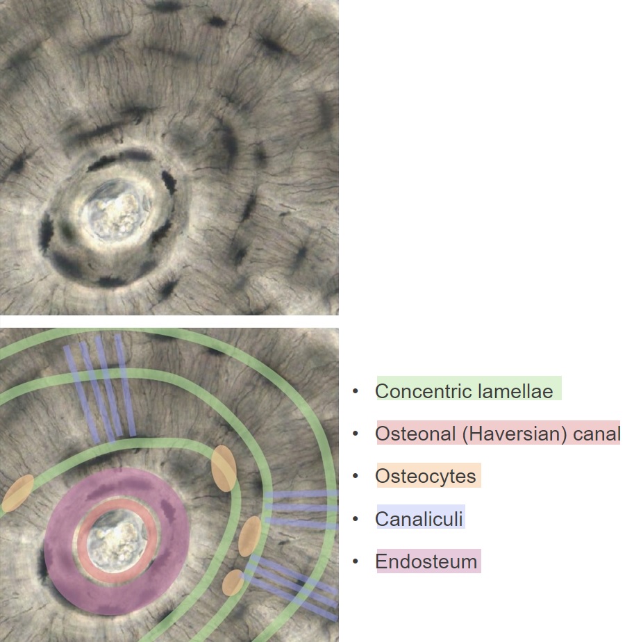
In long bones, the majority of cells and matrix are arranged in functional units known as osteons.
Osteons:
An osteon (also known as a haversian system) is a cylinder of cells and matrix running longitudinally within compact bone. An osteon is made up of a longitudinal central canal, which is surrounded by concentric rings of osteocytesOsteocytesMature osteoblasts that have become embedded in the bone matrix. They occupy a small cavity, called lacuna, in the matrix and are connected to adjacent osteocytes via protoplasmic projections called canaliculi.Bones: Development and Ossification, and bone matrix known as lamellae.
Provides nutrients and removes waste from osteocytesOsteocytesMature osteoblasts that have become embedded in the bone matrix. They occupy a small cavity, called lacuna, in the matrix and are connected to adjacent osteocytes via protoplasmic projections called canaliculi.Bones: Development and Ossification immediately adjacent to the central canal
Perforating canals:
Contain neurovasculature
Run perpendicular to the central canals, connecting the canals to:
External nerves and vessels
One another
Lamellae:
Concentric rings of calcified matrix
OsteocytesOsteocytesMature osteoblasts that have become embedded in the bone matrix. They occupy a small cavity, called lacuna, in the matrix and are connected to adjacent osteocytes via protoplasmic projections called canaliculi.Bones: Development and Ossification are located between lamellae in spaces known as lacunaeLacunaeBones: Development and Ossification.
Canaliculi (tiny canals in the matrix) allow thin, finger-like projections of the osteocytesOsteocytesMature osteoblasts that have become embedded in the bone matrix. They occupy a small cavity, called lacuna, in the matrix and are connected to adjacent osteocytes via protoplasmic projections called canaliculi.Bones: Development and Ossification to connect to one another via gap junctionsGap JunctionsConnections between cells which allow passage of small molecules and electric current. Gap junctions were first described anatomically as regions of close apposition between cells with a narrow (1-2 nm) gap between cell membranes. The variety in the properties of gap junctions is reflected in the number of connexins, the family of proteins which form the junctions.The Cell: Cell Junctions:
Allows for nutrient delivery and waste removal from osteocytesOsteocytesMature osteoblasts that have become embedded in the bone matrix. They occupy a small cavity, called lacuna, in the matrix and are connected to adjacent osteocytes via protoplasmic projections called canaliculi.Bones: Development and Ossification in outer rings, without being immediately adjacent to vasculature
CollagenCollagenA polypeptide substance comprising about one third of the total protein in mammalian organisms. It is the main constituent of skin; connective tissue; and the organic substance of bones (bone and bones) and teeth (tooth).Connective Tissue: Histology:
Fibers “corkscrew” down the matrix in a given lamella
Different helicalHelicalComputed tomography where there is continuous x-ray exposure to the patient while being transported in a spiral or helical pattern through the beam of irradiation. This provides improved three-dimensional contrast and spatial resolution compared to conventional computed tomography, where data is obtained and computed from individual sequential exposures.Computed Tomography (CT) arrangements are present within adjacent lamellae:
Right-handed coils vs. left-handed coils
Varying tightness of coils
Creates a “crisscrossing” network of collagenCollagenA polypeptide substance comprising about one third of the total protein in mammalian organisms. It is the main constituent of skin; connective tissue; and the organic substance of bones (bone and bones) and teeth (tooth).Connective Tissue: Histology → significantly ↑ strength to resist bending and compressionCompressionBlunt Chest Trauma
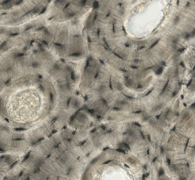
A microscopic image of an osteon
Image by Lecturio.
Cross section of bone demonstrating the structure of an osteon: Concentric lamellae form a ring around the central osteonal canals and contain osteocytes. Within the central canal, blood vessels deliver nutrients to the neighboring osteocytes. The osteocytes have long, thin, “finger-like” projections branching into canaliculi (channels within the matrix). The projections allow the osteocytes to be connected to one another via gap junctions, providing a mechanism for nutrient delivery and waste removal from osteocytes beyond the central canal.
Image by Lecturio.
Circumferential lamellae:
Lamellae running parallel to the bone surface around the entire circumference of the bone
Not part of an osteon functional unit
Locations:
Immediately within the osteogenic layer of periosteum
Remnants of old osteons partially broken down during bone remodelingBone remodelingThe continuous turnover of bone matrix and mineral that involves first an increase in bone resorption (osteoclastic activity) and later, reactive bone formation (osteoblastic activity). The process of bone remodeling takes place in the adult skeleton at discrete foci. The process ensures the mechanical integrity of the skeleton throughout life and plays an important role in calcium homeostasis. An imbalance in the regulation of bone remodeling’s two contrasting events, bone resorption and bone formation, results in many of the metabolic bone diseases, such as osteoporosis.Bones: Remodeling and Healing
Microscopic structure of compact bone
Image: “Cross-sectional view of compact bone showing the basic structural unit, the osteon” by OpenStax College. License: CC BY 4.0
Microscopic structure of spongy bone
In spongy bone, rather than forming concentric rings within osteons, lamellae form concentric rings, which create the trabeculae.
Similarities between spongy and compact bone:
Lamellae form concentric rings
OsteocytesOsteocytesMature osteoblasts that have become embedded in the bone matrix. They occupy a small cavity, called lacuna, in the matrix and are connected to adjacent osteocytes via protoplasmic projections called canaliculi.Bones: Development and Ossification live within lacuna between lamellae
OsteocytesOsteocytesMature osteoblasts that have become embedded in the bone matrix. They occupy a small cavity, called lacuna, in the matrix and are connected to adjacent osteocytes via protoplasmic projections called canaliculi.Bones: Development and Ossification are connected to each other via canaliculi
Differences between spongy and compact bone:
Trabeculae are arranged along lines of force (osteons are parallel to one another in compact bone).
Trabeculae form a lattice-like network, creating space within the bone filled with bone marrowBone marrowThe soft tissue filling the cavities of bones. Bone marrow exists in two types, yellow and red. Yellow marrow is found in the large cavities of large bones and consists mostly of fat cells and a few primitive blood cells. Red marrow is a hematopoietic tissue and is the site of production of erythrocytes and granular leukocytes. Bone marrow is made up of a framework of connective tissue containing branching fibers with the frame being filled with marrow cells.Bone Marrow: Composition and Hematopoiesis.
No central canals → not needed because no osteocytesOsteocytesMature osteoblasts that have become embedded in the bone matrix. They occupy a small cavity, called lacuna, in the matrix and are connected to adjacent osteocytes via protoplasmic projections called canaliculi.Bones: Development and Ossification are far from the blood supply (surrounding marrow)
Microscopic structure of spongy bone
Image: “Spongy bone is composed of trabeculae containing the osteocytes. Red marrow fills the spaces in some bones.” by OpenStax College. License: CC BY 4.0
Bone marrowBone marrowThe soft tissue filling the cavities of bones. Bone marrow exists in two types, yellow and red. Yellow marrow is found in the large cavities of large bones and consists mostly of fat cells and a few primitive blood cells. Red marrow is a hematopoietic tissue and is the site of production of erythrocytes and granular leukocytes. Bone marrow is made up of a framework of connective tissue containing branching fibers with the frame being filled with marrow cells.Bone Marrow: Composition and Hematopoiesis: a general term for hematopoietic soft tissueSoft TissueSoft Tissue Abscess occupying the spaces within bone.
Red marrow: myeloid tissue (can produce blood cells)
Yellow marrow: fatty marrow (no longer produces blood cells)
Locations of bone marrowBone marrowThe soft tissue filling the cavities of bones. Bone marrow exists in two types, yellow and red. Yellow marrow is found in the large cavities of large bones and consists mostly of fat cells and a few primitive blood cells. Red marrow is a hematopoietic tissue and is the site of production of erythrocytes and granular leukocytes. Bone marrow is made up of a framework of connective tissue containing branching fibers with the frame being filled with marrow cells.Bone Marrow: Composition and Hematopoiesis
The medullary cavities of long bones
Diploë of flat bones
Trabecular cavities of spongy bone
Marrow changes during the lifespan
In infants and children: Nearly all bone cavities contain red marrow.
In young to middle-aged adults:
Most red marrow has become yellow marrow
Red marrow exists in:
Vertebrae
RibsRibsA set of twelve curved bones which connect to the vertebral column posteriorly, and terminate anteriorly as costal cartilage. Together, they form a protective cage around the internal thoracic organs.Chest Wall: Anatomy
SternumSternumA long, narrow, and flat bone commonly known as breastbone occurring in the midsection of the anterior thoracic segment or chest region, which stabilizes the rib cage and serves as the point of origin for several muscles that move the arms, head, and neck.Chest Wall: Anatomy
Parts of the pelvic girdle
Proximal head of the humerusHead of The HumerusThe upper rounded extremity of the humerus fitting into the glenoid cavity of the scapula.Arm: Anatomy and femur
Yellow marrow can revert to red marrow in severe or chronic anemiaAnemiaAnemia is a condition in which individuals have low Hb levels, which can arise from various causes. Anemia is accompanied by a reduced number of RBCs and may manifest with fatigue, shortness of breath, pallor, and weakness. Subtypes are classified by the size of RBCs, chronicity, and etiology. Anemia: Overview and Types.
Clinical Relevance
OsteoporosisOsteoporosisOsteoporosis refers to a decrease in bone mass and density leading to an increased number of fractures. There are 2 forms of osteoporosis: primary, which is commonly postmenopausal or senile; and secondary, which is a manifestation of immobilization, underlying medical disorders, or long-term use of certain medications. Osteoporosis: a decrease in bone massMassThree-dimensional lesion that occupies a space within the breastImaging of the Breast and density leading to an increased number of fractures. OsteoporosisOsteoporosisOsteoporosis refers to a decrease in bone mass and density leading to an increased number of fractures. There are 2 forms of osteoporosis: primary, which is commonly postmenopausal or senile; and secondary, which is a manifestation of immobilization, underlying medical disorders, or long-term use of certain medications. Osteoporosis is most commonly caused by a loss of protective estrogenEstrogenCompounds that interact with estrogen receptors in target tissues to bring about the effects similar to those of estradiol. Estrogens stimulate the female reproductive organs, and the development of secondary female sex characteristics. Estrogenic chemicals include natural, synthetic, steroidal, or non-steroidal compounds.Ovaries: Anatomy and/or testosteroneTestosteroneA potent androgenic steroid and major product secreted by the leydig cells of the testis. Its production is stimulated by luteinizing hormone from the pituitary gland. In turn, testosterone exerts feedback control of the pituitary LH and FSH secretion. Depending on the tissues, testosterone can be further converted to dihydrotestosterone or estradiol.Androgens and Antiandrogens later in life, immobilizationImmobilizationDelirium, underlying medical disorders, or long-term use of certain medications. OsteoporosisOsteoporosisOsteoporosis refers to a decrease in bone mass and density leading to an increased number of fractures. There are 2 forms of osteoporosis: primary, which is commonly postmenopausal or senile; and secondary, which is a manifestation of immobilization, underlying medical disorders, or long-term use of certain medications. Osteoporosis most often presents clinically with frequent fractures and loss of vertebral height. Diagnosis is established by measuring bone mineral density. Management includes lifestyle modifications, maintaining adequate levels of calciumCalciumA basic element found in nearly all tissues. It is a member of the alkaline earth family of metals with the atomic symbol ca, atomic number 20, and atomic weight 40. Calcium is the most abundant mineral in the body and combines with phosphorus to form calcium phosphate in the bones and teeth. It is essential for the normal functioning of nerves and muscles and plays a role in blood coagulation (as factor IV) and in many enzymatic processes.Electrolytes and vitamin DVitamin DA vitamin that includes both cholecalciferols and ergocalciferols, which have the common effect of preventing or curing rickets in animals. It can also be viewed as a hormone since it can be formed in skin by action of ultraviolet rays upon the precursors, 7-dehydrocholesterol and ergosterol, and acts on vitamin D receptors to regulate calcium in opposition to parathyroid hormone.Fat-soluble Vitamins and their Deficiencies, and the use of bisphosphonatesBisphosphonatesBisphosphonates are pyrophosphate analogs most well-known for treating osteoporosis by preventing bone loss. Bisphosphonates end in the suffix “-dronate” or “-dronic acid” (e.g., alendronate, risedronate, pamidronate) and bind to hydroxyapatite crystals in bone, inhibiting osteoclast-induced bone resorption.Bisphosphonates.
OsteomalaciaOsteomalaciaDisorder caused by an interruption of the mineralization of organic bone matrix leading to bone softening, bone pain, and weakness. It is the adult form of rickets resulting from disruption of vitamin d; phosphorus; or calcium homeostasis.Osteomalacia and Rickets and ricketsRicketsDisorders caused by interruption of bone mineralization manifesting as osteomalacia in adults and characteristic deformities in infancy and childhood due to disturbances in normal bone formation. The mineralization process may be interrupted by disruption of vitamin d; phosphorus; or calcium homeostasis, resulting from dietary deficiencies, or acquired, or inherited metabolic, or hormonal disturbances.Osteomalacia and Rickets: disorders of decreased bone mineralizationBone mineralizationCalcium (Ca2+) and phosphate (PO43–) combine to form hydroxyapatite crystals on the bone matrix.Bones: Development and Ossification. RicketsRicketsDisorders caused by interruption of bone mineralization manifesting as osteomalacia in adults and characteristic deformities in infancy and childhood due to disturbances in normal bone formation. The mineralization process may be interrupted by disruption of vitamin d; phosphorus; or calcium homeostasis, resulting from dietary deficiencies, or acquired, or inherited metabolic, or hormonal disturbances.Osteomalacia and Rickets affects the cartilageCartilageCartilage is a type of connective tissue derived from embryonic mesenchyme that is responsible for structural support, resilience, and the smoothness of physical actions. Perichondrium (connective tissue membrane surrounding cartilage) compensates for the absence of vasculature in cartilage by providing nutrition and support. Cartilage: Histology of the epiphyseal growth platesGrowth PlatesThe area between the epiphysis and the diaphysis within which bone growth occurs.Osteosarcoma in children. OsteomalaciaOsteomalaciaDisorder caused by an interruption of the mineralization of organic bone matrix leading to bone softening, bone pain, and weakness. It is the adult form of rickets resulting from disruption of vitamin d; phosphorus; or calcium homeostasis.Osteomalacia and Rickets affects the sites of bone turnover in children and adults. Both disorders are most commonly caused by vitamin D deficiencyVitamin D DeficiencyA nutritional condition produced by a deficiency of vitamin D in the diet, insufficient production of vitamin D in the skin, inadequate absorption of vitamin D from the diet, or abnormal conversion of vitamin D to its bioactive metabolites. It is manifested clinically as rickets in children and osteomalacia in adults.Fat-soluble Vitamins and their Deficiencies. RicketsRicketsDisorders caused by interruption of bone mineralization manifesting as osteomalacia in adults and characteristic deformities in infancy and childhood due to disturbances in normal bone formation. The mineralization process may be interrupted by disruption of vitamin d; phosphorus; or calcium homeostasis, resulting from dietary deficiencies, or acquired, or inherited metabolic, or hormonal disturbances.Osteomalacia and Rickets commonly presents with skeletal deformities and growth abnormalities. OsteomalaciaOsteomalaciaDisorder caused by an interruption of the mineralization of organic bone matrix leading to bone softening, bone pain, and weakness. It is the adult form of rickets resulting from disruption of vitamin d; phosphorus; or calcium homeostasis.Osteomalacia and Rickets can present with bone painPainAn unpleasant sensation induced by noxious stimuli which are detected by nerve endings of nociceptive neurons.Pain: Types and Pathways, difficulty with ambulation, and pathologic fractures. Treatment includes vitamin DVitamin DA vitamin that includes both cholecalciferols and ergocalciferols, which have the common effect of preventing or curing rickets in animals. It can also be viewed as a hormone since it can be formed in skin by action of ultraviolet rays upon the precursors, 7-dehydrocholesterol and ergosterol, and acts on vitamin D receptors to regulate calcium in opposition to parathyroid hormone.Fat-soluble Vitamins and their Deficiencies, calciumCalciumA basic element found in nearly all tissues. It is a member of the alkaline earth family of metals with the atomic symbol ca, atomic number 20, and atomic weight 40. Calcium is the most abundant mineral in the body and combines with phosphorus to form calcium phosphate in the bones and teeth. It is essential for the normal functioning of nerves and muscles and plays a role in blood coagulation (as factor IV) and in many enzymatic processes.Electrolytes, and phosphorus supplementation.
Paget disease of bone: a focal disorder of bone metabolismBone metabolismBone is the primary storage site of calcium in the body; thus, bone metabolism plays a critical role in maintaining normal calcium levels. Bone metabolism (and thus calcium levels) are primarily regulated by 3 hormones, namely, calcitonin, parathyroid hormone (PTH), and vitamin D. Calcium Hemostasis and Bone Metabolism commonly affecting the skullSkullThe skull (cranium) is the skeletal structure of the head supporting the face and forming a protective cavity for the brain. The skull consists of 22 bones divided into the viscerocranium (facial skeleton) and the neurocranium.Skull: Anatomy, spineSpineThe human spine, or vertebral column, is the most important anatomical and functional axis of the human body. It consists of 7 cervical vertebrae, 12 thoracic vertebrae, and 5 lumbar vertebrae and is limited cranially by the skull and caudally by the sacrum.Vertebral Column: Anatomy, pelvisPelvisThe pelvis consists of the bony pelvic girdle, the muscular and ligamentous pelvic floor, and the pelvic cavity, which contains viscera, vessels, and multiple nerves and muscles. The pelvic girdle, composed of 2 “hip” bones and the sacrum, is a ring-like bony structure of the axial skeleton that links the vertebral column with the lower extremities.Pelvis: Anatomy, and long bones of the lower extremity. The main clinical manifestations of Paget disease are bone painPainAn unpleasant sensation induced by noxious stimuli which are detected by nerve endings of nociceptive neurons.Pain: Types and Pathways and the consequences of bone deformities (e.g., fractures, osteoarthritisOsteoarthritisOsteoarthritis (OA) is the most common form of arthritis, and is due to cartilage destruction and changes of the subchondral bone. The risk of developing this disorder increases with age, obesity, and repetitive joint use or trauma. Patients develop gradual joint pain, stiffness lasting < 30 minutes, and decreased range of motion. Osteoarthritis, nerve impingement). Management of Paget disease includes bisphosphonatesBisphosphonatesBisphosphonates are pyrophosphate analogs most well-known for treating osteoporosis by preventing bone loss. Bisphosphonates end in the suffix “-dronate” or “-dronic acid” (e.g., alendronate, risedronate, pamidronate) and bind to hydroxyapatite crystals in bone, inhibiting osteoclast-induced bone resorption.Bisphosphonates, calcitoninCalcitoninA peptide hormone that lowers calcium concentration in the blood. In humans, it is released by thyroid cells and acts to decrease the formation and absorptive activity of osteoclasts. Its role in regulating plasma calcium is much greater in children and in certain diseases than in normal adults.Other Antiresorptive Drugs, and surgery to manage fractures, deformities, or complications.
HyperparathyroidismHyperparathyroidismHyperparathyroidism is a condition associated with elevated blood levels of parathyroid hormone (PTH). Depending on the pathogenesis of this condition, hyperparathyroidism can be defined as primary, secondary or tertiary. Hyperparathyroidism: a condition associated with elevated blood levels of parathyroidParathyroidThe parathyroid glands are 2 pairs of small endocrine glands found in close proximity to the thyroid gland. The superior parathyroid glands are lodged within the parenchyma of the upper poles of the right and left thyroid lobes; the inferior parathyroid glands are close to the inferior tips or poles of the lobes.Parathyroid Glands: Anatomy hormone (PTH). HyperparathyroidismHyperparathyroidismHyperparathyroidism is a condition associated with elevated blood levels of parathyroid hormone (PTH). Depending on the pathogenesis of this condition, hyperparathyroidism can be defined as primary, secondary or tertiary. Hyperparathyroidism may be due to an inherent disease within the parathyroidParathyroidThe parathyroid glands are 2 pairs of small endocrine glands found in close proximity to the thyroid gland. The superior parathyroid glands are lodged within the parenchyma of the upper poles of the right and left thyroid lobes; the inferior parathyroid glands are close to the inferior tips or poles of the lobes.Parathyroid Glands: Anatomy gland or abnormalities of calciumCalciumA basic element found in nearly all tissues. It is a member of the alkaline earth family of metals with the atomic symbol ca, atomic number 20, and atomic weight 40. Calcium is the most abundant mineral in the body and combines with phosphorus to form calcium phosphate in the bones and teeth. It is essential for the normal functioning of nerves and muscles and plays a role in blood coagulation (as factor IV) and in many enzymatic processes.Electrolytes metabolism. Individuals classically present with “stones (nephrolithiasisNephrolithiasisNephrolithiasis is the formation of a stone, or calculus, anywhere along the urinary tract caused by precipitations of solutes in the urine. The most common type of kidney stone is the calcium oxalate stone, but other types include calcium phosphate, struvite (ammonium magnesium phosphate), uric acid, and cystine stones.Nephrolithiasis), bones (↓ bone mineral density), abdominal groans (nonspecific abdominal painAbdominal PainAcute Abdomen), and psychiatric overtones (neuropsychiatric symptoms).” Diagnosis is based on laboratory assessment of serum PTH, calciumCalciumA basic element found in nearly all tissues. It is a member of the alkaline earth family of metals with the atomic symbol ca, atomic number 20, and atomic weight 40. Calcium is the most abundant mineral in the body and combines with phosphorus to form calcium phosphate in the bones and teeth. It is essential for the normal functioning of nerves and muscles and plays a role in blood coagulation (as factor IV) and in many enzymatic processes.Electrolytes and phosphatePhosphateInorganic salts of phosphoric acid.Electrolytes levels, and urinary calciumCalciumA basic element found in nearly all tissues. It is a member of the alkaline earth family of metals with the atomic symbol ca, atomic number 20, and atomic weight 40. Calcium is the most abundant mineral in the body and combines with phosphorus to form calcium phosphate in the bones and teeth. It is essential for the normal functioning of nerves and muscles and plays a role in blood coagulation (as factor IV) and in many enzymatic processes.Electrolytes. Management is typically surgical and the treatment of any underlying conditions.
Bone fracturesBone fracturesBreaks in bones.Bones: Remodeling and Healing: a partial or complete interruption in the continuity of a bone (periosteum and/or cortex) resulting from mechanical stress (typically injuries or metabolic disorders of the bone). Clinical presentation varies depending on the cause and location of the injury. Presentation generally includes deformityDeformityExamination of the Upper Limbs, painPainAn unpleasant sensation induced by noxious stimuli which are detected by nerve endings of nociceptive neurons.Pain: Types and Pathways, edemaEdemaEdema is a condition in which excess serous fluid accumulates in the body cavity or interstitial space of connective tissues. Edema is a symptom observed in several medical conditions. It can be categorized into 2 types, namely, peripheral (in the extremities) and internal (in an organ or body cavity). Edema, and inflammationInflammationInflammation is a complex set of responses to infection and injury involving leukocytes as the principal cellular mediators in the body’s defense against pathogenic organisms. Inflammation is also seen as a response to tissue injury in the process of wound healing. The 5 cardinal signs of inflammation are pain, heat, redness, swelling, and loss of function. Inflammation. Diagnosis is made clinically and confirmed with imaging. Management may be splinting or surgery.
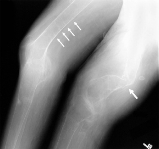
Osteomalacia
Image: “A radiograph of the distal femurs shows further evidence of badly malformed bones secondary to severe osteomalacia (large arrow), as well as several additional pseodofractures (small arrows)” by Gamache L et al. License: CC BY 3.0
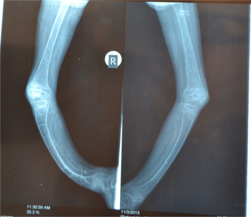
Rickets
Image: “X-rays of both lower limbs showing severe bowing of the legs and diffuse osteopenia. It also shows dense transverse lines in the tibia suggestive of looser’s zones indicative of rickets” by Al-Sharafi BA et al. License: CC BY 4.0
References
Clarke, B. (2023). Normal bone anatomy and physiology. Clinical Journal of the American Society of Nephrology, 18(8), 1153-1168.
Florencio-Silva, R., Sasso, G. R., Sasso-Cerri, E., Simões, M. J., & Cerri, P. S. (2023). Biology of bone tissue: structure, function, and factors that influence bone cells. BioMed Research International, 2023.
Gasser, J. A., & Kneissel, M. (2024). Bone physiology and biology. In J. P. Bilezikian (Ed.), Primer on the metabolic bone diseases and disorders of mineral metabolism (10th ed., pp. 3-19). Wiley-Blackwell.
Hendrickx, G., Boudin, E., & Van Hul, W. (2022). A look behind the scenes: The risk and pathogenesis of primary osteoporosis. Nature Reviews Rheumatology, 17(4), 213-230.
Kenkre, J. S., & Bassett, J. (2022). The bone remodelling cycle. Annals of Clinical Biochemistry, 59(5), 394-404.
Marsell, R., & Einhorn, T. A. (2023). The biology of fracture healing. Injury, 54(6), 1801-1808.
Mohseni, E., Planell, J. A., Mata, A., & Engel, E. (2023). Novel advances in understanding the molecular basis of bone development, diseases and regeneration. International Materials Reviews, 68(5), 522-554.
Plotkin, L. I., & Bruzzaniti, A. (2023). Molecular signaling in bone cells: Regulation of cell differentiation and survival. Advances in Protein Chemistry and Structural Biology, 129, 91-119.
Prideaux, M., Findlay, D. M., & Atkins, G. J. (2022). Osteocytes: The master cells in bone remodelling. Current Opinion in Pharmacology, 47, 24-31.
Weaver, C. M., & Peacock, M. (2022). Calcium. In A. C. Ross, B. Caballero, R. J. Cousins, K. L. Tucker, & T. R. Ziegler (Eds.), Modern nutrition in health and disease (12th ed., pp. 133-149). Lippincott Williams & Wilkins.
Create your free account or log in to continue reading!