Cell junctions are proteinaceous structures that physically hold 2 surfaces (cell-to-cell or cell-to-matrix) together. Cell junctions aid in communicationCommunicationThe exchange or transmission of ideas, attitudes, or beliefs between individuals or groups.Decision-making Capacity and Legal Competence and structural support and act as a barrier. They are classified as occluding (tight junctions), anchoring (adherens, desmosomesDesmosomesA type of junction that attaches one cell to its neighbor. One of a number of differentiated regions which occur, for example, where the cytoplasmic membranes of adjacent epithelial cells are closely apposed. It consists of a circular region of each membrane together with associated intracellular microfilaments and an intercellular material which may include, for example, mucopolysaccharides.Bullous Pemphigoid and Pemphigus Vulgaris and hemidesmosomes), and communicating (gap junctions). Type II hypersensitivity has been noticed with autoantibody production against the components of anchoring junctions, resulting in pathology such as pemphigus vulgarisPemphigus vulgarisBullous pemphigoid and pemphigus vulgaris are two different blistering autoimmune diseases. In pemphigus vulgaris, autoantibodies attack the desmosomal proteins, which connect the keratinocytes to one another. This attack results in a more severe, potentially fatal condition with fragile, flaccid blisters, usually with significant mucosal involvement. Bullous Pemphigoid and Pemphigus Vulgaris and bullous pemphigoidBullous pemphigoidBullous pemphigoid and pemphigus vulgaris are two different blistering autoimmune diseases. In bullous pemphigoid, autoantibodies attack the hemidesmosomes, which connect epidermal keratinocytes to the basement membrane. This attack results in large, tense subepidermal blisters. Bullous Pemphigoid and Pemphigus Vulgaris.
Tight junctions (occluding junctions or zonula occludens) are intercellular adhesionAdhesionThe process whereby platelets adhere to something other than platelets, e.g., collagen; basement membrane; microfibrils; or other ‘foreign’ surfaces.Coagulation Studies complexes composed of proteinsProteinsLinear polypeptides that are synthesized on ribosomes and may be further modified, crosslinked, cleaved, or assembled into complex proteins with several subunits. The specific sequence of amino acids determines the shape the polypeptide will take, during protein folding, and the function of the protein.Energy Homeostasis whose primary role is regulating the passage of water and solutes between epithelial cells (paracellularParacellularRenal Potassium Regulation permeability).
Location
Located at the most apical aspect of the lateral membrane
Mainly present in gastric mucosaGastric mucosaLining of the stomach, consisting of an inner epithelium, a middle lamina propria, and an outer muscularis mucosae. The surface cells produce mucus that protects the stomach from attack by digestive acid and enzymes. When the epithelium invaginates into the lamina propria at various region of the stomach (cardia; gastric fundus; and pylorus), different tubular gastric glands are formed. These glands consist of cells that secrete mucus, enzymes, hydrochloric acid, or hormones.Stomach: Anatomy, renal tubules, brainBrainThe part of central nervous system that is contained within the skull (cranium). Arising from the neural tube, the embryonic brain is comprised of three major parts including prosencephalon (the forebrain); mesencephalon (the midbrain); and rhombencephalon (the hindbrain). The developed brain consists of cerebrum; cerebellum; and other structures in the brain stem.Nervous System: Anatomy, Structure, and ClassificationcapillariesCapillariesCapillaries are the primary structures in the circulatory system that allow the exchange of gas, nutrients, and other materials between the blood and the extracellular fluid (ECF). Capillaries are the smallest of the blood vessels. Because a capillary diameter is so small, only 1 RBC may pass through at a time.Capillaries: Histology
Composition
Cell junctions are branching networks of sealing strands, each formed by multiple transmembrane proteinsProteinsLinear polypeptides that are synthesized on ribosomes and may be further modified, crosslinked, cleaved, or assembled into complex proteins with several subunits. The specific sequence of amino acids determines the shape the polypeptide will take, during protein folding, and the function of the protein.Energy Homeostasis and associated intracellular proteinsProteinsLinear polypeptides that are synthesized on ribosomes and may be further modified, crosslinked, cleaved, or assembled into complex proteins with several subunits. The specific sequence of amino acids determines the shape the polypeptide will take, during protein folding, and the function of the protein.Energy Homeostasis.
Transmembrane proteinsProteinsLinear polypeptides that are synthesized on ribosomes and may be further modified, crosslinked, cleaved, or assembled into complex proteins with several subunits. The specific sequence of amino acids determines the shape the polypeptide will take, during protein folding, and the function of the protein.Energy Homeostasis:
Embedded in the plasma membranePlasma membraneA cell membrane (also known as the plasma membrane or plasmalemma) is a biological membrane that separates the cell contents from the outside environment. A cell membrane is composed of a phospholipid bilayer and proteins that function to protect cellular DNA and mediate the exchange of ions and molecules.The Cell: Cell Membrane of 2 adjacent cells: Extracellular domains of proteinsProteinsLinear polypeptides that are synthesized on ribosomes and may be further modified, crosslinked, cleaved, or assembled into complex proteins with several subunits. The specific sequence of amino acids determines the shape the polypeptide will take, during protein folding, and the function of the protein.Energy Homeostasis in 1 cell are continuous with those of transmembrane proteinsProteinsLinear polypeptides that are synthesized on ribosomes and may be further modified, crosslinked, cleaved, or assembled into complex proteins with several subunits. The specific sequence of amino acids determines the shape the polypeptide will take, during protein folding, and the function of the protein.Energy Homeostasis in the opposing cell.
Occludin proteinsProteinsLinear polypeptides that are synthesized on ribosomes and may be further modified, crosslinked, cleaved, or assembled into complex proteins with several subunits. The specific sequence of amino acids determines the shape the polypeptide will take, during protein folding, and the function of the protein.Energy Homeostasis have extra- and intracellular loops that regulate paracellularParacellularRenal Potassium Regulation permeability.
Influence intracellular activity such as geneGeneA category of nucleic acid sequences that function as units of heredity and which code for the basic instructions for the development, reproduction, and maintenance of organisms.Basic Terms of Genetics expression and energy metabolism
Have 9 major domains, which are distributed intra- and extracellularly
Stabilize tight junctions
Support the barrier function of tight junctions
Claudin proteinsProteinsLinear polypeptides that are synthesized on ribosomes and may be further modified, crosslinked, cleaved, or assembled into complex proteins with several subunits. The specific sequence of amino acids determines the shape the polypeptide will take, during protein folding, and the function of the protein.Energy Homeostasis are considered the backbone of tight junctions.
4 transmembrane domains, with both ends lying within the cytoplasm
Have the ability to bindBINDHyperbilirubinemia of the Newborn to scaffolding proteinsProteinsLinear polypeptides that are synthesized on ribosomes and may be further modified, crosslinked, cleaved, or assembled into complex proteins with several subunits. The specific sequence of amino acids determines the shape the polypeptide will take, during protein folding, and the function of the protein.Energy Homeostasis
Junctional adhesionAdhesionThe process whereby platelets adhere to something other than platelets, e.g., collagen; basement membrane; microfibrils; or other ‘foreign’ surfaces.Coagulation Studies molecules regulate the paracellularParacellularRenal Potassium Regulation pathway.
Have only 1 transmembrane domain
Help maintain cell polarity
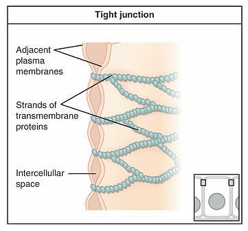
Illustration depicting strands of transmembrane proteins (claudins and occludins) tightly binding adjacent plasma membranes
Image: “Types of Cell Junctions” by OpenStax College. License: CC BY 3.0, edited by Lecturio.
Function
DiffusionDiffusionThe tendency of a gas or solute to pass from a point of higher pressure or concentration to a point of lower pressure or concentration and to distribute itself throughout the available space. Diffusion, especially facilitated diffusion, is a major mechanism of biological transport.Peritoneal Dialysis and Hemodialysis barrier between adjacent cells
Prevent passage of ions and molecules between cells
Molecules are selectively allowed based upon size and charge.
Generally, cationsCationsPositively charged atoms, radicals or groups of atoms which travel to the cathode or negative pole during electrolysis.Electrolytes are preferentially allowed in transit.
Separate tissue compartments into apical and basal sides and maintain the polarity of cells
Prevent lateral diffusionDiffusionThe tendency of a gas or solute to pass from a point of higher pressure or concentration to a point of lower pressure or concentration and to distribute itself throughout the available space. Diffusion, especially facilitated diffusion, is a major mechanism of biological transport.Peritoneal Dialysis and Hemodialysis of proteinsProteinsLinear polypeptides that are synthesized on ribosomes and may be further modified, crosslinked, cleaved, or assembled into complex proteins with several subunits. The specific sequence of amino acids determines the shape the polypeptide will take, during protein folding, and the function of the protein.Energy Homeostasis between the apical and basolateral surfaces
Preserve the functioning of specialized activities such as receptor-mediated endocytosisEndocytosisCellular uptake of extracellular materials within membrane-limited vacuoles or microvesicles. Endosomes play a central role in endocytosis.The Cell: Cell Membrane
Maintain osmotic balance
Generally, cells are classified as being tight or leaky. Tight junctions have an important role in determining the permeability of epithelia.
Tight epithelial cells include distal convoluted tubuleDistal convoluted tubuleThe portion of renal tubule that begins from the enlarged segment of the ascending limb of the loop of henle. It reenters the kidney cortex and forms the convoluted segments of the distal tubule.Gitelman Syndrome cells in the kidney and bileBileAn emulsifying agent produced in the liver and secreted into the duodenum. Its composition includes bile acids and salts; cholesterol; and electrolytes. It aids digestion of fats in the duodenum.Gallbladder and Biliary Tract: Anatomy ducts and the cells composing the blood–brain barrierBlood–Brain BarrierMeningitis in Children (BBBBBBSpecialized non-fenestrated tightly-joined endothelial cells with tight junctions that form a transport barrier for certain substances between the cerebral capillaries and the brain tissue.Nervous System: Histology).
Leaky epithelial cells include cells in the proximal tubuleProximal tubuleThe renal tubule portion that extends from the bowman capsule in the kidney cortex into the kidney medulla. The proximal tubule consists of a convoluted proximal segment in the cortex, and a distal straight segment descending into the medulla where it forms the u-shaped loop of henle.Tubular System of the kidney. These cells contain fewer tight junctions, which contributes to their leakiness.
Adherens are junctions between cells that are linked to the actinActinFilamentous proteins that are the main constituent of the thin filaments of muscle fibers. The filaments (known also as filamentous or f-actin) can be dissociated into their globular subunits; each subunit is composed of a single polypeptide 375 amino acids long. This is known as globular or g-actin. In conjunction with myosins, actin is responsible for the contraction and relaxation of muscle.Skeletal Muscle ContractioncytoskeletonCytoskeletonThe network of filaments, tubules, and interconnecting filamentous bridges which give shape, structure, and organization to the cytoplasm.The Cell: Cytosol and Cytoskeleton. These junctions are also known as zonula adherens, intermediate junctions, or belt desmosomesDesmosomesA type of junction that attaches one cell to its neighbor. One of a number of differentiated regions which occur, for example, where the cytoplasmic membranes of adjacent epithelial cells are closely apposed. It consists of a circular region of each membrane together with associated intracellular microfilaments and an intercellular material which may include, for example, mucopolysaccharides.Bullous Pemphigoid and Pemphigus Vulgaris.
Location:
Between adjacent epithelial cells
Mainly seen in endothelial and epithelial cells
More basal than tight junctions
Composition:
ActinActinFilamentous proteins that are the main constituent of the thin filaments of muscle fibers. The filaments (known also as filamentous or f-actin) can be dissociated into their globular subunits; each subunit is composed of a single polypeptide 375 amino acids long. This is known as globular or g-actin. In conjunction with myosins, actin is responsible for the contraction and relaxation of muscle.Skeletal Muscle Contraction filaments
Intracellular
Part of the cytoskeletonCytoskeletonThe network of filaments, tubules, and interconnecting filamentous bridges which give shape, structure, and organization to the cytoplasm.The Cell: Cytosol and Cytoskeleton
E-cadherins
Transmembrane adhesionAdhesionThe process whereby platelets adhere to something other than platelets, e.g., collagen; basement membrane; microfibrils; or other ‘foreign’ surfaces.Coagulation Studies protein
CalciumCalciumA basic element found in nearly all tissues. It is a member of the alkaline earth family of metals with the atomic symbol ca, atomic number 20, and atomic weight 40. Calcium is the most abundant mineral in the body and combines with phosphorus to form calcium phosphate in the bones and teeth. It is essential for the normal functioning of nerves and muscles and plays a role in blood coagulation (as factor IV) and in many enzymatic processes.Electrolytes dependent
Form homodimers
ActinActinFilamentous proteins that are the main constituent of the thin filaments of muscle fibers. The filaments (known also as filamentous or f-actin) can be dissociated into their globular subunits; each subunit is composed of a single polypeptide 375 amino acids long. This is known as globular or g-actin. In conjunction with myosins, actin is responsible for the contraction and relaxation of muscle.Skeletal Muscle Contraction filaments and E-cadherins are connected by vinculin and catenin.
Catenins bindBINDHyperbilirubinemia of the NewborncadherinCadherinCalcium-dependent cell adhesion proteins. They are important in the formation of adherens junctions between cells. Cadherins are classified by their distinct immunological and tissue specificities, either by letters (e- for epithelial, n- for neural, and p- for placental cadherins) or by numbers (cadherin-12 or n-cadherin 2 for brain-cadherin). Cadherins promote cell adhesion via a homophilic mechanism as in the construction of tissues and of the whole animal body.Gastric Cancer.
Catenins may also bindBINDHyperbilirubinemia of the Newborn to the actinActinFilamentous proteins that are the main constituent of the thin filaments of muscle fibers. The filaments (known also as filamentous or f-actin) can be dissociated into their globular subunits; each subunit is composed of a single polypeptide 375 amino acids long. This is known as globular or g-actin. In conjunction with myosins, actin is responsible for the contraction and relaxation of muscle.Skeletal Muscle ContractioncytoskeletonCytoskeletonThe network of filaments, tubules, and interconnecting filamentous bridges which give shape, structure, and organization to the cytoplasm.The Cell: Cytosol and Cytoskeleton.
Function:
Maintaining cells in a belt shape
Anchoring cells
Providing strength
Maintaining cell shape (may serve a role in the actinActinFilamentous proteins that are the main constituent of the thin filaments of muscle fibers. The filaments (known also as filamentous or f-actin) can be dissociated into their globular subunits; each subunit is composed of a single polypeptide 375 amino acids long. This is known as globular or g-actin. In conjunction with myosins, actin is responsible for the contraction and relaxation of muscle.Skeletal Muscle Contraction contractile ring)
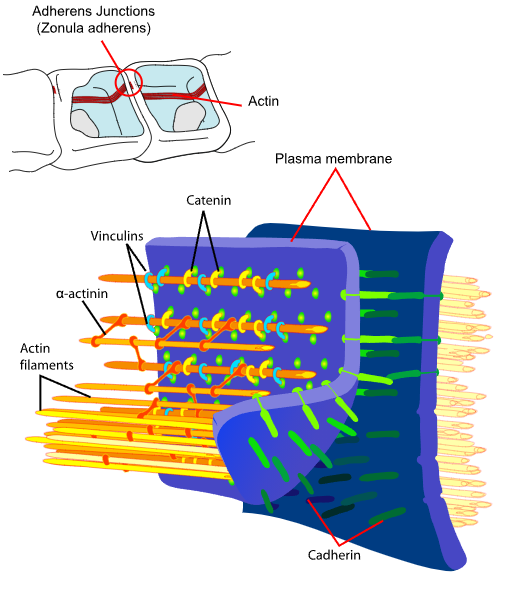
Adherens junctions interact with actin filaments through their proteins, such as cadherin and catenin.
Image: “Principal interactions of structural proteins at cadherin-based adherens junction” by Mariana Ruiz . License: Public Domain
Demosome
Definition:
DesmosomesDesmosomesA type of junction that attaches one cell to its neighbor. One of a number of differentiated regions which occur, for example, where the cytoplasmic membranes of adjacent epithelial cells are closely apposed. It consists of a circular region of each membrane together with associated intracellular microfilaments and an intercellular material which may include, for example, mucopolysaccharides.Bullous Pemphigoid and Pemphigus Vulgaris are strong structures that assist in cellular adhesionAdhesionThe process whereby platelets adhere to something other than platelets, e.g., collagen; basement membrane; microfibrils; or other ‘foreign’ surfaces.Coagulation Studies, tethering intermediate filamentsIntermediate filamentsCytoplasmic filaments intermediate in diameter (about 10 nanometers) between the microfilaments and the microtubules. They may be composed of any of a number of different proteins and form a ring around the cell nucleus.The Cell: Cytosol and Cytoskeleton to the plasma membranePlasma membraneA cell membrane (also known as the plasma membrane or plasmalemma) is a biological membrane that separates the cell contents from the outside environment. A cell membrane is composed of a phospholipid bilayer and proteins that function to protect cellular DNA and mediate the exchange of ions and molecules.The Cell: Cell Membrane. These structures are also known as maculaMaculaAn oval area in the retina, 3 to 5 mm in diameter, usually located temporal to the posterior pole of the eye and slightly below the level of the optic disk. It is characterized by the presence of a yellow pigment diffusely permeating the inner layers, contains the fovea centralis in its center, and provides the best phototropic visual acuity. It is devoid of retinal blood vessels, except in its periphery, and receives nourishment from the choriocapillaris of the choroid.Eye: Anatomy adherens or spot desmosomesDesmosomesA type of junction that attaches one cell to its neighbor. One of a number of differentiated regions which occur, for example, where the cytoplasmic membranes of adjacent epithelial cells are closely apposed. It consists of a circular region of each membrane together with associated intracellular microfilaments and an intercellular material which may include, for example, mucopolysaccharides.Bullous Pemphigoid and Pemphigus Vulgaris.
Location:
Located between adjacent cells
Found in epithelial cells, cardiomyocytes (intercalated discsIntercalated discsIrregular, transverse, thick parts of the sarcolemma at the terminal ends of the cell branches.Muscle Tissue: Histology)
Generally, found in cells that are under large amounts of mechanical stress
Made of cadherinCadherinCalcium-dependent cell adhesion proteins. They are important in the formation of adherens junctions between cells. Cadherins are classified by their distinct immunological and tissue specificities, either by letters (e- for epithelial, n- for neural, and p- for placental cadherins) or by numbers (cadherin-12 or n-cadherin 2 for brain-cadherin). Cadherins promote cell adhesion via a homophilic mechanism as in the construction of tissues and of the whole animal body.Gastric CancerproteinsProteinsLinear polypeptides that are synthesized on ribosomes and may be further modified, crosslinked, cleaved, or assembled into complex proteins with several subunits. The specific sequence of amino acids determines the shape the polypeptide will take, during protein folding, and the function of the protein.Energy Homeostasis
Desmoglein: a cellular adhesionAdhesionThe process whereby platelets adhere to something other than platelets, e.g., collagen; basement membrane; microfibrils; or other ‘foreign’ surfaces.Coagulation Studies protein
Desmocollin: a cellular adhesionAdhesionThe process whereby platelets adhere to something other than platelets, e.g., collagen; basement membrane; microfibrils; or other ‘foreign’ surfaces.Coagulation Studies protein
Located on the cytoplasmic side of the cell membraneCell MembraneA cell membrane (also known as the plasma membrane or plasmalemma) is a biological membrane that separates the cell contents from the outside environment. A cell membrane is composed of a phospholipid bilayer and proteins that function to protect cellular DNA and mediate the exchange of ions and molecules. The Cell: Cell Membrane
KeratinKeratinA class of fibrous proteins or scleroproteins that represents the principal constituent of epidermis; hair; nails; horny tissues, and the organic matrix of tooth enamel. Two major conformational groups have been characterized, alpha-keratin, whose peptide backbone forms a coiled-coil alpha helical structure consisting of type I keratin and a type II keratin, and beta-keratin, whose backbone forms a zigzag or pleated sheet structure. Alpha-keratins have been classified into at least 20 subtypes. In addition multiple isoforms of subtypes have been found which may be due to gene duplication.Seborrheic Keratosisintermediate filamentsIntermediate filamentsCytoplasmic filaments intermediate in diameter (about 10 nanometers) between the microfilaments and the microtubules. They may be composed of any of a number of different proteins and form a ring around the cell nucleus.The Cell: Cytosol and Cytoskeleton
Desmin filaments
Function:
Structural support
Maintains cell structure against mechanical force
Disease:
Several diseases are associated with desmosomesDesmosomesA type of junction that attaches one cell to its neighbor. One of a number of differentiated regions which occur, for example, where the cytoplasmic membranes of adjacent epithelial cells are closely apposed. It consists of a circular region of each membrane together with associated intracellular microfilaments and an intercellular material which may include, for example, mucopolysaccharides.Bullous Pemphigoid and Pemphigus Vulgaris. CardiomyopathyCardiomyopathyCardiomyopathy refers to a group of myocardial diseases associated with structural changes of the heart muscles (myocardium) and impaired systolic and/or diastolic function in the absence of other heart disorders (coronary artery disease, hypertension, valvular disease, and congenital heart disease). Cardiomyopathy: Overview and Types and blistering conditions have been linked with mutations in the desmosome family of proteinsProteinsLinear polypeptides that are synthesized on ribosomes and may be further modified, crosslinked, cleaved, or assembled into complex proteins with several subunits. The specific sequence of amino acids determines the shape the polypeptide will take, during protein folding, and the function of the protein.Energy Homeostasis.
Arrhythmogenic cardiomyopathyCardiomyopathyCardiomyopathy refers to a group of myocardial diseases associated with structural changes of the heart muscles (myocardium) and impaired systolic and/or diastolic function in the absence of other heart disorders (coronary artery disease, hypertension, valvular disease, and congenital heart disease). Cardiomyopathy: Overview and Types (CM)
Pemphigus vulgarisPemphigus vulgarisBullous pemphigoid and pemphigus vulgaris are two different blistering autoimmune diseases. In pemphigus vulgaris, autoantibodies attack the desmosomal proteins, which connect the keratinocytes to one another. This attack results in a more severe, potentially fatal condition with fragile, flaccid blisters, usually with significant mucosal involvement. Bullous Pemphigoid and Pemphigus Vulgaris
Pemphigus foliaceus
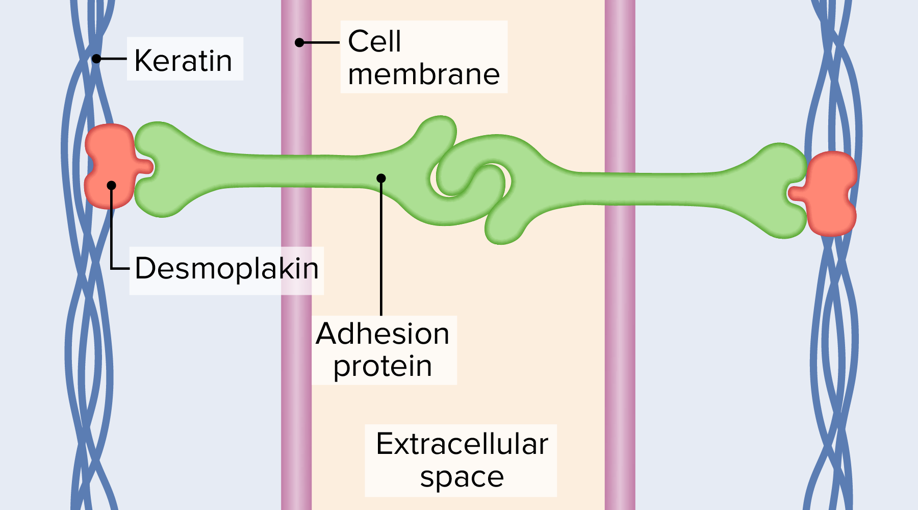
Desmosomes linking together adjacent cells
Image by Lecturio.
Hemidesmosome
Definition:
Hemidesmosomes are small specialized structures that function to connect a cell to the extracellular matrixExtracellular matrixA meshwork-like substance found within the extracellular space and in association with the basement membrane of the cell surface. It promotes cellular proliferation and provides a supporting structure to which cells or cell lysates in culture dishes adhere.Hypertrophic and Keloid Scars.
Location:
Found on the basal side of the epithelial cell
Located between the cell and the extracellular matrixExtracellular matrixA meshwork-like substance found within the extracellular space and in association with the basement membrane of the cell surface. It promotes cellular proliferation and provides a supporting structure to which cells or cell lysates in culture dishes adhere.Hypertrophic and Keloid Scars, connecting basal epithelial cells to the lamina lucida
Mainly found in keratinocytesKeratinocytesEpidermal cells which synthesize keratin and undergo characteristic changes as they move upward from the basal layers of the epidermis to the cornified (horny) layer of the skin. Successive stages of differentiation of the keratinocytes forming the epidermal layers are basal cell, spinous or prickle cell, and the granular cell.Skin: Structure and Functions of the skinSkinThe skin, also referred to as the integumentary system, is the largest organ of the body. The skin is primarily composed of the epidermis (outer layer) and dermis (deep layer). The epidermis is primarily composed of keratinocytes that undergo rapid turnover, while the dermis contains dense layers of connective tissue.Skin: Structure and Functions
Located in stratified and pseudostratified epitheliumEpitheliumThe epithelium is a complex of specialized cellular organizations arranged into sheets and lining cavities and covering the surfaces of the body. The cells exhibit polarity, having an apical and a basal pole. Structures important for the epithelial integrity and function involve the basement membrane, the semipermeable sheet on which the cells rest, and interdigitations, as well as cellular junctions. Surface Epithelium: Histology
Contain 5 main proteinsProteinsLinear polypeptides that are synthesized on ribosomes and may be further modified, crosslinked, cleaved, or assembled into complex proteins with several subunits. The specific sequence of amino acids determines the shape the polypeptide will take, during protein folding, and the function of the protein.Energy Homeostasis
Type 2: contain fewer proteinsProteinsLinear polypeptides that are synthesized on ribosomes and may be further modified, crosslinked, cleaved, or assembled into complex proteins with several subunits. The specific sequence of amino acids determines the shape the polypeptide will take, during protein folding, and the function of the protein.Energy Homeostasis
Multiprotein complex:
Integrin (transmembrane linker)
KeratinKeratinA class of fibrous proteins or scleroproteins that represents the principal constituent of epidermis; hair; nails; horny tissues, and the organic matrix of tooth enamel. Two major conformational groups have been characterized, alpha-keratin, whose peptide backbone forms a coiled-coil alpha helical structure consisting of type I keratin and a type II keratin, and beta-keratin, whose backbone forms a zigzag or pleated sheet structure. Alpha-keratins have been classified into at least 20 subtypes. In addition multiple isoforms of subtypes have been found which may be due to gene duplication.Seborrheic Keratosis filaments
Basement membraneBasement membraneA darkly stained mat-like extracellular matrix (ecm) that separates cell layers, such as epithelium from endothelium or a layer of connective tissue. The ecm layer that supports an overlying epithelium or endothelium is called basal lamina. Basement membrane (bm) can be formed by the fusion of either two adjacent basal laminae or a basal lamina with an adjacent reticular lamina of connective tissue. Bm, composed mainly of type IV collagen; glycoprotein laminin; and proteoglycan, provides barriers as well as channels between interacting cell layers.Thin Basement Membrane Nephropathy (TBMN) (lamininLamininLarge, noncollagenous glycoprotein with antigenic properties. It is localized in the basement membrane lamina lucida and functions to bind epithelial cells to the basement membrane. Evidence suggests that the protein plays a role in tumor invasion.Connective Tissue: Histology, collagenCollagenA polypeptide substance comprising about one third of the total protein in mammalian organisms. It is the main constituent of skin; connective tissue; and the organic substance of bones (bone and bones) and teeth (tooth).Connective Tissue: Histology)
IntegrinsIntegrinsA family of transmembrane glycoproteins (membrane glycoproteins) consisting of noncovalent heterodimers. They interact with a wide variety of ligands including extracellular matrix proteins; complement, and other cells, while their intracellular domains interact with the cytoskeleton. The integrins consist of at least three identified families: the cytoadhesin receptors(receptors, cytoadhesin), the leukocyte adhesion receptors (receptors, leukocyte adhesion), and the very late antigen receptors. Each family contains a common beta-subunit (integrin beta chains) combined with one or more distinct alpha-subunits (integrin alpha chains). These receptors participate in cell-matrix and cell-cell adhesion in many physiologically important processes, including embryological development; hemostasis; thrombosis; wound healing; immune and nonimmune defense mechanisms; and oncogenic transformation.Leukocyte Adhesion Deficiency Type 1 integrate the intracellular cytoskeletonCytoskeletonThe network of filaments, tubules, and interconnecting filamentous bridges which give shape, structure, and organization to the cytoplasm.The Cell: Cytosol and Cytoskeleton (keratinKeratinA class of fibrous proteins or scleroproteins that represents the principal constituent of epidermis; hair; nails; horny tissues, and the organic matrix of tooth enamel. Two major conformational groups have been characterized, alpha-keratin, whose peptide backbone forms a coiled-coil alpha helical structure consisting of type I keratin and a type II keratin, and beta-keratin, whose backbone forms a zigzag or pleated sheet structure. Alpha-keratins have been classified into at least 20 subtypes. In addition multiple isoforms of subtypes have been found which may be due to gene duplication.Seborrheic Keratosis) to the basement membraneBasement membraneA darkly stained mat-like extracellular matrix (ecm) that separates cell layers, such as epithelium from endothelium or a layer of connective tissue. The ecm layer that supports an overlying epithelium or endothelium is called basal lamina. Basement membrane (bm) can be formed by the fusion of either two adjacent basal laminae or a basal lamina with an adjacent reticular lamina of connective tissue. Bm, composed mainly of type IV collagen; glycoprotein laminin; and proteoglycan, provides barriers as well as channels between interacting cell layers.Thin Basement Membrane Nephropathy (TBMN).
Firm adhesionAdhesionThe process whereby platelets adhere to something other than platelets, e.g., collagen; basement membrane; microfibrils; or other ‘foreign’ surfaces.Coagulation Studies of cells to basement membraneBasement membraneA darkly stained mat-like extracellular matrix (ecm) that separates cell layers, such as epithelium from endothelium or a layer of connective tissue. The ecm layer that supports an overlying epithelium or endothelium is called basal lamina. Basement membrane (bm) can be formed by the fusion of either two adjacent basal laminae or a basal lamina with an adjacent reticular lamina of connective tissue. Bm, composed mainly of type IV collagen; glycoprotein laminin; and proteoglycan, provides barriers as well as channels between interacting cell layers.Thin Basement Membrane Nephropathy (TBMN)
Disease:
Hemidesmosomes are important for keeping keratinocytesKeratinocytesEpidermal cells which synthesize keratin and undergo characteristic changes as they move upward from the basal layers of the epidermis to the cornified (horny) layer of the skin. Successive stages of differentiation of the keratinocytes forming the epidermal layers are basal cell, spinous or prickle cell, and the granular cell.Skin: Structure and Functions attached to the basal laminaBasal LaminaCapillaries: Histology. Disease of these structures leads to blistering conditions (epidermolysis bullosaEpidermolysis bullosaGroup of genetically determined disorders characterized by the blistering of skin and mucosae. There are four major forms: acquired, simple, junctional, and dystrophic. Each of the latter three has several varieties.Squamous Cell Carcinoma (SCC))
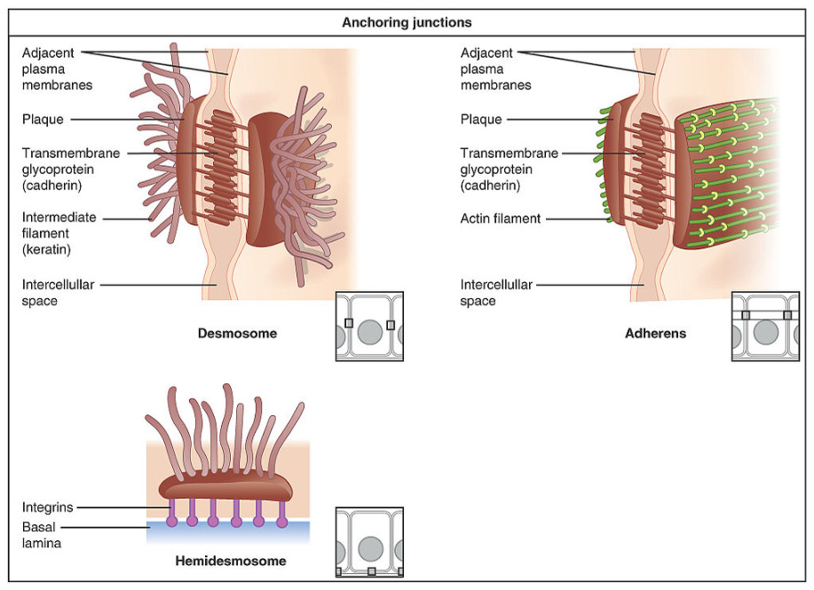
Illustration of the 3 types of anchoring junctions that maintain cell shape
Image: “Types of Cell Junctions” by OpenStax College. License: CC BY 3.0, edited by Lecturio.
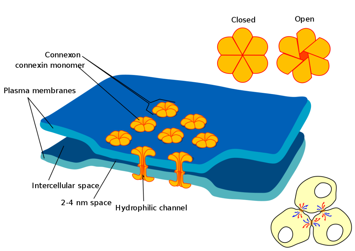
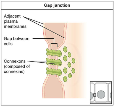
Gap Junctions
Definition
Gap junctions are protein channelsChannelsThe Cell: Cell Membrane that connect the cytoplasm of 2 cells to allow for molecular passage. This structure may also be called a nexus or maculaMaculaAn oval area in the retina, 3 to 5 mm in diameter, usually located temporal to the posterior pole of the eye and slightly below the level of the optic disk. It is characterized by the presence of a yellow pigment diffusely permeating the inner layers, contains the fovea centralis in its center, and provides the best phototropic visual acuity. It is devoid of retinal blood vessels, except in its periphery, and receives nourishment from the choriocapillaris of the choroid.Eye: Anatomy communicans.
Gap junctions between 2 adjacent cells with multiple connexons
Image: “Gap cell junction” by Mariana Ruiz . License: Public Domain, edited by Lecturio.
Location
Located between adjacent epithelial cells
Found in any nonmotile cell in the body (not found in motile cells such as sperm)
Most often found in cardiac cells and retinal cells
Composition
Connexons make up the gap junction.
Made of 6 connexin proteinsProteinsLinear polypeptides that are synthesized on ribosomes and may be further modified, crosslinked, cleaved, or assembled into complex proteins with several subunits. The specific sequence of amino acids determines the shape the polypeptide will take, during protein folding, and the function of the protein.Energy Homeostasis that span transmembranes
Form a tube with pores on either end
A pair of connexons connect within the intercellular space.
The intercellular space is between 2 and 4 nm.
Hemichannels consisting of the same proteinsProteinsLinear polypeptides that are synthesized on ribosomes and may be further modified, crosslinked, cleaved, or assembled into complex proteins with several subunits. The specific sequence of amino acids determines the shape the polypeptide will take, during protein folding, and the function of the protein.Energy Homeostasis are called homomeric.
Hemichannels consisting of different proteinsProteinsLinear polypeptides that are synthesized on ribosomes and may be further modified, crosslinked, cleaved, or assembled into complex proteins with several subunits. The specific sequence of amino acids determines the shape the polypeptide will take, during protein folding, and the function of the protein.Energy Homeostasis are called heteromeric.
Illustration of a gap junction
Image: “Types of Cell Junctions” by OpenStax College. License: CC BY 3.0, edited by Lecturio.
Regulatory molecules (cytokinesCytokinesNon-antibody proteins secreted by inflammatory leukocytes and some non-leukocytic cells, that act as intercellular mediators. They differ from classical hormones in that they are produced by a number of tissue or cell types rather than by specialized glands. They generally act locally in a paracrine or autocrine rather than endocrine manner.Adaptive Immune Response)
Metabolites
Allow for coupling of the electrical and metabolic functions between cells
Mediate activation of 2nd messengers and their impacts on cellular function
MetastasisMetastasisThe transfer of a neoplasm from one organ or part of the body to another remote from the primary site.Grading, Staging, and Metastasis: loss of E-cadherinE-cadherinCalcium-dependent cell adhesion proteins. They are important in the formation of adherens junctions between cells. Cadherins are classified by their distinct immunological and tissue specificities, either by letters (e- for epithelial, n- for neural, and p- for placental cadherins) or by numbers (cadherin-12 or n-cadherin 2 for brain-cadherin). Cadherins promote cell adhesion via a homophilic mechanism as in the construction of tissues and of the whole animal body.Gastric Cancer function results in weakening in the anchoring of cells via the adherens junction. The loss of E-cadherinE-cadherinCalcium-dependent cell adhesion proteins. They are important in the formation of adherens junctions between cells. Cadherins are classified by their distinct immunological and tissue specificities, either by letters (e- for epithelial, n- for neural, and p- for placental cadherins) or by numbers (cadherin-12 or n-cadherin 2 for brain-cadherin). Cadherins promote cell adhesion via a homophilic mechanism as in the construction of tissues and of the whole animal body.Gastric Cancer has been shown to increase cancer (CACACondylomata acuminata are a clinical manifestation of genital HPV infection. Condylomata acuminata are described as raised, pearly, flesh-colored, papular, cauliflower-like lesions seen in the anogenital region that may cause itching, pain, or bleeding.Condylomata Acuminata (Genital Warts)) cell invasion. Cancer cells may migrate, causing metastasisMetastasisThe transfer of a neoplasm from one organ or part of the body to another remote from the primary site.Grading, Staging, and Metastasis without the functionality of the adherens junction.
Pemphigus vulgarisPemphigus vulgarisBullous pemphigoid and pemphigus vulgaris are two different blistering autoimmune diseases. In pemphigus vulgaris, autoantibodies attack the desmosomal proteins, which connect the keratinocytes to one another. This attack results in a more severe, potentially fatal condition with fragile, flaccid blisters, usually with significant mucosal involvement. Bullous Pemphigoid and Pemphigus Vulgaris: a disease that is a type II hypersensitivity reactionType II hypersensitivity reactionType II hypersensitivity, also known as antibody-mediated cytotoxic hypersensitivity, is caused by immunoglobulin G (IgG) and IgM antibodies directed against antigens on cells or extracellular materials. The reaction leads to cytotoxic processes involving antibodies and the complement system. Type II Hypersensitivity Reaction whereby autoantibodiesAutoantibodiesAntibodies that react with self-antigens (autoantigens) of the organism that produced them.Blotting Techniques target the desmosome complex (desmogelin), which results in the loss of structural integrity, especially during shear forces on epidermisEpidermisThe external, nonvascular layer of the skin. It is made up, from within outward, of five layers of epithelium: (1) basal layer (stratum basale epidermidis); (2) spinous layer (stratum spinosum epidermidis); (3) granular layer (stratum granulosum epidermidis); (4) clear layer (stratum lucidum epidermidis); and (5) horny layer (stratum corneum epidermidis).Skin: Structure and Functions, causing the dermal layer to slide off, forming bullaeBullaeErythema Multiforme. Despite being a rare condition, pemphigus vulgarisPemphigus vulgarisBullous pemphigoid and pemphigus vulgaris are two different blistering autoimmune diseases. In pemphigus vulgaris, autoantibodies attack the desmosomal proteins, which connect the keratinocytes to one another. This attack results in a more severe, potentially fatal condition with fragile, flaccid blisters, usually with significant mucosal involvement. Bullous Pemphigoid and Pemphigus Vulgaris is the most common form of pemphigus. The condition is progressive without treatment. The primary treatment is a corticosteroid.
Bullous pemphigoidBullous pemphigoidBullous pemphigoid and pemphigus vulgaris are two different blistering autoimmune diseases. In bullous pemphigoid, autoantibodies attack the hemidesmosomes, which connect epidermal keratinocytes to the basement membrane. This attack results in large, tense subepidermal blisters. Bullous Pemphigoid and Pemphigus Vulgaris: a type II hypersensitivity reactionType II hypersensitivity reactionType II hypersensitivity, also known as antibody-mediated cytotoxic hypersensitivity, is caused by immunoglobulin G (IgG) and IgM antibodies directed against antigens on cells or extracellular materials. The reaction leads to cytotoxic processes involving antibodies and the complement system. Type II Hypersensitivity Reaction whereby autoantibodiesAutoantibodiesAntibodies that react with self-antigens (autoantigens) of the organism that produced them.Blotting Techniques target the hemidesmosome complex (desmogelin), which results in the loss of connection of the epidermisEpidermisThe external, nonvascular layer of the skin. It is made up, from within outward, of five layers of epithelium: (1) basal layer (stratum basale epidermidis); (2) spinous layer (stratum spinosum epidermidis); (3) granular layer (stratum granulosum epidermidis); (4) clear layer (stratum lucidum epidermidis); and (5) horny layer (stratum corneum epidermidis).Skin: Structure and Functions to the dermal basement membraneBasement membraneA darkly stained mat-like extracellular matrix (ecm) that separates cell layers, such as epithelium from endothelium or a layer of connective tissue. The ecm layer that supports an overlying epithelium or endothelium is called basal lamina. Basement membrane (bm) can be formed by the fusion of either two adjacent basal laminae or a basal lamina with an adjacent reticular lamina of connective tissue. Bm, composed mainly of type IV collagen; glycoprotein laminin; and proteoglycan, provides barriers as well as channels between interacting cell layers.Thin Basement Membrane Nephropathy (TBMN). Diagnosis requires a classic clinical presentation and confirmatory findings on biopsyBiopsyRemoval and pathologic examination of specimens from the living body.Ewing Sarcoma. PatientsPatientsIndividuals participating in the health care system for the purpose of receiving therapeutic, diagnostic, or preventive procedures.Clinician–Patient Relationship present with tense bullaeBullaeErythema Multiforme that rupture. Topical steroidsSteroidsA group of polycyclic compounds closely related biochemically to terpenes. They include cholesterol, numerous hormones, precursors of certain vitamins, bile acids, alcohols (sterols), and certain natural drugs and poisons. Steroids have a common nucleus, a fused, reduced 17-carbon atom ring system, cyclopentanoperhydrophenanthrene. Most steroids also have two methyl groups and an aliphatic side-chain attached to the nucleus.Benign Liver Tumors are the main treatment modality.
References
(2016). Guyton and Hall textbook of medical physiology (13th edition.). Philadelphia, PA: Elsevier.
Meng, W., & Takeichi, M. (2009). Adherens junction: Molecular architecture and regulation. Cold Spring Harbor Perspectives in Biology, 1(6), a002899. https://doi.org/10.1101/cshperspect.a002899