The human eye is a sensory Sensory Neurons which conduct nerve impulses to the central nervous system. Nervous System: Histology organ whose primary function is vision Vision Ophthalmic Exam. The eye has a spheroidal shape and is structured in 3 layers: a supporting outer fibrous Fibrous Fibrocystic Change layer, a middle vascular layer, and an inner neural layer. The eye can also be subdivided into 3 compartments: the anterior, posterior, and vitreous chambers. Surrounding the eyeball itself are the extraocular muscles, the lacrimal apparatus, various nerves and vessels, and the bony structure of the orbit. Light travels through the compartments of the eye to focus on the retina, which is the location where photoreceptors convert the stimulus into a neural impulse that is carried by the optic nerve Optic nerve The 2nd cranial nerve which conveys visual information from the retina to the brain. The nerve carries the axons of the retinal ganglion cells which sort at the optic chiasm and continue via the optic tracts to the brain. The largest projection is to the lateral geniculate nuclei; other targets include the superior colliculi and the suprachiasmatic nuclei. Though known as the second cranial nerve, it is considered part of the central nervous system. The 12 Cranial Nerves: Overview and Functions to the brain Brain The part of central nervous system that is contained within the skull (cranium). Arising from the neural tube, the embryonic brain is comprised of three major parts including prosencephalon (the forebrain); mesencephalon (the midbrain); and rhombencephalon (the hindbrain). The developed brain consists of cerebrum; cerebellum; and other structures in the brain stem. Nervous System: Anatomy, Structure, and Classification.
Last updated: Dec 15, 2025
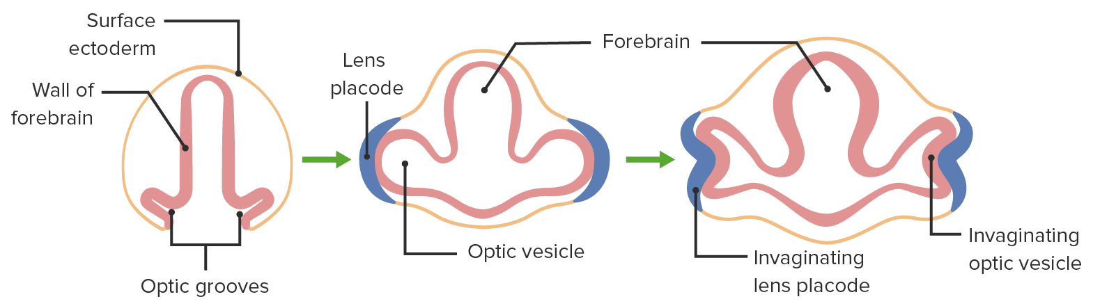
Embryologic development of the eye
Image by Lecturio.The adult eye is a complex organ contained within the orbital cavity (composed of 7 bones). Each eye has multiple layers and chambers and is surrounded by 6 extraocular muscles.

Anatomy of the eye
Image by Lecturio.The eye is composed of 3 layers ( fibrous Fibrous Fibrocystic Change, vascular, neural) and a transparent connective tissue Connective tissue Connective tissues originate from embryonic mesenchyme and are present throughout the body except inside the brain and spinal cord. The main function of connective tissues is to provide structural support to organs. Connective tissues consist of cells and an extracellular matrix. Connective Tissue: Histology covering (conjunctiva).
Conjunctiva:
Fibrous Fibrous Fibrocystic Change layer:
Vascular layer:
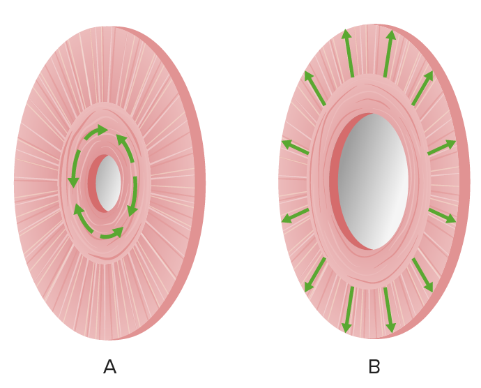
The sphincter muscles of the iris are responsible for constricting (A) and dilating (B) the pupil.
Image by Lecturio.Neural layer:
Retina:
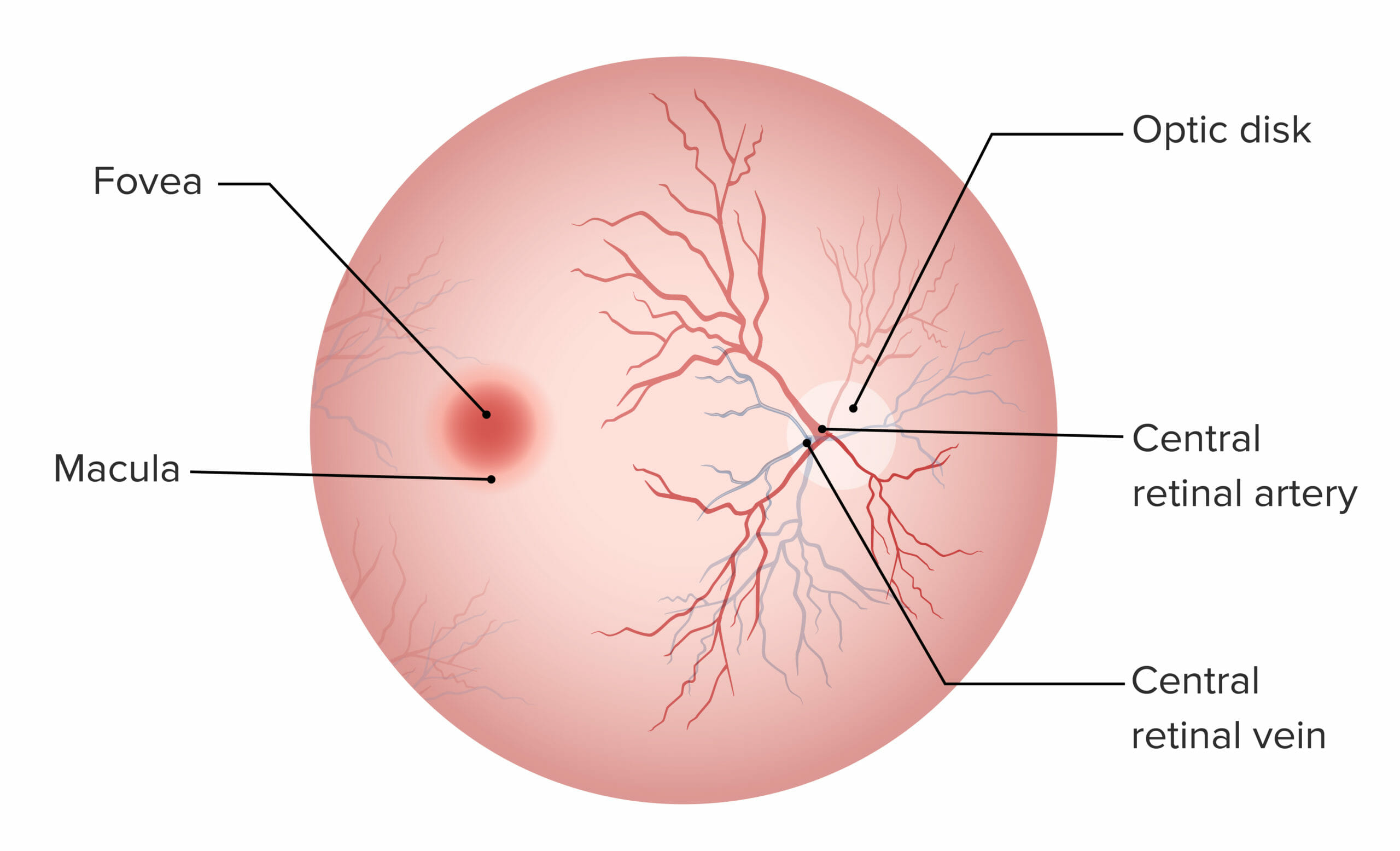
The fovea and macula lack blood vessels and are responsible for high-acuity vision. The optic disc is the entrance point for the vasculature of the eye and does not contain photoreceptors.
Characteristics of the retina and its layers:
| Layer of the retina (external to internal) | Characteristics |
|---|---|
| Pigment epithelium Epithelium The epithelium is a complex of specialized cellular organizations arranged into sheets and lining cavities and covering the surfaces of the body. The cells exhibit polarity, having an apical and a basal pole. Structures important for the epithelial integrity and function involve the basement membrane, the semipermeable sheet on which the cells rest, and interdigitations, as well as cellular junctions. Surface Epithelium: Histology |
|
| Layer of rods and cones |
|
| External limiting membrane | Supports the photoreceptor cells |
| Outer nuclear membrane | Photoreceptors cell nuclei ( 1st-order neuron 1st-order neuron Originates in the hypothalamus and descends to the first synapse in the cervical spinal cord, located at levels C8–T2 (also called the ciliospinal center of Budge). Horner Syndrome) |
| Outer plexiform layer | 1st synapse Synapse The junction between 2 neurons is called a synapse. The synapse allows a neuron to pass an electrical or chemical signal to another neuron or target effector cell. Synapses and Neurotransmission, between cones and rods and bipolar Bipolar Nervous System: Histology cells |
| Middle limiting membrane | Supporting membrane |
| Inner nuclear layer | Cell bodies and nuclei of bipolar Bipolar Nervous System: Histology cells ( 2nd-order neuron 2nd-order neuron Preganglionic pupillomotor fibers exit the spinal cord at T1, travel through the brachial plexus, over the lung apex, ascending to the superior cervical ganglion located near the angle of the mandible and the bifurcation of the common carotid artery. Horner Syndrome), transmit information to ganglion cells Ganglion Cells The Visual Pathway and Related Disorders |
| Inner plexiform layer | Second synapse Synapse The junction between 2 neurons is called a synapse. The synapse allows a neuron to pass an electrical or chemical signal to another neuron or target effector cell. Synapses and Neurotransmission, between bipolar Bipolar Nervous System: Histology cells and ganglion cells Ganglion Cells The Visual Pathway and Related Disorders |
| Ganglion cell layer |
|
| Optic nerve fibers Nerve Fibers Slender processes of neurons, including the axons and their glial envelopes (myelin sheath). Nerve fibers conduct nerve impulses to and from the central nervous system. Nervous System: Histology | Axons Axons Nerve fibers that are capable of rapidly conducting impulses away from the neuron cell body. Nervous System: Histology of ganglion cells Ganglion Cells The Visual Pathway and Related Disorders |
| Internal limiting membrane | Most internal layer, closest to the vitreous humor Humor Defense Mechanisms |
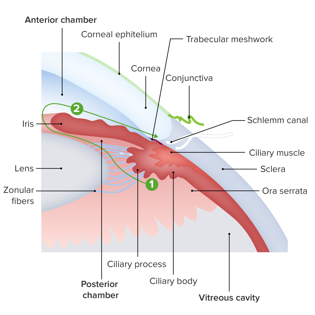
Diagram of the structure of the limbal region:
The aqueous humor is produced by the ciliary processes (1), circulates through the pupil of the iris and into the anterior chamber (2), and finally through the Schlemm canal into the scleral venous sinus.
The entire arterial supply of the eye is provided by branches of the ophthalmic artery and drained by a system of veins Veins Veins are tubular collections of cells, which transport deoxygenated blood and waste from the capillary beds back to the heart. Veins are classified into 3 types: small veins/venules, medium veins, and large veins. Each type contains 3 primary layers: tunica intima, tunica media, and tunica adventitia. Veins: Histology that unite to form the central vein of the retina.
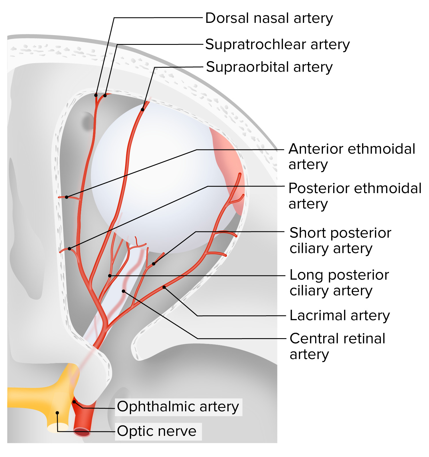
Arterial supply of the eye
Image by Lecturio.
Venous drainage of the eye
Image by Lecturio.