The development of the brainBrainThe part of central nervous system that is contained within the skull (cranium). Arising from the neural tube, the embryonic brain is comprised of three major parts including prosencephalon (the forebrain); mesencephalon (the midbrain); and rhombencephalon (the hindbrain). The developed brain consists of cerebrum; cerebellum; and other structures in the brain stem.Nervous System: Anatomy, Structure, and Classification, spinal cordSpinal cordThe spinal cord is the major conduction pathway connecting the brain to the body; it is part of the CNS. In cross section, the spinal cord is divided into an H-shaped area of gray matter (consisting of synapsing neuronal cell bodies) and a surrounding area of white matter (consisting of ascending and descending tracts of myelinated axons). Spinal Cord: Anatomy, and face involve several complex processes that occur simultaneously to achieve correct organ development. Beginning with neurulationNeurulationAn early embryonic developmental process of chordates that is characterized by morphogenic movements of ectoderm resulting in the formation of the neural plate; the neural crest; and the neural tube. Improper closure of the neural groove results in congenital neural tube defects.Gastrulation and Neurulation, the neural tubeNeural tubeA tube of ectodermal tissue in an embryo that will give rise to the central nervous system, including the spinal cord and the brain. Lumen within the neural tube is called neural canal which gives rise to the central canal of the spinal cord and the ventricles of the brain.Gastrulation and Neurulation and neural crest cellsNeural crest cellsGastrulation and Neurulation form the central and peripheral nervous systems. Beginning at the 4th week, the face begins to develop as well, and through the creation of frontonasal, medial, lateral, and mandibular prominence, recognizable facial features can be observed from the 14th week onward.
The development of the brainBrainThe part of central nervous system that is contained within the skull (cranium). Arising from the neural tube, the embryonic brain is comprised of three major parts including prosencephalon (the forebrain); mesencephalon (the midbrain); and rhombencephalon (the hindbrain). The developed brain consists of cerebrum; cerebellum; and other structures in the brain stem.Nervous System: Anatomy, Structure, and Classification is a specific part of gastrulationGastrulationBoth gastrulation and neurulation are critical events that occur during the 3rd week of embryonic development. Gastrulation is the process by which the bilaminar disc differentiates into a trilaminar disc, made up of the 3 primary germ layers: the ectoderm, mesoderm, and endoderm.Gastrulation and Neurulation, called neurulationNeurulationAn early embryonic developmental process of chordates that is characterized by morphogenic movements of ectoderm resulting in the formation of the neural plate; the neural crest; and the neural tube. Improper closure of the neural groove results in congenital neural tube defects.Gastrulation and Neurulation, that creates the cells of the nervous systemNervous systemThe nervous system is a small and complex system that consists of an intricate network of neural cells (or neurons) and even more glial cells (for support and insulation). It is divided according to its anatomical components as well as its functional characteristics. The brain and spinal cord are referred to as the central nervous system, and the branches of nerves from these structures are referred to as the peripheral nervous system.Nervous System: Anatomy, Structure, and Classification.
NeurulationNeurulationAn early embryonic developmental process of chordates that is characterized by morphogenic movements of ectoderm resulting in the formation of the neural plate; the neural crest; and the neural tube. Improper closure of the neural groove results in congenital neural tube defects.Gastrulation and Neurulation
Note: This animation does not have sound.
Day 16 after fertilizationFertilizationTo undergo fertilization, the sperm enters the uterus, travels towards the ampulla of the fallopian tube, and encounters the oocyte. The zona pellucida (the outer layer of the oocyte) deteriorates along with the zygote, which travels towards the uterus and eventually forms a blastocyst, allowing for implantation to occur. Fertilization and First Week → embryonal cells belong to 1 of 3 germ cell layers:
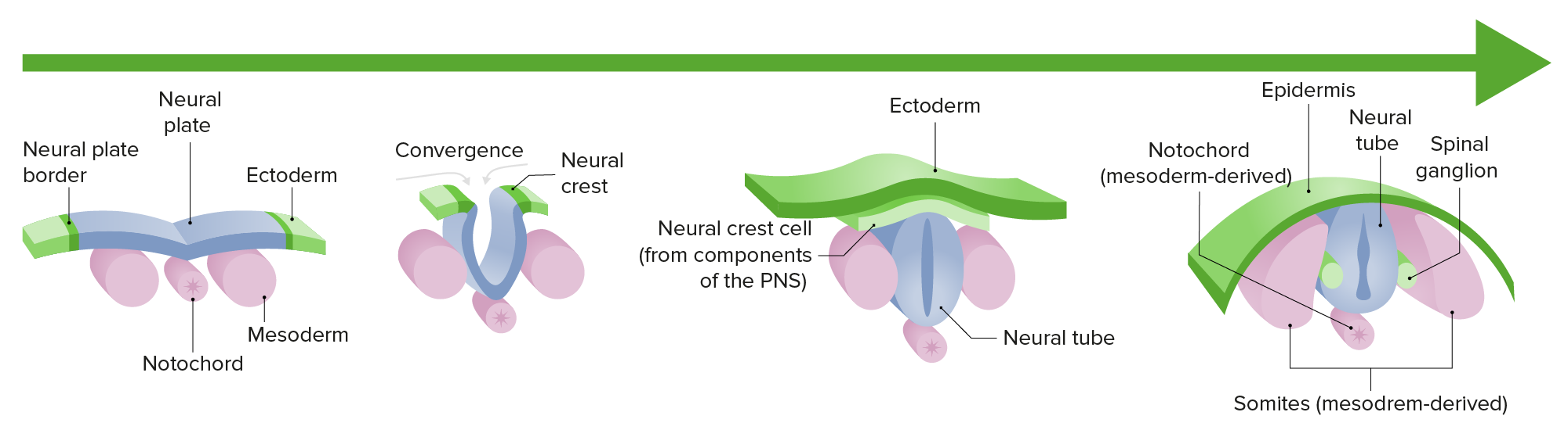
Differentiates into the neuroectoderm, creating the neural plateNeural plateThe region in the dorsal ectoderm of a chordate embryo that gives rise to the future central nervous system. Tissue in the neural plate is called the neuroectoderm, often used as a synonym of neural plate.Gastrulation and Neurulation
Cell replication in the neural plateNeural plateThe region in the dorsal ectoderm of a chordate embryo that gives rise to the future central nervous system. Tissue in the neural plate is called the neuroectoderm, often used as a synonym of neural plate.Gastrulation and Neurulation gives rise to 2 ridges (neural crests).
The depression between the crests is known as the neural fold.
MesodermMesodermThe middle germ layer of an embryo derived from three paired mesenchymal aggregates along the neural tube.Gastrulation and Neurulation:
Differentiates and transforms in a tube structure called the notochordNotochordA cartilaginous rod of mesodermal cells at the dorsal midline of all chordate embryos. In lower vertebrates, notochord is the backbone of support. In the higher vertebrates, notochord is a transient structure, and segments of the vertebral column will develop around it. Notochord is also a source of midline signals that pattern surrounding tissues including the neural tube development.Gastrulation and Neurulation.
The notochordNotochordA cartilaginous rod of mesodermal cells at the dorsal midline of all chordate embryos. In lower vertebrates, notochord is the backbone of support. In the higher vertebrates, notochord is a transient structure, and segments of the vertebral column will develop around it. Notochord is also a source of midline signals that pattern surrounding tissues including the neural tube development.Gastrulation and Neurulationsignals the neural fold to enlarge on either side of the neural grooveNeural grooveGastrulation and Neurulation, creating the neural tubeNeural tubeA tube of ectodermal tissue in an embryo that will give rise to the central nervous system, including the spinal cord and the brain. Lumen within the neural tube is called neural canal which gives rise to the central canal of the spinal cord and the ventricles of the brain.Gastrulation and Neurulation.
EndodermEndodermThe inner of the three germ layers of an embryo.Gastrulation and Neurulation: will give rise to the lining of the GI and respiratory systems
Neurulation: the differentiation and growth of the neural plate into the neural tube during the first trimester of gestation
Image by Lecturio.
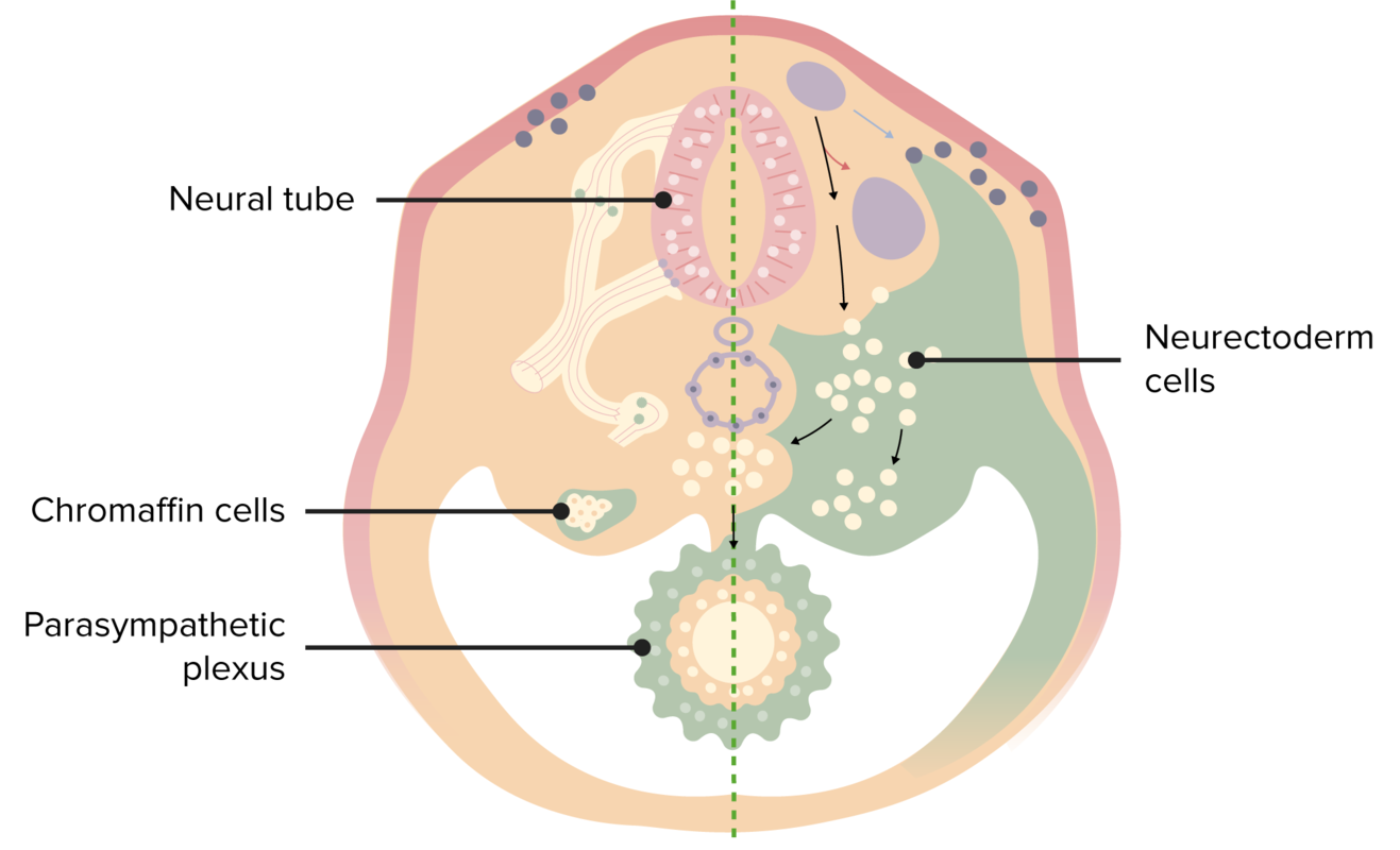
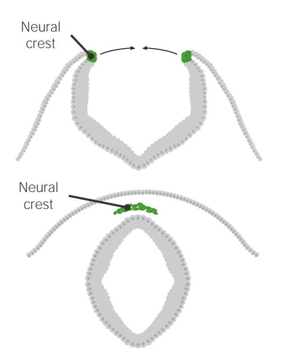
Neural crestNeural crestThe two longitudinal ridges along the primitive streak appearing near the end of gastrulation during development of nervous system (neurulation). The ridges are formed by folding of neural plate. Between the ridges is a neural groove which deepens as the fold become elevated. When the folds meet at midline, the groove becomes a closed tube, the neural tube.Hirschsprung Disease cell migration
Neuroectoderm cells migrate in waves from the neural crests to create peripheral nervous systemNervous systemThe nervous system is a small and complex system that consists of an intricate network of neural cells (or neurons) and even more glial cells (for support and insulation). It is divided according to its anatomical components as well as its functional characteristics. The brain and spinal cord are referred to as the central nervous system, and the branches of nerves from these structures are referred to as the peripheral nervous system.Nervous System: Anatomy, Structure, and Classification structures:
The 1st wave of migration creates:
Sympathetic ganglia
Parasympathetic ganglia
Chromaffin cellsChromaffin cellsCells that store epinephrine secretory vesicles. During times of stress, the nervous system signals the vesicles to secrete their hormonal content. Their name derives from their ability to stain a brownish color with chromic salts. Characteristically, they are located in the adrenal medulla and paraganglia of the sympathetic nervous system.Adrenal Hormones (sympathetic ganglia migrated into the adrenal medullaAdrenal MedullaThe inner portion of the adrenal gland. Derived from ectoderm, adrenal medulla consists mainly of chromaffin cells that produces and stores a number of neurotransmitters, mainly adrenaline (epinephrine) and norepinephrine. The activity of the adrenal medulla is regulated by the sympathetic nervous system.Adrenal Glands: Anatomy)
The 2nd wave of migration creates:
Posterior root ganglia
Schwann cells
Satellite cellsSatellite CellsThe non-neuronal cells that surround the neuronal cell bodies of the ganglia. They are distinguished from the perineuronal satellite oligodendrocytes (oligodendroglia) found in the central nervous system.Nervous System: Histology
The 3rd wave of migration creates:
MelanocytesMelanocytesMammalian pigment cells that produce melanins, pigments found mainly in the epidermis, but also in the eyes and the hair, by a process called melanogenesis. Coloration can be altered by the number of melanocytes or the amount of pigment produced and stored in the organelles called melanosomes. The large non-mammalian melanin-containing cells are called melanophores.Skin: Structure and Functions
Make up secondary ganglia of cranial nervesCranial nervesThere are 12 pairs of cranial nerves (CNs), which run from the brain to various parts of the head, neck, and trunk. The CNs can be sensory or motor or both. The CNs are named and numbered in Roman numerals according to their location, from the front to the back of the brain.The 12 Cranial Nerves: Overview and Functions 5, 7, 9, and 10
Migrate into the pharyngeal archesPharyngeal archesThe branchial arches, also known as pharyngeal or visceral arches, are embryonic structures seen in the development of vertebrates that serve as precursors for many structures of the face, neck, and head. These arches are composed of a central core of mesoderm, which is covered externally by ectoderm and internally by endoderm.Branchial Apparatus and Aortic Arches of the head, neckNeckThe part of a human or animal body connecting the head to the rest of the body.Peritonsillar Abscess, and face to create mesenchyme, which contributes to boneBoneBone is a compact type of hardened connective tissue composed of bone cells, membranes, an extracellular mineralized matrix, and central bone marrow. The 2 primary types of bone are compact and spongy. Bones: Structure and Types and connective tissueConnective tissueConnective tissues originate from embryonic mesenchyme and are present throughout the body except inside the brain and spinal cord. The main function of connective tissues is to provide structural support to organs. Connective tissues consist of cells and an extracellular matrix.Connective Tissue: Histology
During the embryonic development of the central nervous system (CNS), neuroectodermal cells migrate from the neural crests in sequential waves to form specialized structures of the peripheral nervous system, including sympathetic and parasympathetic ganglia, chromaffin cells, and Schwann cells.
Image by Lecturio.
Neural crest cells arise from the edges of the neural plates that fold to form the neural tube. The neural tube gives rise to the CNS, whereas neural crest cells migrate and proliferate around the developing embryo to give rise to pigment cells, autonomic and sensory nerve cells, Schwann cells, and endocrine cells (chromaffin cells in the medulla).
Image by Lecturio.
Mnemonics
To quickly recall that each Schwann cellSchwann CellNeuroglial cells of the peripheral nervous system which form the insulating myelin sheaths of peripheral axons.Nervous System: Histology myelinates only 1 axon of the peripheral nervous systemPeripheral nervous systemThe nervous system outside of the brain and spinal cord. The peripheral nervous system has autonomic and somatic divisions. The autonomic nervous system includes the enteric, parasympathetic, and sympathetic subdivisions. The somatic nervous system includes the cranial and spinal nerves and their ganglia and the peripheral sensory receptors.Nervous System: Anatomy, Structure, and Classification, remember: “Schwone” = 1 axon.
To quickly recall where oligodendrocyteOligodendrocyteA class of large neuroglial (macroglial) cells in the central nervous system. Oligodendroglia may be called interfascicular, perivascular, or perineuronal (not the same as satellite cells, perineuronal of ganglia) according to their location. They form the insulating myelin sheath of axons in the central nervous system.Nervous System: Histology and Schwann cells are located, remember: COPS: CNS = Oligodendrocyte; PNS = Schwann cells.
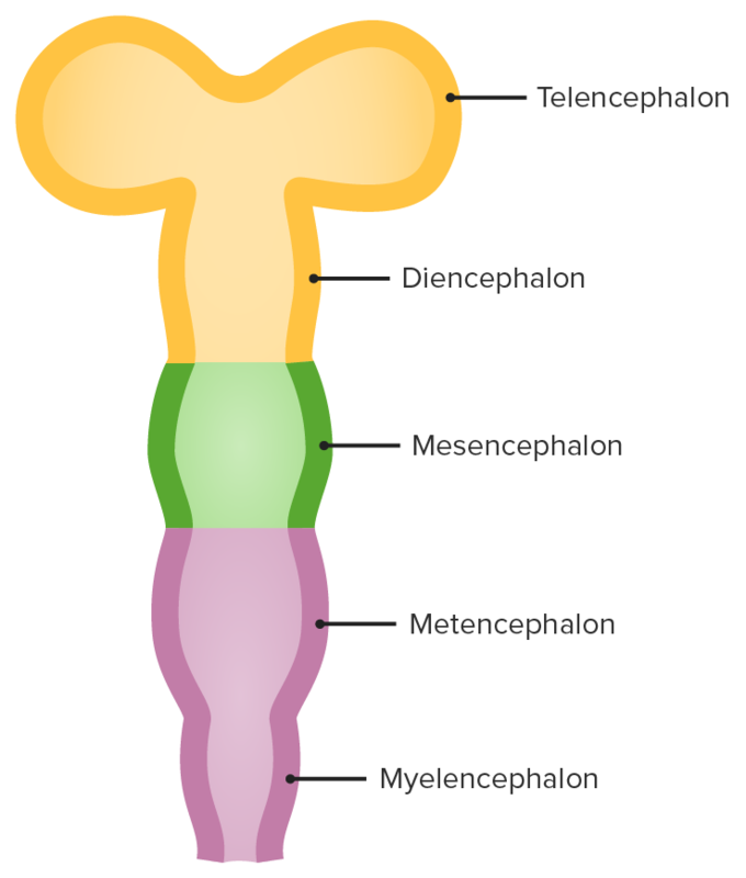
The neural tubeNeural tubeA tube of ectodermal tissue in an embryo that will give rise to the central nervous system, including the spinal cord and the brain. Lumen within the neural tube is called neural canal which gives rise to the central canal of the spinal cord and the ventricles of the brain.Gastrulation and Neurulation develops 3 bulges (primary brainBrainThe part of central nervous system that is contained within the skull (cranium). Arising from the neural tube, the embryonic brain is comprised of three major parts including prosencephalon (the forebrain); mesencephalon (the midbrain); and rhombencephalon (the hindbrain). The developed brain consists of cerebrum; cerebellum; and other structures in the brain stem.Nervous System: Anatomy, Structure, and ClassificationvesiclesVesiclesFemale Genitourinary Examination) at the cranial end:
Prosencephalon (forebrain) splits, giving rise to:
The telencephalon, which becomes the cerebral cortexCerebral cortexThe cerebral cortex is the largest and most developed part of the human brain and CNS. Occupying the upper part of the cranial cavity, the cerebral cortex has 4 lobes and is divided into 2 hemispheres that are joined centrally by the corpus callosum. Cerebral Cortex: Anatomy. This portion of the neural canal becomes the lateral ventricles and 3rd ventricle.
The diencephalon, which becomes the thalamusThalamusThe thalamus is a large, ovoid structure in the dorsal part of the diencephalon that is located between the cerebral cortex and midbrain. It consists of several interconnected nuclei of grey matter separated by the laminae of white matter. The thalamus is the main conductor of information that passes between the cerebral cortex and the periphery, spinal cord, or brain stem.Thalamus: Anatomy, hypothalamusHypothalamusThe hypothalamus is a collection of various nuclei within the diencephalon in the center of the brain. The hypothalamus plays a vital role in endocrine regulation as the primary regulator of the pituitary gland, and it is the major point of integration between the central nervous and endocrine systems.Hypothalamus, and pineal glandPineal glandA light-sensitive neuroendocrine organ attached to the roof of the third ventricle of the brain. The pineal gland secretes melatonin, other biogenic amines and neuropeptides.Hormones: Overview and Types
Mesencephalon (midbrainMidbrainThe middle of the three primitive cerebral vesicles of the embryonic brain. Without further subdivision, midbrain develops into a short, constricted portion connecting the pons and the diencephalon. Midbrain contains two major parts, the dorsal tectum mesencephali and the ventral tegmentum mesencephali, housing components of auditory, visual, and other sensorimotor systems.Brain Stem: Anatomy): also contains the cerebral aqueductCerebral aqueductNarrow channel in the mesencephalon that connects the third and fourth cerebral ventricles.Ventricular System: Anatomy
Rhombencephalon (hindbrain) splits to become:
The metencephalon, which becomes the ponsPonsThe front part of the hindbrain (rhombencephalon) that lies between the medulla and the midbrain (mesencephalon) ventral to the cerebellum. It is composed of two parts, the dorsal and the ventral. The pons serves as a relay station for neural pathways between the cerebellum to the cerebrum.Brain Stem: Anatomy and cerebellumCerebellumThe cerebellum, Latin for “little brain,” is located in the posterior cranial fossa, dorsal to the pons and midbrain, and its principal role is in the coordination of movements. The cerebellum consists of 3 lobes on either side of its 2 hemispheres and is connected in the middle by the vermis. Cerebellum: Anatomy
The myelencephalon, which becomes the medulla oblongataMedulla OblongataThe lower portion of the brain stem. It is inferior to the pons and anterior to the cerebellum. Medulla oblongata serves as a relay station between the brain and the spinal cord, and contains centers for regulating respiratory, vasomotor, cardiac, and reflex activities.Brain Stem: Anatomy
The remnant of the neural canal present around the metencephalon and myelencephalon, which becomes the 4th ventricle
Cerebrospinal fluidCerebrospinal FluidA watery fluid that is continuously produced in the choroid plexus and circulates around the surface of the brain; spinal cord; and in the cerebral ventricles.Ventricular System: Anatomy (CSF) circulatory system
Ependymal cellsEpendymal CellsThe macroglial cells of ependyma. They are characterized by bipolar cell body shape and processes that contact basal lamina around blood vessels and/or the pia mater and the cerebral ventricles. Muller cells of the retina are included based on similar microenvironmental contacts and morphology.Nervous System: Histology:
Line the central canal and the core of the neural tubeNeural tubeA tube of ectodermal tissue in an embryo that will give rise to the central nervous system, including the spinal cord and the brain. Lumen within the neural tube is called neural canal which gives rise to the central canal of the spinal cord and the ventricles of the brain.Gastrulation and Neurulation
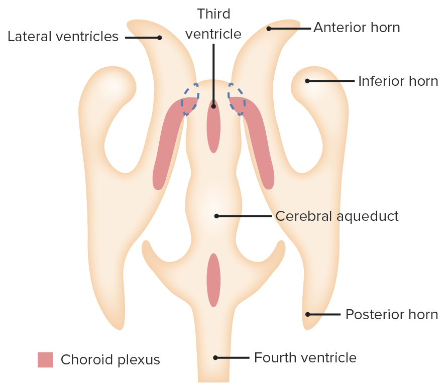
In lateral, 3rd, and 4th ventricles, ependymal cellsEpendymal CellsThe macroglial cells of ependyma. They are characterized by bipolar cell body shape and processes that contact basal lamina around blood vessels and/or the pia mater and the cerebral ventricles. Muller cells of the retina are included based on similar microenvironmental contacts and morphology.Nervous System: Histology become the choroidChoroidThe thin, highly vascular membrane covering most of the posterior of the eye between the retina and sclera.Eye: Anatomy plexus.
ChoroidChoroidThe thin, highly vascular membrane covering most of the posterior of the eye between the retina and sclera.Eye: Anatomy plexus: filters blood and releases CSF into the ventricular systemVentricular SystemThe ventricular system is an extension of the subarachnoid space into the brain consisting of a series of interconnecting spaces and channels. Four chambers are filled with cerebrospinal fluid (CSF): the paired lateral ventricles, the unpaired 3rd ventricle, and the unpaired 4th ventricle. Ventricular System: Anatomy
Table: Stages of embryonic development
Neural tubeNeural tubeA tube of ectodermal tissue in an embryo that will give rise to the central nervous system, including the spinal cord and the brain. Lumen within the neural tube is called neural canal which gives rise to the central canal of the spinal cord and the ventricles of the brain.Gastrulation and Neurulation
Anterior neural tubeNeural tubeA tube of ectodermal tissue in an embryo that will give rise to the central nervous system, including the spinal cord and the brain. Lumen within the neural tube is called neural canal which gives rise to the central canal of the spinal cord and the ventricles of the brain.Gastrulation and Neurulation
Prosencephalon
Telencephalon
Cerebrum
Lateral ventriclesLateral ventriclesCavity in each of the cerebral hemispheres derived from the cavity of the embryonic neural tube. They are separated from each other by the septum pellucidum, and each communicates with the third ventricle by the foramen of monro, through which also the choroid plexuses (choroid plexus) of the lateral ventricles become continuous with that of the third ventricle.Ventricular System: Anatomy
Anterior neural tubeNeural tubeA tube of ectodermal tissue in an embryo that will give rise to the central nervous system, including the spinal cord and the brain. Lumen within the neural tube is called neural canal which gives rise to the central canal of the spinal cord and the ventricles of the brain.Gastrulation and Neurulation
Prosencephalon
Diencephalon
Diencephalon
3rd ventricle
Anterior neural tubeNeural tubeA tube of ectodermal tissue in an embryo that will give rise to the central nervous system, including the spinal cord and the brain. Lumen within the neural tube is called neural canal which gives rise to the central canal of the spinal cord and the ventricles of the brain.Gastrulation and Neurulation
Mesencephalon
Mesencephalon
MidbrainMidbrainThe middle of the three primitive cerebral vesicles of the embryonic brain. Without further subdivision, midbrain develops into a short, constricted portion connecting the pons and the diencephalon. Midbrain contains two major parts, the dorsal tectum mesencephali and the ventral tegmentum mesencephali, housing components of auditory, visual, and other sensorimotor systems.Brain Stem: Anatomy
Anterior neural tubeNeural tubeA tube of ectodermal tissue in an embryo that will give rise to the central nervous system, including the spinal cord and the brain. Lumen within the neural tube is called neural canal which gives rise to the central canal of the spinal cord and the ventricles of the brain.Gastrulation and Neurulation
Rhombencephalon
Metencephalon
PonsPonsThe front part of the hindbrain (rhombencephalon) that lies between the medulla and the midbrain (mesencephalon) ventral to the cerebellum. It is composed of two parts, the dorsal and the ventral. The pons serves as a relay station for neural pathways between the cerebellum to the cerebrum.Brain Stem: AnatomycerebellumCerebellumThe cerebellum, Latin for “little brain,” is located in the posterior cranial fossa, dorsal to the pons and midbrain, and its principal role is in the coordination of movements. The cerebellum consists of 3 lobes on either side of its 2 hemispheres and is connected in the middle by the vermis. Cerebellum: Anatomy
4th ventricle
Anterior neural tubeNeural tubeA tube of ectodermal tissue in an embryo that will give rise to the central nervous system, including the spinal cord and the brain. Lumen within the neural tube is called neural canal which gives rise to the central canal of the spinal cord and the ventricles of the brain.Gastrulation and Neurulation
Rhombencephalon
Myelencephalon
Medulla
4th ventricle
FlexionFlexionExamination of the Upper Limbs of the neural tubeNeural tubeA tube of ectodermal tissue in an embryo that will give rise to the central nervous system, including the spinal cord and the brain. Lumen within the neural tube is called neural canal which gives rise to the central canal of the spinal cord and the ventricles of the brain.Gastrulation and Neurulation
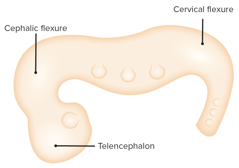
The neural tubeNeural tubeA tube of ectodermal tissue in an embryo that will give rise to the central nervous system, including the spinal cord and the brain. Lumen within the neural tube is called neural canal which gives rise to the central canal of the spinal cord and the ventricles of the brain.Gastrulation and Neurulation develops a series of bends in the sagittal planeSagittal planeAnterior Abdominal Wall: Anatomy:
At 3-vesicle stage:
Cervical flexure: between spinal cordSpinal cordThe spinal cord is the major conduction pathway connecting the brain to the body; it is part of the CNS. In cross section, the spinal cord is divided into an H-shaped area of gray matter (consisting of synapsing neuronal cell bodies) and a surrounding area of white matter (consisting of ascending and descending tracts of myelinated axons). Spinal Cord: Anatomy and rhombencephalon
Cephalic flexure: between prosencephalon and mesencephalon
As the neural tubeNeural tubeA tube of ectodermal tissue in an embryo that will give rise to the central nervous system, including the spinal cord and the brain. Lumen within the neural tube is called neural canal which gives rise to the central canal of the spinal cord and the ventricles of the brain.Gastrulation and Neurulation develops further: pontine flexure (between myelencephalon and metencephalon)
The prosencephalon, mesencephalon, and rhombencephalon form 5 secondary brain vesicles. The telencephalon becomes the left and right cerebral cortex; the diencephalon becomes the thalamus, hypothalamus, and pineal gland; the mesencephalon becomes the midbrain; the metencephalon becomes the pons and cerebellum; and the myelencephalon becomes the medulla oblongata. The rest of the neural tube develops into the spinal cord.
Image by Lecturio.
The choroid plexus (yellow) is developed from the ependymal lining of the lateral, 3rd, and 4th ventricles. The function of the choroid plexus is filtration of the blood to release ultrafiltrate into ventricles in the form of CSF.
Image by Lecturio.
As the neural tube grows, it develops into a series of bends in the sagittal plane. Cervical flexure occurs between the spinal cord and the rhombencephalon; cephalic flexure occurs between the prosencephalon and the mesencephalon.
SpineSpineThe human spine, or vertebral column, is the most important anatomical and functional axis of the human body. It consists of 7 cervical vertebrae, 12 thoracic vertebrae, and 5 lumbar vertebrae and is limited cranially by the skull and caudally by the sacrum.Vertebral Column: Anatomy
As the cephalic portion of the neural tubeNeural tubeA tube of ectodermal tissue in an embryo that will give rise to the central nervous system, including the spinal cord and the brain. Lumen within the neural tube is called neural canal which gives rise to the central canal of the spinal cord and the ventricles of the brain.Gastrulation and Neurulation becomes the brainBrainThe part of central nervous system that is contained within the skull (cranium). Arising from the neural tube, the embryonic brain is comprised of three major parts including prosencephalon (the forebrain); mesencephalon (the midbrain); and rhombencephalon (the hindbrain). The developed brain consists of cerebrum; cerebellum; and other structures in the brain stem.Nervous System: Anatomy, Structure, and Classification, the rest becomes the spinal cordSpinal cordThe spinal cord is the major conduction pathway connecting the brain to the body; it is part of the CNS. In cross section, the spinal cord is divided into an H-shaped area of gray matter (consisting of synapsing neuronal cell bodies) and a surrounding area of white matter (consisting of ascending and descending tracts of myelinated axons). Spinal Cord: Anatomy.
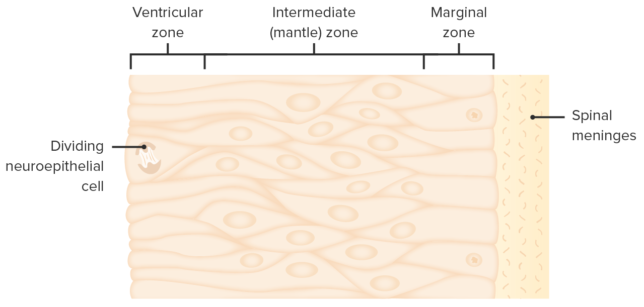
Proliferation of neuroepithelial cells
As they push outward, intermediate and marginal zones are created.
Marginal zoneMarginal zoneMALT Lymphoma: comes in contact with sclerotomal mesenchyme that will form the meningesMeningesThe brain and the spinal cord are enveloped by 3 overlapping layers of connective tissue called the meninges. The layers are, from the most external layer to the most internal layer, the dura mater, arachnoid mater, and pia mater. Between these layers are 3 potential spaces called the epidural, subdural, and subarachnoid spaces. Meninges: Anatomy
Intermediate and marginal zones fill the space inside the neural canal.
The cells in the marginal and intermediate zones will differentiate into neuronsNeuronsThe basic cellular units of nervous tissue. Each neuron consists of a body, an axon, and dendrites. Their purpose is to receive, conduct, and transmit impulses in the nervous system.Nervous System: Histology.
The neuroepithelial tissue of the spineSpineThe human spine, or vertebral column, is the most important anatomical and functional axis of the human body. It consists of 7 cervical vertebrae, 12 thoracic vertebrae, and 5 lumbar vertebrae and is limited cranially by the skull and caudally by the sacrum.Vertebral Column: Anatomy has a regional specialization:
AxonsAxonsNerve fibers that are capable of rapidly conducting impulses away from the neuron cell body.Nervous System: Histology extend to the brainBrainThe part of central nervous system that is contained within the skull (cranium). Arising from the neural tube, the embryonic brain is comprised of three major parts including prosencephalon (the forebrain); mesencephalon (the midbrain); and rhombencephalon (the hindbrain). The developed brain consists of cerebrum; cerebellum; and other structures in the brain stem.Nervous System: Anatomy, Structure, and Classification.
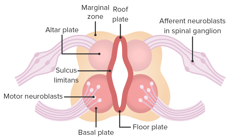
NeuronsNeuronsThe basic cellular units of nervous tissue. Each neuron consists of a body, an axon, and dendrites. Their purpose is to receive, conduct, and transmit impulses in the nervous system.Nervous System: Histology in dorsal root ganglia (originally from neural crest cellsNeural crest cellsGastrulation and Neurulation) extend to the skinSkinThe skin, also referred to as the integumentary system, is the largest organ of the body. The skin is primarily composed of the epidermis (outer layer) and dermis (deep layer). The epidermis is primarily composed of keratinocytes that undergo rapid turnover, while the dermis contains dense layers of connective tissue.Skin: Structure and Functions and back to the Alar plate.
Basal plate (motorMotorNeurons which send impulses peripherally to activate muscles or secretory cells.Nervous System: Histology): NeuronsNeuronsThe basic cellular units of nervous tissue. Each neuron consists of a body, an axon, and dendrites. Their purpose is to receive, conduct, and transmit impulses in the nervous system.Nervous System: Histology innervate myotomeMyotomeDevelopment of the Limbs.
Sulcus limitans: separates alar plate from the basal plate
Top and bottom of the neural tubeNeural tubeA tube of ectodermal tissue in an embryo that will give rise to the central nervous system, including the spinal cord and the brain. Lumen within the neural tube is called neural canal which gives rise to the central canal of the spinal cord and the ventricles of the brain.Gastrulation and Neurulation are closed by the roof plate and floor plate.
Central canal at the core of neural tubeNeural tubeA tube of ectodermal tissue in an embryo that will give rise to the central nervous system, including the spinal cord and the brain. Lumen within the neural tube is called neural canal which gives rise to the central canal of the spinal cord and the ventricles of the brain.Gastrulation and Neurulation: lined by ependymal cellsEpendymal CellsThe macroglial cells of ependyma. They are characterized by bipolar cell body shape and processes that contact basal lamina around blood vessels and/or the pia mater and the cerebral ventricles. Muller cells of the retina are included based on similar microenvironmental contacts and morphology.Nervous System: Histology
The spinal cordSpinal cordThe spinal cord is the major conduction pathway connecting the brain to the body; it is part of the CNS. In cross section, the spinal cord is divided into an H-shaped area of gray matter (consisting of synapsing neuronal cell bodies) and a surrounding area of white matter (consisting of ascending and descending tracts of myelinated axons). Spinal Cord: Anatomy fills the bony spineSpineThe human spine, or vertebral column, is the most important anatomical and functional axis of the human body. It consists of 7 cervical vertebrae, 12 thoracic vertebrae, and 5 lumbar vertebrae and is limited cranially by the skull and caudally by the sacrum.Vertebral Column: Anatomy as the fetus develops.
Week 8: Spinal cordSpinal cordThe spinal cord is the major conduction pathway connecting the brain to the body; it is part of the CNS. In cross section, the spinal cord is divided into an H-shaped area of gray matter (consisting of synapsing neuronal cell bodies) and a surrounding area of white matter (consisting of ascending and descending tracts of myelinated axons). Spinal Cord: Anatomy extends along the entire length of the vertebral columnVertebral columnThe human spine, or vertebral column, is the most important anatomical and functional axis of the human body. It consists of 7 cervical vertebrae, 12 thoracic vertebrae, and 5 lumbar vertebrae and is limited cranially by the skull and caudally by the sacrum. Vertebral Column: Anatomy.
Inferior end reaches:
L3 level at birth
L1 level in adulthood
Spinal nerve roots that exit below L1–L3 create the cauda equinaCauda EquinaThe lower part of the spinal cord consisting of the lumbar, sacral, and coccygeal nerve roots.Spinal Cord Injuries
Conus medullarisConus MedullarisSpinal Cord Injuries: tapered end of the spinal cordSpinal cordThe spinal cord is the major conduction pathway connecting the brain to the body; it is part of the CNS. In cross section, the spinal cord is divided into an H-shaped area of gray matter (consisting of synapsing neuronal cell bodies) and a surrounding area of white matter (consisting of ascending and descending tracts of myelinated axons). Spinal Cord: Anatomy
The brainstem resembles the spinal cordSpinal cordThe spinal cord is the major conduction pathway connecting the brain to the body; it is part of the CNS. In cross section, the spinal cord is divided into an H-shaped area of gray matter (consisting of synapsing neuronal cell bodies) and a surrounding area of white matter (consisting of ascending and descending tracts of myelinated axons). Spinal Cord: Anatomy in embryological organization. The basal plate gives rise to motorMotorNeurons which send impulses peripherally to activate muscles or secretory cells.Nervous System: Histology nuclei, while the alar plate gives rise to sensorySensoryNeurons which conduct nerve impulses to the central nervous system.Nervous System: Histology nuclei.
MotorMotorNeurons which send impulses peripherally to activate muscles or secretory cells.Nervous System: Histology nuclei are ventral.
Cranial medulla
Roof plate is more opened up (“open book” appearance).
The basal plate is more medially located.
The alar plate is more laterally located.
Some sensorySensoryNeurons which conduct nerve impulses to the central nervous system.Nervous System: HistologyneuronsNeuronsThe basic cellular units of nervous tissue. Each neuron consists of a body, an axon, and dendrites. Their purpose is to receive, conduct, and transmit impulses in the nervous system.Nervous System: Histology migrate anteriorly to form the olivary nucleusNucleusWithin a eukaryotic cell, a membrane-limited body which contains chromosomes and one or more nucleoli (cell nucleolus). The nuclear membrane consists of a double unit-type membrane which is perforated by a number of pores; the outermost membrane is continuous with the endoplasmic reticulum. A cell may contain more than one nucleus.The Cell: Organelles later in development.
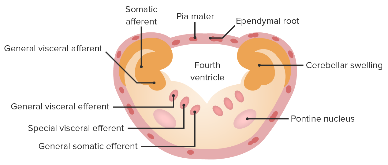
PonsPonsThe front part of the hindbrain (rhombencephalon) that lies between the medulla and the midbrain (mesencephalon) ventral to the cerebellum. It is composed of two parts, the dorsal and the ventral. The pons serves as a relay station for neural pathways between the cerebellum to the cerebrum.Brain Stem: Anatomy
Alar plate migrates to take a spot anterior to the basal plate and gives rise to the pontine nuclei.
Basal plate is now more posterior, but still gives rise to all the motorMotorNeurons which send impulses peripherally to activate muscles or secretory cells.Nervous System: Histology nuclei.
CerebellumCerebellumThe cerebellum, Latin for “little brain,” is located in the posterior cranial fossa, dorsal to the pons and midbrain, and its principal role is in the coordination of movements. The cerebellum consists of 3 lobes on either side of its 2 hemispheres and is connected in the middle by the vermis. Cerebellum: Anatomy develops directly posterior to the ponsPonsThe front part of the hindbrain (rhombencephalon) that lies between the medulla and the midbrain (mesencephalon) ventral to the cerebellum. It is composed of two parts, the dorsal and the ventral. The pons serves as a relay station for neural pathways between the cerebellum to the cerebrum.Brain Stem: Anatomy from neuroepithelial cells.
The marginal zone is the area adjacent to the mantle zone. The cells proliferate from the ventricular zone to fill up the gap so that the area for the spinal cord will be limited.
Image by Lecturio.
The basal plate becomes the anterior horn of the spinal cord, and its neurons have a motor function. The neural tube is closed by a roof plate posteriorly and a floor plate anteriorly.
Image by Lecturio.
In the cranial medulla, the roof plate is more opened out and has an “open book” appearance. Sulcus limitans still separates the alar and basal plates, but they are now located laterally (alar plate) and medially (basal plate).
Image by Lecturio.
As the cranial medulla develops, the olivary nucleus appears; it is a distinctive structure in the brain stem, as it acts as a sensory relay area.
Image by Lecturio.
Pontine nuclei are going to be sensory in origin, they migrate anterior to the basal plate.
Image by Lecturio.
The pons sits ventrally and anteriorly. The cerebellum develops as an extension of the neuroepithelium.
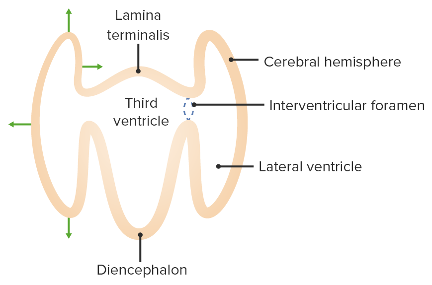
The neural canal at this level develops into the left and right ventricles.
Interventricular foramenInterventricular foramenVentricular System: Anatomy: connects lateral ventriclesLateral ventriclesCavity in each of the cerebral hemispheres derived from the cavity of the embryonic neural tube. They are separated from each other by the septum pellucidum, and each communicates with the third ventricle by the foramen of monro, through which also the choroid plexuses (choroid plexus) of the lateral ventricles become continuous with that of the third ventricle.Ventricular System: Anatomy to the 3rd ventricle
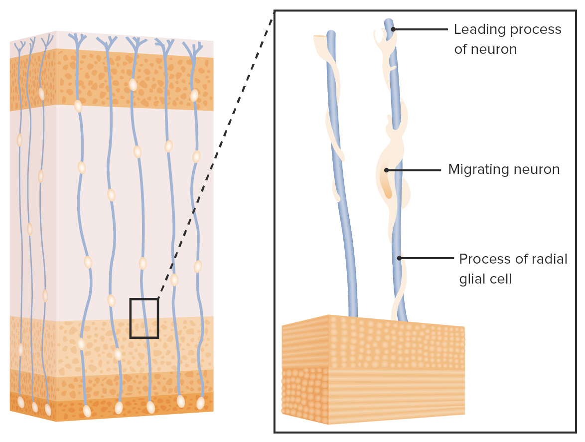
The development of lobes of the brainBrainThe part of central nervous system that is contained within the skull (cranium). Arising from the neural tube, the embryonic brain is comprised of three major parts including prosencephalon (the forebrain); mesencephalon (the midbrain); and rhombencephalon (the hindbrain). The developed brain consists of cerebrum; cerebellum; and other structures in the brain stem.Nervous System: Anatomy, Structure, and Classification occurs from the ventricles outward:
Neuron precursor cells near the ventricles replicate rapidly.
Glial cellsGlial CellsThe non-neuronal cells of the nervous system. They not only provide physical support, but also respond to injury, regulate the ionic and chemical composition of the extracellular milieu, participate in the blood-brain barrier and blood-retinal barrier, form the myelin insulation of nervous pathways, guide neuronal migration during development, and exchange metabolites with neurons. Neuroglia have high-affinity transmitter uptake systems, voltage-dependent and transmitter-gated ion channels, and can release transmitters, but their role in signaling (as in many other functions) is unclear.Nervous System: Histology extend radial processes to provide a pathway for the neuronsNeuronsThe basic cellular units of nervous tissue. Each neuron consists of a body, an axon, and dendrites. Their purpose is to receive, conduct, and transmit impulses in the nervous system.Nervous System: Histology.
Neuroepithelial cells migrate laterally, passing through subventricular zone → intermediate zone → cortical plate → marginal zoneMarginal zoneMALT Lymphoma
Different kinds of neuronsNeuronsThe basic cellular units of nervous tissue. Each neuron consists of a body, an axon, and dendrites. Their purpose is to receive, conduct, and transmit impulses in the nervous system.Nervous System: Histology stop at different points, giving rise to specialized layers.
6 months: Distinctive lobes start to appear.
Corpus callosum:
Becomes more defined as neuronsNeuronsThe basic cellular units of nervous tissue. Each neuron consists of a body, an axon, and dendrites. Their purpose is to receive, conduct, and transmit impulses in the nervous system.Nervous System: Histology from 1 cortex migrate to the other
Nervous structure that allows 1 side of the brainBrainThe part of central nervous system that is contained within the skull (cranium). Arising from the neural tube, the embryonic brain is comprised of three major parts including prosencephalon (the forebrain); mesencephalon (the midbrain); and rhombencephalon (the hindbrain). The developed brain consists of cerebrum; cerebellum; and other structures in the brain stem.Nervous System: Anatomy, Structure, and Classification to communicate with another side
Gyri and sulci are not evident until near term.
9 months: BrainBrainThe part of central nervous system that is contained within the skull (cranium). Arising from the neural tube, the embryonic brain is comprised of three major parts including prosencephalon (the forebrain); mesencephalon (the midbrain); and rhombencephalon (the hindbrain). The developed brain consists of cerebrum; cerebellum; and other structures in the brain stem.Nervous System: Anatomy, Structure, and Classification looks like a smaller version of the adult brainBrainThe part of central nervous system that is contained within the skull (cranium). Arising from the neural tube, the embryonic brain is comprised of three major parts including prosencephalon (the forebrain); mesencephalon (the midbrain); and rhombencephalon (the hindbrain). The developed brain consists of cerebrum; cerebellum; and other structures in the brain stem.Nervous System: Anatomy, Structure, and Classification.
The interventricular foramen connects the lateral ventricles to the 3rd ventricle.
Image by Lecturio.
Neurons travel from the ventricular zone to the marginal zone by following glial cells that extend processes throughout the entire length.
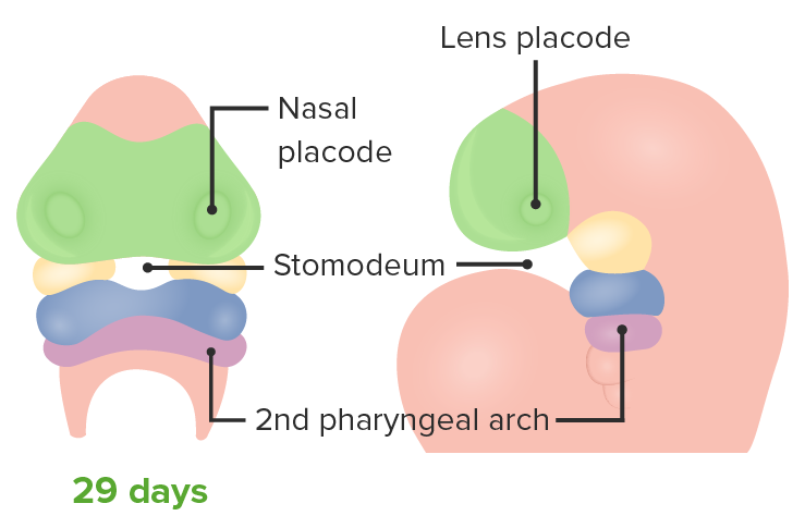
End of the 4th week: 1st facial structures are visible.
Centrally: stomodeum (early mouth)
Inferiorly: mandibular prominence
Laterally: 2 maxillary prominences
Superiorly: frontonasal prominence with nasal placodes
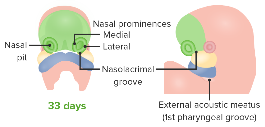
5th week: Nasal placodes deepen into nasal pits surrounded by nasal prominences.
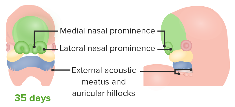
6th and 7th weeks:
Mandibular prominences fuse → jawJawThe jaw is made up of the mandible, which comprises the lower jaw, and the maxilla, which comprises the upper jaw. The mandible articulates with the temporal bone via the temporomandibular joint (TMJ). The 4 muscles of mastication produce the movements of the TMJ to ensure the efficient chewing of food. Jaw and Temporomandibular Joint: Anatomy formed
Eyes are visible on the lateral side of the face (as frontonasal prominence narrows, eyes move medially).
Nasolacrimal groove forms:junction between frontonasal prominence and maxillary prominence, future nasolacrimal ductNasolacrimal DuctNasolacrimal duct.Dacryocystitis
Medial nasal prominences:
Grow together and fuse at the midline → stretch inferiorly
Fuse with maxillary prominence → form the upper lipUpper LipMelasma
Medial and lateral nasal prominences: fuse with the maxillary prominence → form cheek and upper lipUpper LipMelasma
Frontonasal prominence → becomes the foreheadForeheadThe part of the face above the eyes.Melasma, noseNoseThe nose is the human body’s primary organ of smell and functions as part of the upper respiratory system. The nose may be best known for inhaling oxygen and exhaling carbon dioxide, but it also contributes to other important functions, such as tasting. The anatomy of the nose can be divided into the external nose and the nasal cavity. Nose Anatomy (External & Internal), and philtrum
Maxillary prominence → cheek
Mandibular prominence → mandibleMandibleThe largest and strongest bone of the face constituting the lower jaw. It supports the lower teeth.Jaw and Temporomandibular Joint: Anatomy and area anterior to the ear
Note: This animation does not have sound.
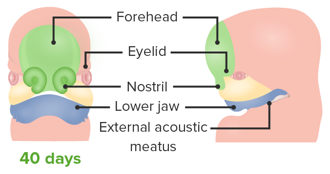
Stomodeum is surrounded by mandibular prominence (blue) inferiorly, maxillary prominences (yellow) laterally, and frontonasal prominence (green) superiorly.
Image by Lecturio.
Nasolacrimal marks the area of the junction between the frontonasal prominence (green) and the maxillary prominence (yellow).
Image by Lecturio.
After there is contact between the median nasal prominences (green) and the maxillary prominence (yellow), they stretch inferiorly and fuse with the maxillary prominences to form the cheek and upper lip.
Image by Lecturio.
Median nasal (green) and maxillary (yellow) prominences spread outward to form the upper lip. This process is followed by the fusion of right and left medial nasal prominences to form the middle of the nose and the philtrum along the midline of the upper lip.
Image by Lecturio.
Final image of the developed face. Mandibular prominence (blue) forms the mandible and the area anterior to the ear.
The following are pathological conditions that can arise as a result of errors in the development of the brainBrainThe part of central nervous system that is contained within the skull (cranium). Arising from the neural tube, the embryonic brain is comprised of three major parts including prosencephalon (the forebrain); mesencephalon (the midbrain); and rhombencephalon (the hindbrain). The developed brain consists of cerebrum; cerebellum; and other structures in the brain stem.Nervous System: Anatomy, Structure, and Classification, spinal cordSpinal cordThe spinal cord is the major conduction pathway connecting the brain to the body; it is part of the CNS. In cross section, the spinal cord is divided into an H-shaped area of gray matter (consisting of synapsing neuronal cell bodies) and a surrounding area of white matter (consisting of ascending and descending tracts of myelinated axons). Spinal Cord: Anatomy, and face:
HydrocephalusHydrocephalusExcessive accumulation of cerebrospinal fluid within the cranium which may be associated with dilation of cerebral ventricles, intracranial.Subarachnoid Hemorrhage: blockage of the ventricular systemVentricular SystemThe ventricular system is an extension of the subarachnoid space into the brain consisting of a series of interconnecting spaces and channels. Four chambers are filled with cerebrospinal fluid (CSF): the paired lateral ventricles, the unpaired 3rd ventricle, and the unpaired 4th ventricle. Ventricular System: Anatomy that causes swellingSwellingInflammation and pressure exerted on the brainBrainThe part of central nervous system that is contained within the skull (cranium). Arising from the neural tube, the embryonic brain is comprised of three major parts including prosencephalon (the forebrain); mesencephalon (the midbrain); and rhombencephalon (the hindbrain). The developed brain consists of cerebrum; cerebellum; and other structures in the brain stem.Nervous System: Anatomy, Structure, and Classification. In adults, the skullSkullThe skull (cranium) is the skeletal structure of the head supporting the face and forming a protective cavity for the brain. The skull consists of 22 bones divided into the viscerocranium (facial skeleton) and the neurocranium.Skull: Anatomy is already developed, so accumulated fluid presses on the brainBrainThe part of central nervous system that is contained within the skull (cranium). Arising from the neural tube, the embryonic brain is comprised of three major parts including prosencephalon (the forebrain); mesencephalon (the midbrain); and rhombencephalon (the hindbrain). The developed brain consists of cerebrum; cerebellum; and other structures in the brain stem.Nervous System: Anatomy, Structure, and Classification. In neonates, as bones have not yet completely ossified, the head circumferenceHead CircumferencePhysical Examination of the Newborn increases. May be caused congenitally by cerebral aqueductCerebral aqueductNarrow channel in the mesencephalon that connects the third and fourth cerebral ventricles.Ventricular System: AnatomystenosisStenosisHypoplastic Left Heart Syndrome (HLHS).
Posterior fossa malformations (Arnold-Chiari malformations (CM)): Chiari I malformation is a congenital disorder associated with ectopic cerebellar tonsilsTonsilsTonsillitis located inferior to the foramen magnum. Children are usually asymptomatic. Chiari II malformation is caused by herniationHerniationOmphalocele of the cerebellar tonsilsTonsilsTonsillitis, as well as vermis, through the foramen magnum. Chiari II leads to non-communicating hydrocephalusNon-Communicating HydrocephalusHydrocephalus in Children.
Frontonasal dysplasia (cleft lipCleft lipThe embryological development of craniofacial structures is an intricate sequential process involving tissue growth and directed cell apoptosis. Disruption of any step in this process may result in the formation of a cleft lip alone or in combination with a cleft palate. As the most common craniofacial malformation of the newborn, the diagnosis of a cleft is clinical and usually apparent at birth. Cleft Lip and Cleft Palate and cleft palateCleft palateCongenital fissure of the soft and/or hard palate, due to faulty fusion.Cleft Lip and Cleft Palate): Sonic hedgehog overactivity causes accumulation of excessive tissue in the frontonasal prominence area, resulting in a broad noseNoseThe nose is the human body’s primary organ of smell and functions as part of the upper respiratory system. The nose may be best known for inhaling oxygen and exhaling carbon dioxide, but it also contributes to other important functions, such as tasting. The anatomy of the nose can be divided into the external nose and the nasal cavity. Nose Anatomy (External & Internal) and widely separated eyes (hypertelorismHypertelorismAbnormal increase in the interorbital distance due to overdevelopment of the lesser wings of the sphenoid.DiGeorge Syndrome). This disorder may also cause cleft noseNoseThe nose is the human body’s primary organ of smell and functions as part of the upper respiratory system. The nose may be best known for inhaling oxygen and exhaling carbon dioxide, but it also contributes to other important functions, such as tasting. The anatomy of the nose can be divided into the external nose and the nasal cavity. Nose Anatomy (External & Internal) and midline cleft lipCleft lipThe embryological development of craniofacial structures is an intricate sequential process involving tissue growth and directed cell apoptosis. Disruption of any step in this process may result in the formation of a cleft lip alone or in combination with a cleft palate. As the most common craniofacial malformation of the newborn, the diagnosis of a cleft is clinical and usually apparent at birth. Cleft Lip and Cleft Palate due to the failure to fuse medial nasal prominences.
Holoprosencephaly: a disorder caused by decreased activity of the sonic hedgehog geneGeneA category of nucleic acid sequences that function as units of heredity and which code for the basic instructions for the development, reproduction, and maintenance of organisms.Basic Terms of Genetics, resulting in narrowing of the face. More severe cases involve failure of the right and left cerebral cortexes to fully separate, as well as cyclopia.
Neural tubeNeural tubeA tube of ectodermal tissue in an embryo that will give rise to the central nervous system, including the spinal cord and the brain. Lumen within the neural tube is called neural canal which gives rise to the central canal of the spinal cord and the ventricles of the brain.Gastrulation and Neurulation defects: 1 of the most common congenital CNS malformations. The defects develop between the 3rd and 4th week of gestation and are often caused by folic acid deficiencyFolic Acid DeficiencyA nutritional condition produced by a deficiency of folic acid in the diet. Many plant and animal tissues contain folic acid, abundant in green leafy vegetables, yeast, liver, and mushrooms but destroyed by long-term cooking. Alcohol interferes with its intermediate metabolism and absorption. Folic acid deficiency may develop in long-term anticonvulsant therapy or with use of oral contraceptives. This deficiency causes anemia, macrocytic anemia, and megaloblastic anemia. It is indistinguishable from vitamin B 12 deficiency in peripheral blood and bone marrow findings, but the neurologic lesions seen in B 12 deficiency do not occur.Megaloblastic Anemia. The deficiency results in improper closure of the neural plateNeural plateThe region in the dorsal ectoderm of a chordate embryo that gives rise to the future central nervous system. Tissue in the neural plate is called the neuroectoderm, often used as a synonym of neural plate.Gastrulation and Neurulation in the embryoEmbryoThe entity of a developing mammal, generally from the cleavage of a zygote to the end of embryonic differentiation of basic structures. For the human embryo, this represents the first two months of intrauterine development preceding the stages of the fetus.Fertilization and First Week, mainly at the caudal or cranial ends, giving rise to anencephalyAnencephalyA malformation of the nervous system caused by failure of the anterior neuropore to close. Infants are born with intact spinal cords, cerebellums, and brainstems, but lack formation of neural structures above this level. The skull is only partially formed but the eyes are usually normal. This condition may be associated with folate deficiency. Affected infants are only capable of primitive (brain stem) reflexes and usually do not survive for more than two weeks.Neural Tube Defects.
References
Sadler, T. W. (2014). Langman’ Medical Embryology.
Arnold WH, Meiselbach V. (2009). 3-D reconstruction of a human fetus with combined holoprosencephaly and cyclopia. Head Face Med. doi: 10.1186/1746-160X-5-14.