The cerebral cortex is the largest and most developed part of the human brain Brain The part of central nervous system that is contained within the skull (cranium). Arising from the neural tube, the embryonic brain is comprised of three major parts including prosencephalon (the forebrain); mesencephalon (the midbrain); and rhombencephalon (the hindbrain). The developed brain consists of cerebrum; cerebellum; and other structures in the brain stem. Nervous System: Anatomy, Structure, and Classification and central nervous system Central nervous system The main information-processing organs of the nervous system, consisting of the brain, spinal cord, and meninges. Nervous System: Anatomy, Structure, and Classification (CNS). Occupying the upper part of the cranial cavity, the cerebral cortex has 4 lobes and is divided into 2 hemispheres that are joined centrally by the corpus callosum. The cortex is organized in gyri that are separated by sulci. The cerebral cortex provides the neural substrate Substrate A substance upon which the enzyme acts. Basics of Enzymes for the conscious experience of sensory Sensory Neurons which conduct nerve impulses to the central nervous system. Nervous System: Histology stimuli.
Last updated: Dec 15, 2025
The cerebral cortex is the outermost layer of the brain Brain The part of central nervous system that is contained within the skull (cranium). Arising from the neural tube, the embryonic brain is comprised of three major parts including prosencephalon (the forebrain); mesencephalon (the midbrain); and rhombencephalon (the hindbrain). The developed brain consists of cerebrum; cerebellum; and other structures in the brain stem. Nervous System: Anatomy, Structure, and Classification:
The cerebrum is the largest portion of the brain Brain The part of central nervous system that is contained within the skull (cranium). Arising from the neural tube, the embryonic brain is comprised of three major parts including prosencephalon (the forebrain); mesencephalon (the midbrain); and rhombencephalon (the hindbrain). The developed brain consists of cerebrum; cerebellum; and other structures in the brain stem. Nervous System: Anatomy, Structure, and Classification. The cerebrum is composed of gray matter (cerebral cortex) and underlying white matter White Matter The region of central nervous system that appears lighter in color than the other type, gray matter. It mainly consists of myelinated nerve fibers and contains few neuronal cell bodies or dendrites. Brown-Séquard Syndrome structures:

The embryonic development of the cerebrum: note the lineage starting from the neural tube → prosencephalon → telencephalon → cerebrum
Image: “Brain Vesicle” by Phil Schatz. License: CC BY 4.0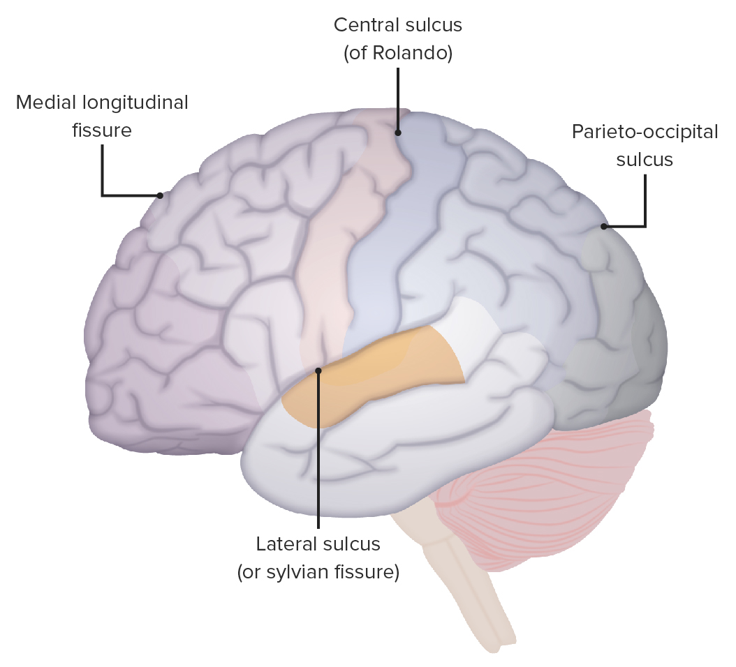
The locations of the lateral sulcus, central sulcus, and parieto-occipital sulcus
Image by Lecturio.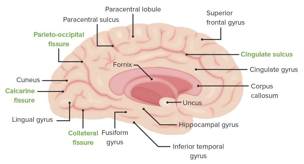
The medial surface of the left cerebral hemisphere: Note the location of the collateral sulcus separating the lingual gyrus from the fusiform gyrus, the calcarine fissure separating the cuneus gyrus from the lingual gyrus, and the parieto-occipital sulcus separating the parietal lobe from the occipital lobe.
Image by Lecturio.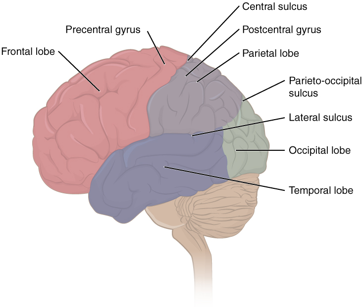
The image demonstrates the location of some key structures, including each of the 4 lobes, the precentral and postcentral gyrus, and the central, lateral, and parieto-occipital sulci.
Image: “Lobes of Cerebral Cortex” by OpenStax Anatomy and Physiology. License: CC BY 4.0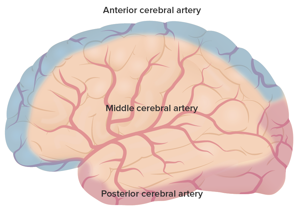
The primary arterial supply throughout the cerebrum: Note the regions covered by the anterior cerebral artery in purple, the middle cerebral artery (MCA) in red, and the posterior cerebral artery (PCA) in pink.
Image by Lecturio.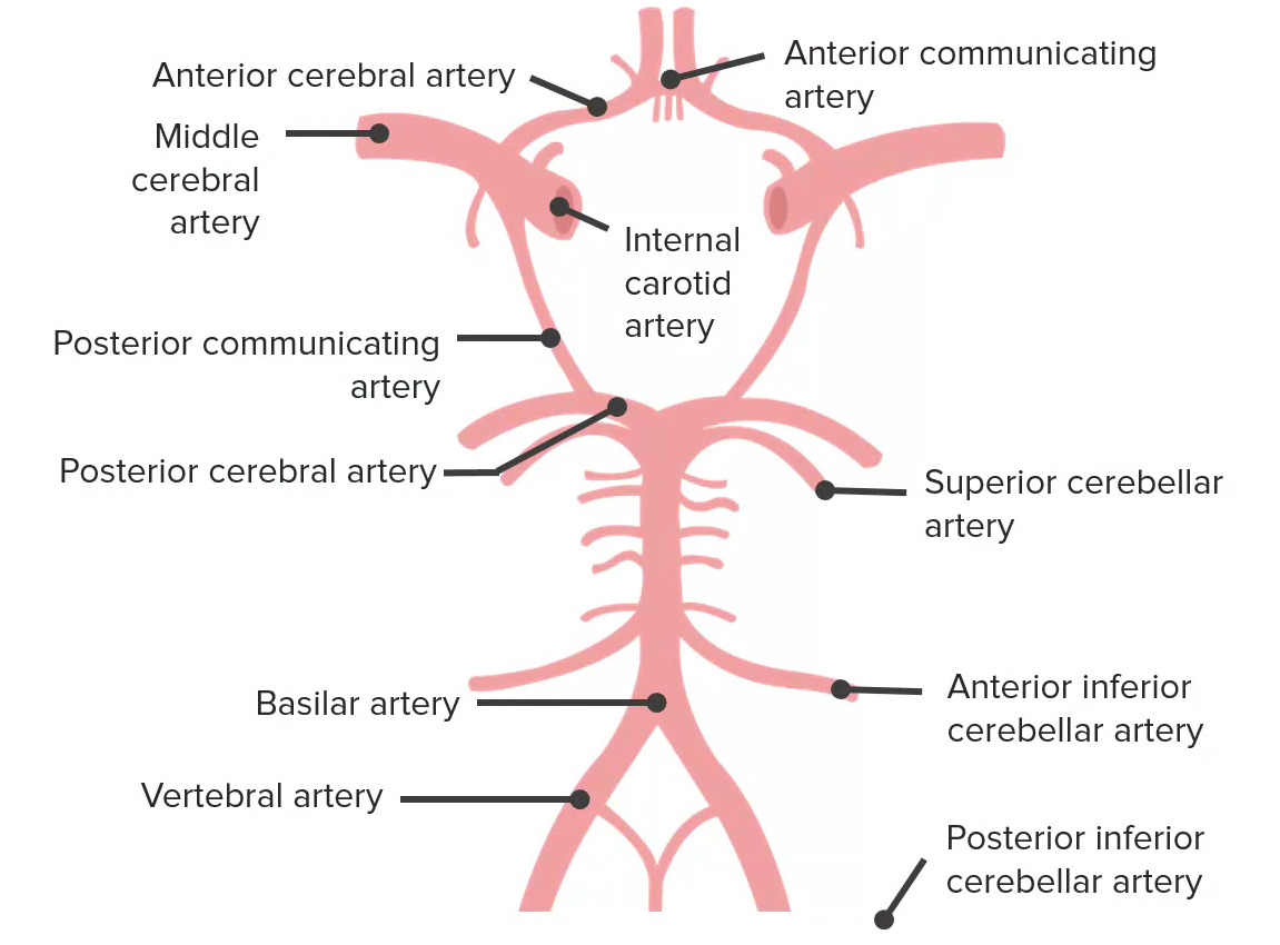
Blood supply to the brain is derived from 2 sources: 1) the internal carotid arteries and 2) the vertebrobasilar system. These sources interconnect to form the circle of Willis, which is depicted here. The circle of Willis has 5 components, which include the anterior communicating artery, the anterior cerebral arteries, the internal carotid artery, the posterior communicating artery, and the posterior cerebral arteries (PCAs).
Image by Lecturio.The frontal Frontal The bone that forms the frontal aspect of the skull. Its flat part forms the forehead, articulating inferiorly with the nasal bone and the cheek bone on each side of the face. Skull: Anatomy lobe is the most anterior/superior aspect of the supratentorial brain Brain The part of central nervous system that is contained within the skull (cranium). Arising from the neural tube, the embryonic brain is comprised of three major parts including prosencephalon (the forebrain); mesencephalon (the midbrain); and rhombencephalon (the hindbrain). The developed brain consists of cerebrum; cerebellum; and other structures in the brain stem. Nervous System: Anatomy, Structure, and Classification. It controls many of the higher-order functions of the brain Brain The part of central nervous system that is contained within the skull (cranium). Arising from the neural tube, the embryonic brain is comprised of three major parts including prosencephalon (the forebrain); mesencephalon (the midbrain); and rhombencephalon (the hindbrain). The developed brain consists of cerebrum; cerebellum; and other structures in the brain stem. Nervous System: Anatomy, Structure, and Classification, including motor Motor Neurons which send impulses peripherally to activate muscles or secretory cells. Nervous System: Histology function, executive thought, and speech.
| Name | Location | Brodmann number |
Function |
|---|---|---|---|
| Primary motor Motor Neurons which send impulses peripherally to activate muscles or secretory cells. Nervous System: Histology cortex | Precentral gyrus | 4 | Dictates contralateral motor Motor Neurons which send impulses peripherally to activate muscles or secretory cells. Nervous System: Histology control |
| Premotor cortex | Anterior to primary motor Motor Neurons which send impulses peripherally to activate muscles or secretory cells. Nervous System: Histology cortex | 6 | Programming of motor Motor Neurons which send impulses peripherally to activate muscles or secretory cells. Nervous System: Histology events; neurons Neurons The basic cellular units of nervous tissue. Each neuron consists of a body, an axon, and dendrites. Their purpose is to receive, conduct, and transmit impulses in the nervous system. Nervous System: Histology activate prior to primary motor Motor Neurons which send impulses peripherally to activate muscles or secretory cells. Nervous System: Histology neurons Neurons The basic cellular units of nervous tissue. Each neuron consists of a body, an axon, and dendrites. Their purpose is to receive, conduct, and transmit impulses in the nervous system. Nervous System: Histology |
| Supplementary motor Motor Neurons which send impulses peripherally to activate muscles or secretory cells. Nervous System: Histology cortex | Midline surface of the hemisphere anterior to leg Leg The lower leg, or just “leg” in anatomical terms, is the part of the lower limb between the knee and the ankle joint. The bony structure is composed of the tibia and fibula bones, and the muscles of the leg are grouped into the anterior, lateral, and posterior compartments by extensions of fascia. Leg: Anatomy representation of primary motor Motor Neurons which send impulses peripherally to activate muscles or secretory cells. Nervous System: Histology cortex | 6 | Planning of complex motor Motor Neurons which send impulses peripherally to activate muscles or secretory cells. Nervous System: Histology movements |
| Prefrontal cortex | Anterior portion of frontal Frontal The bone that forms the frontal aspect of the skull. Its flat part forms the forehead, articulating inferiorly with the nasal bone and the cheek bone on each side of the face. Skull: Anatomy lobe | 8–14, 24, 25, 32, 44–47 | Olfaction Olfaction The sense of smell, or olfaction, begins in a small area on the roof of the nasal cavity, which is covered in specialized mucosa. From there, the olfactory nerve transmits the sensory perception of smell via the olfactory pathway. This pathway is composed of the olfactory cells and bulb, the tractus and striae olfactoriae, and the primary olfactory cortex and amygdala. Olfaction: Anatomy and executive function (problem-solving, judgment Judgment The process of discovering or asserting an objective or intrinsic relation between two objects or concepts; a faculty or power that enables a person to make judgments; the process of bringing to light and asserting the implicit meaning of a concept; a critical evaluation of a person or situation. Psychiatric Assessment, planning, behavior, and emotions) |
| Frontal Frontal The bone that forms the frontal aspect of the skull. Its flat part forms the forehead, articulating inferiorly with the nasal bone and the cheek bone on each side of the face. Skull: Anatomy eye field | Intersection of the middle frontal Frontal The bone that forms the frontal aspect of the skull. Its flat part forms the forehead, articulating inferiorly with the nasal bone and the cheek bone on each side of the face. Skull: Anatomy gyrus with precentral gyrus | 8 |
|
| Broca area | Inferior frontal Frontal The bone that forms the frontal aspect of the skull. Its flat part forms the forehead, articulating inferiorly with the nasal bone and the cheek bone on each side of the face. Skull: Anatomy gyrus of the dominant hemisphere | 44, 45 | Word production ( motor Motor Neurons which send impulses peripherally to activate muscles or secretory cells. Nervous System: Histology speech) |
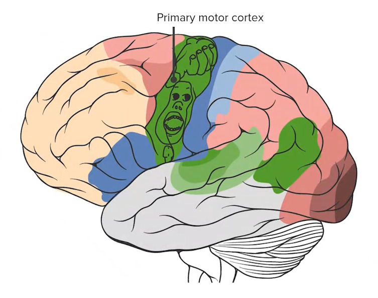
Note the primary motor cortex (the most posterior structure of the frontal lobe) with the overlaid homunculus, detailing the proportions of the cortex dedicated to processing each specific motor function.
Image by Lecturio.The parietal Parietal One of a pair of irregularly shaped quadrilateral bones situated between the frontal bone and occipital bone, which together form the sides of the cranium. Skull: Anatomy lobe lies posterior to the frontal Frontal The bone that forms the frontal aspect of the skull. Its flat part forms the forehead, articulating inferiorly with the nasal bone and the cheek bone on each side of the face. Skull: Anatomy lobe and superior to the occipital Occipital Part of the back and base of the cranium that encloses the foramen magnum. Skull: Anatomy lobe. It is associated with the processes of sensation and language comprehension.
| Name | Location | Brodmann number |
Function |
|---|---|---|---|
| Primary somatosensory cortex | Postcentral gyrus | 3, 1, 2 | Receives contralateral somatosensory input from the ventral posteromedial nucleus Nucleus Within a eukaryotic cell, a membrane-limited body which contains chromosomes and one or more nucleoli (cell nucleolus). The nuclear membrane consists of a double unit-type membrane which is perforated by a number of pores; the outermost membrane is continuous with the endoplasmic reticulum. A cell may contain more than one nucleus. The Cell: Organelles and ventral posterolateral nucleus Nucleus Within a eukaryotic cell, a membrane-limited body which contains chromosomes and one or more nucleoli (cell nucleolus). The nuclear membrane consists of a double unit-type membrane which is perforated by a number of pores; the outermost membrane is continuous with the endoplasmic reticulum. A cell may contain more than one nucleus. The Cell: Organelles of the thalamus Thalamus The thalamus is a large, ovoid structure in the dorsal part of the diencephalon that is located between the cerebral cortex and midbrain. It consists of several interconnected nuclei of grey matter separated by the laminae of white matter. The thalamus is the main conductor of information that passes between the cerebral cortex and the periphery, spinal cord, or brain stem. Thalamus: Anatomy |
| Parietal Parietal One of a pair of irregularly shaped quadrilateral bones situated between the frontal bone and occipital bone, which together form the sides of the cranium. Skull: Anatomy association areas | Posterior parietal Parietal One of a pair of irregularly shaped quadrilateral bones situated between the frontal bone and occipital bone, which together form the sides of the cranium. Skull: Anatomy | 5, 7 | Stereognosis Stereognosis Perception of shape and form of objects by touch, via tactile stimuli. Neurological Examination and awareness of contralateral self and surroundings |
| Wernicke area | Supramarginal gyrus of the dominant hemisphere (in addition to the superior temporal gyrus)* | 22, 40 | Comprehension of language |
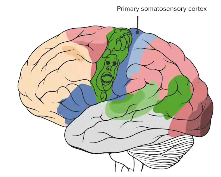
The primary somatosensory cortex (labeled in dark blue) marks the anterior-most region of the parietal lobe.
Image by Lecturio.The occipital Occipital Part of the back and base of the cranium that encloses the foramen magnum. Skull: Anatomy lobe is the most posterior lobe Posterior lobe Cerebellum: Anatomy of the supratentorial brain Brain The part of central nervous system that is contained within the skull (cranium). Arising from the neural tube, the embryonic brain is comprised of three major parts including prosencephalon (the forebrain); mesencephalon (the midbrain); and rhombencephalon (the hindbrain). The developed brain consists of cerebrum; cerebellum; and other structures in the brain stem. Nervous System: Anatomy, Structure, and Classification. It is primarily involved with visual processing.
| Name | Location | Brodmann number |
Function |
|---|---|---|---|
| Primary visual cortex Primary Visual Cortex The Visual Pathway and Related Disorders | Posterior occipital Occipital Part of the back and base of the cranium that encloses the foramen magnum. Skull: Anatomy lobe | 17 | Vision Vision Ophthalmic Exam and acuity (input from lateral geniculate nucleus Lateral Geniculate Nucleus The Visual Pathway and Related Disorders via optic radiations) |
| Visual association cortex | Extrastriate cortex | 18, 19 | Processes inputs related to form, color, motion, depth, and spatial relationships |
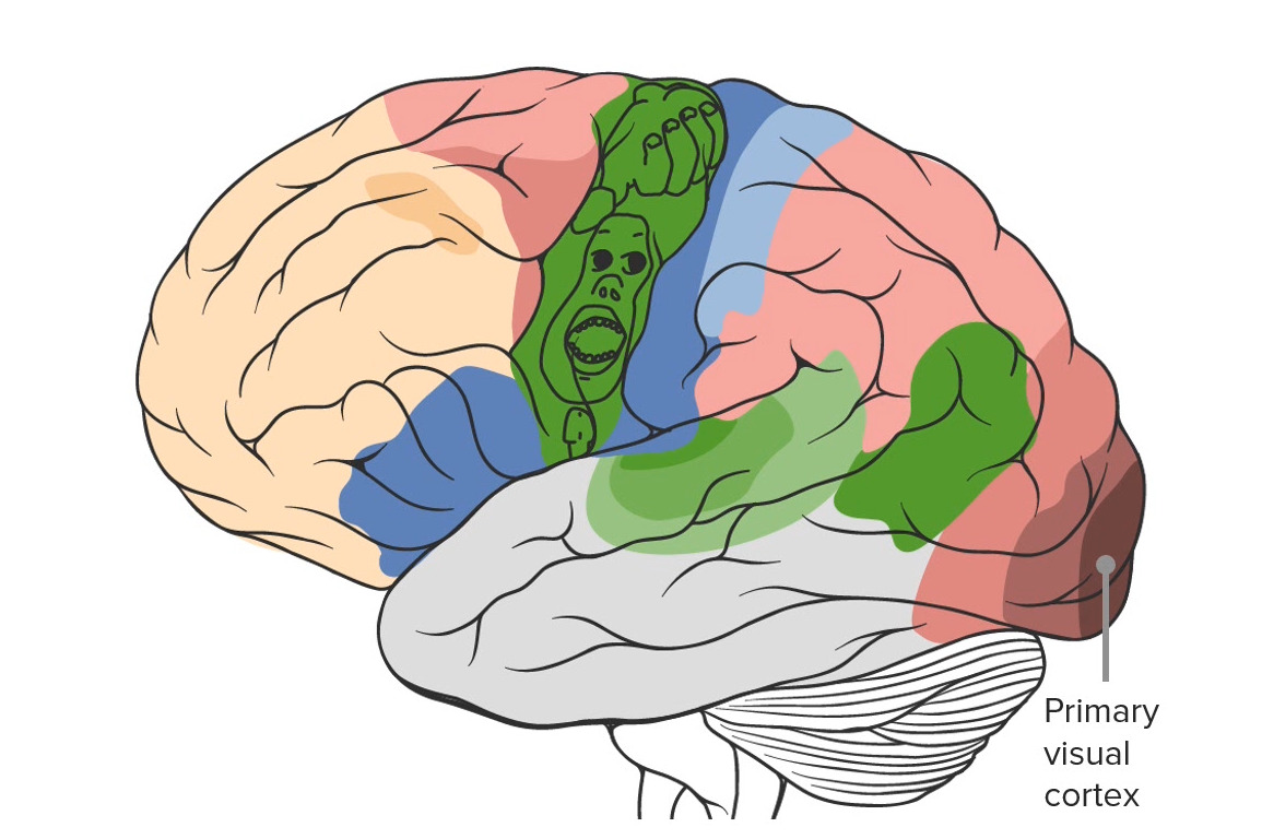
Note the location of the primary visual cortex in the most posterior region of the brain in the occipital lobe.
Image by Lecturio.The temporal lobe is the most anterior/inferior aspect of the supratentorial brain Brain The part of central nervous system that is contained within the skull (cranium). Arising from the neural tube, the embryonic brain is comprised of three major parts including prosencephalon (the forebrain); mesencephalon (the midbrain); and rhombencephalon (the hindbrain). The developed brain consists of cerebrum; cerebellum; and other structures in the brain stem. Nervous System: Anatomy, Structure, and Classification. It is involved with the processes of hearing, olfaction Olfaction The sense of smell, or olfaction, begins in a small area on the roof of the nasal cavity, which is covered in specialized mucosa. From there, the olfactory nerve transmits the sensory perception of smell via the olfactory pathway. This pathway is composed of the olfactory cells and bulb, the tractus and striae olfactoriae, and the primary olfactory cortex and amygdala. Olfaction: Anatomy, and memory Memory Complex mental function having four distinct phases: (1) memorizing or learning, (2) retention, (3) recall, and (4) recognition. Clinically, it is usually subdivided into immediate, recent, and remote memory. Psychiatric Assessment.
| Name | Location | Brodmann number |
Function |
|---|---|---|---|
| Primary auditory cortex Auditory cortex The region of the cerebral cortex that receives the auditory radiation from the medial geniculate body. Auditory and Vestibular Pathways: Anatomy | Superior temporal plane of temporal lobes within lateral sulcus | 40, 41 | Hearing |
| Middle and inferior temporal gyri | Middle and inferior temporal lobe | 20, 21 | Long-term memory Memory Complex mental function having four distinct phases: (1) memorizing or learning, (2) retention, (3) recall, and (4) recognition. Clinically, it is usually subdivided into immediate, recent, and remote memory. Psychiatric Assessment |
| Parahippocampal gyrus | Medially located in the inferior temporo-occipital cortex | 34 | Short-term memory Memory Complex mental function having four distinct phases: (1) memorizing or learning, (2) retention, (3) recall, and (4) recognition. Clinically, it is usually subdivided into immediate, recent, and remote memory. Psychiatric Assessment |
| Uncus | Continuous with the hippocampal gyrus | 35 | Olfaction Olfaction The sense of smell, or olfaction, begins in a small area on the roof of the nasal cavity, which is covered in specialized mucosa. From there, the olfactory nerve transmits the sensory perception of smell via the olfactory pathway. This pathway is composed of the olfactory cells and bulb, the tractus and striae olfactoriae, and the primary olfactory cortex and amygdala. Olfaction: Anatomy |
| Fusiform Gyrus | Occipitotemporal Medial Gyrus | 37 | Facial recognition |
| Wernicke area | Superior temporal gyrus of the dominant hemisphere (in addition to the supramarginal gyrus of the parietal Parietal One of a pair of irregularly shaped quadrilateral bones situated between the frontal bone and occipital bone, which together form the sides of the cranium. Skull: Anatomy lobe)* | 22, 40 | Comprehension of language |
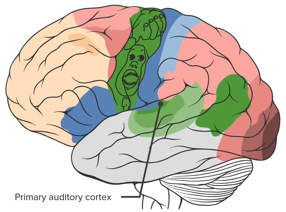
Note the primary auditory cortex located in the temporal lobe
Image by Lecturio.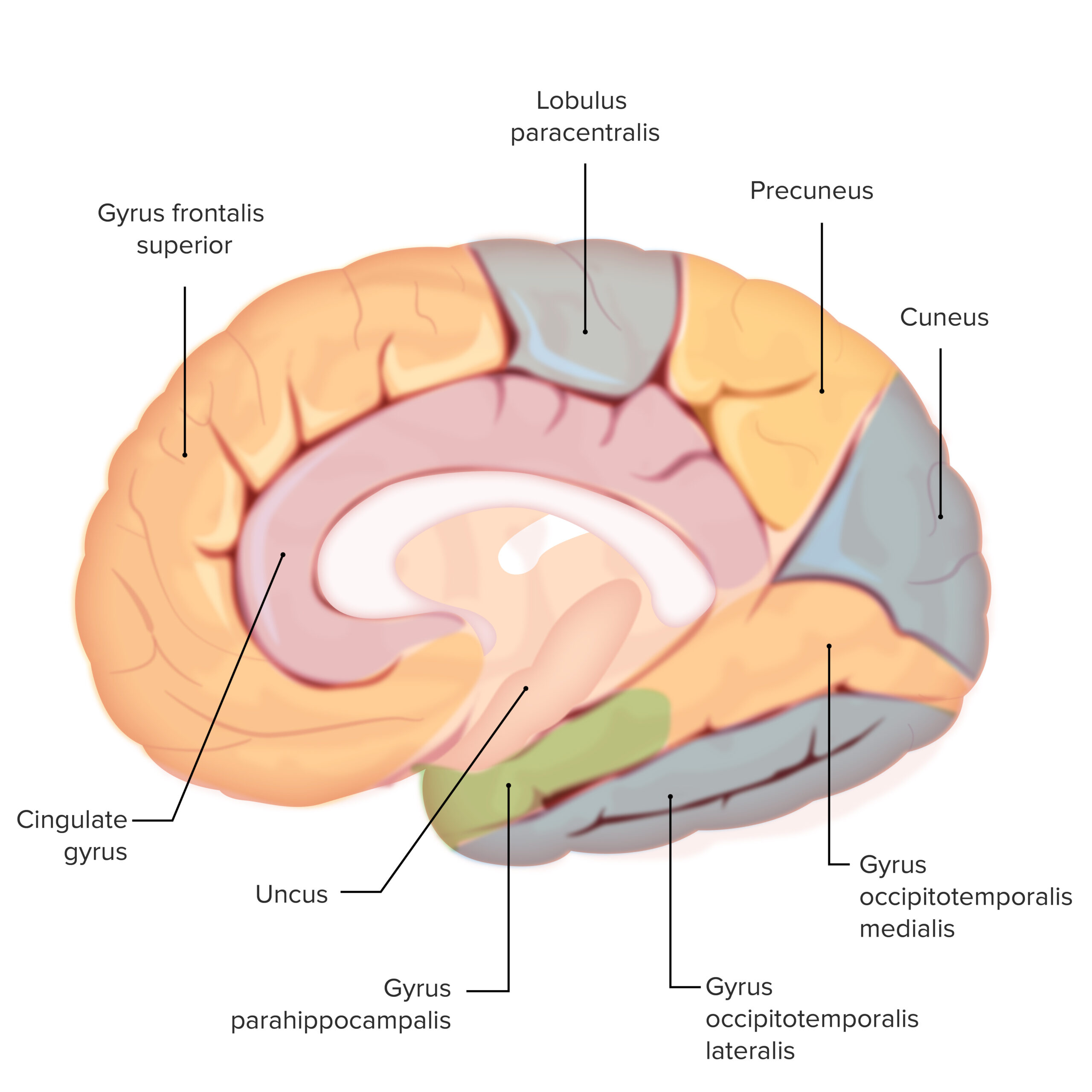
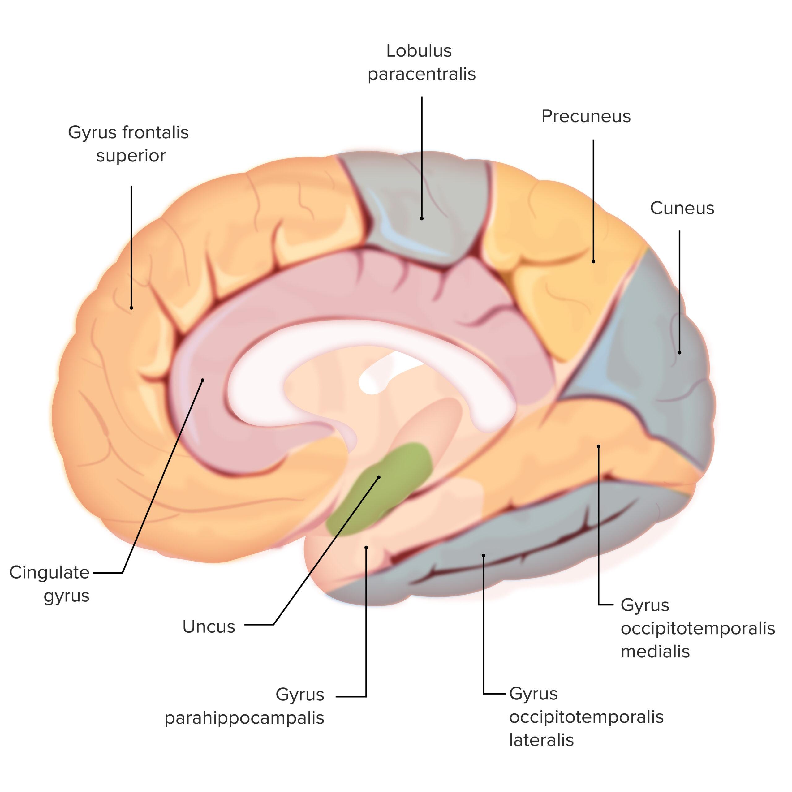
The various gyri throughout the brain: Note the parahippocampal gyrus (shaded in green)
This structure is important for short-term memory formation
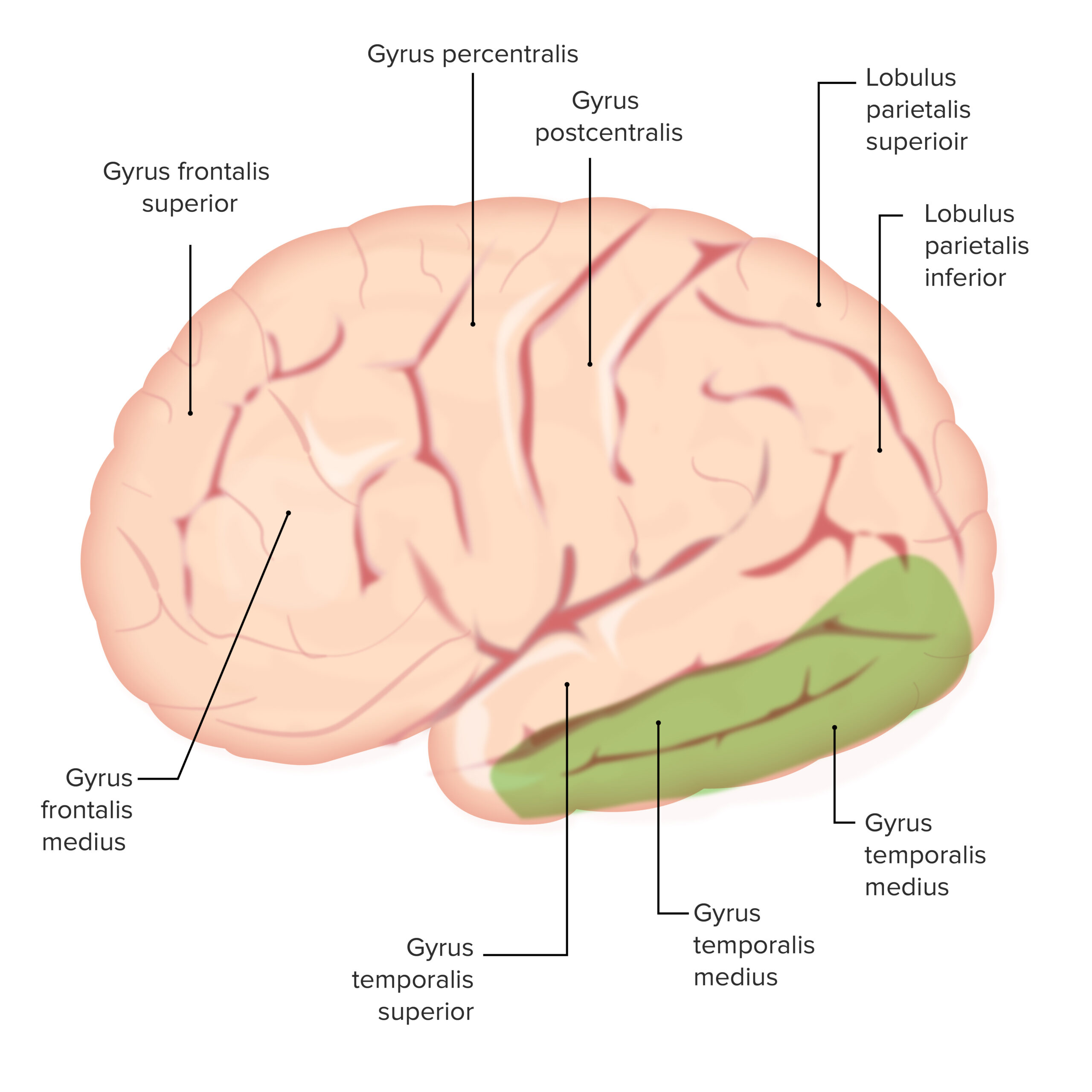
The various gyri throughout the brain: Note the medial and inferior temporal gyri (both shaded in green).
These structures are important for long-term memory
The various gyri throughout the brain: Note the uncus (shaded in green)
This is an important olfactory structure
The following structures are closely related to the cerebral cortex by location or function: