Blood supply to the brainBrainThe part of central nervous system that is contained within the skull (cranium). Arising from the neural tube, the embryonic brain is comprised of three major parts including prosencephalon (the forebrain); mesencephalon (the midbrain); and rhombencephalon (the hindbrain). The developed brain consists of cerebrum; cerebellum; and other structures in the brain stem.Nervous System: Anatomy, Structure, and Classification can be divided into an anterior and a posterior circulationCirculationThe movement of the blood as it is pumped through the cardiovascular system.ABCDE Assessment, which interconnect to form the circle of WillisCircle of WillisA polygonal anastomosis at the base of the brain formed by the internal carotid, proximal parts of the anterior, middle, and posterior cerebral arteries, the anterior communicating artery and the posterior communicating arteries.Subarachnoid Hemorrhage. The anterior circulationCirculationThe movement of the blood as it is pumped through the cardiovascular system.ABCDE Assessment is derived from the internal carotid arteriesCarotid ArteriesEither of the two principal arteries on both sides of the neck that supply blood to the head and neck; each divides into two branches, the internal carotid artery and the external carotid artery.Carotid Arterial System: Anatomy and consists mainly of the anterior and middle cerebral arteriesArteriesArteries are tubular collections of cells that transport oxygenated blood and nutrients from the heart to the tissues of the body. The blood passes through the arteries in order of decreasing luminal diameter, starting in the largest artery (the aorta) and ending in the small arterioles. Arteries are classified into 3 types: large elastic arteries, medium muscular arteries, and small arteries and arterioles. Arteries: Histology. The posterior circulationCirculationThe movement of the blood as it is pumped through the cardiovascular system.ABCDE Assessment is derived from the vertebral arteriesArteriesArteries are tubular collections of cells that transport oxygenated blood and nutrients from the heart to the tissues of the body. The blood passes through the arteries in order of decreasing luminal diameter, starting in the largest artery (the aorta) and ending in the small arterioles. Arteries are classified into 3 types: large elastic arteries, medium muscular arteries, and small arteries and arterioles. Arteries: Histology and consists primarily of the cerebellar and posterior cerebral arteriesArteriesArteries are tubular collections of cells that transport oxygenated blood and nutrients from the heart to the tissues of the body. The blood passes through the arteries in order of decreasing luminal diameter, starting in the largest artery (the aorta) and ending in the small arterioles. Arteries are classified into 3 types: large elastic arteries, medium muscular arteries, and small arteries and arterioles. Arteries: Histology. The primary venous drainage of the brainBrainThe part of central nervous system that is contained within the skull (cranium). Arising from the neural tube, the embryonic brain is comprised of three major parts including prosencephalon (the forebrain); mesencephalon (the midbrain); and rhombencephalon (the hindbrain). The developed brain consists of cerebrum; cerebellum; and other structures in the brain stem.Nervous System: Anatomy, Structure, and Classification occurs via the internal jugular veinInternal jugular veinParapharyngeal Abscess.
The arterial supply of the brainBrainThe part of central nervous system that is contained within the skull (cranium). Arising from the neural tube, the embryonic brain is comprised of three major parts including prosencephalon (the forebrain); mesencephalon (the midbrain); and rhombencephalon (the hindbrain). The developed brain consists of cerebrum; cerebellum; and other structures in the brain stem.Nervous System: Anatomy, Structure, and Classification is derived from 2 arterial systems:
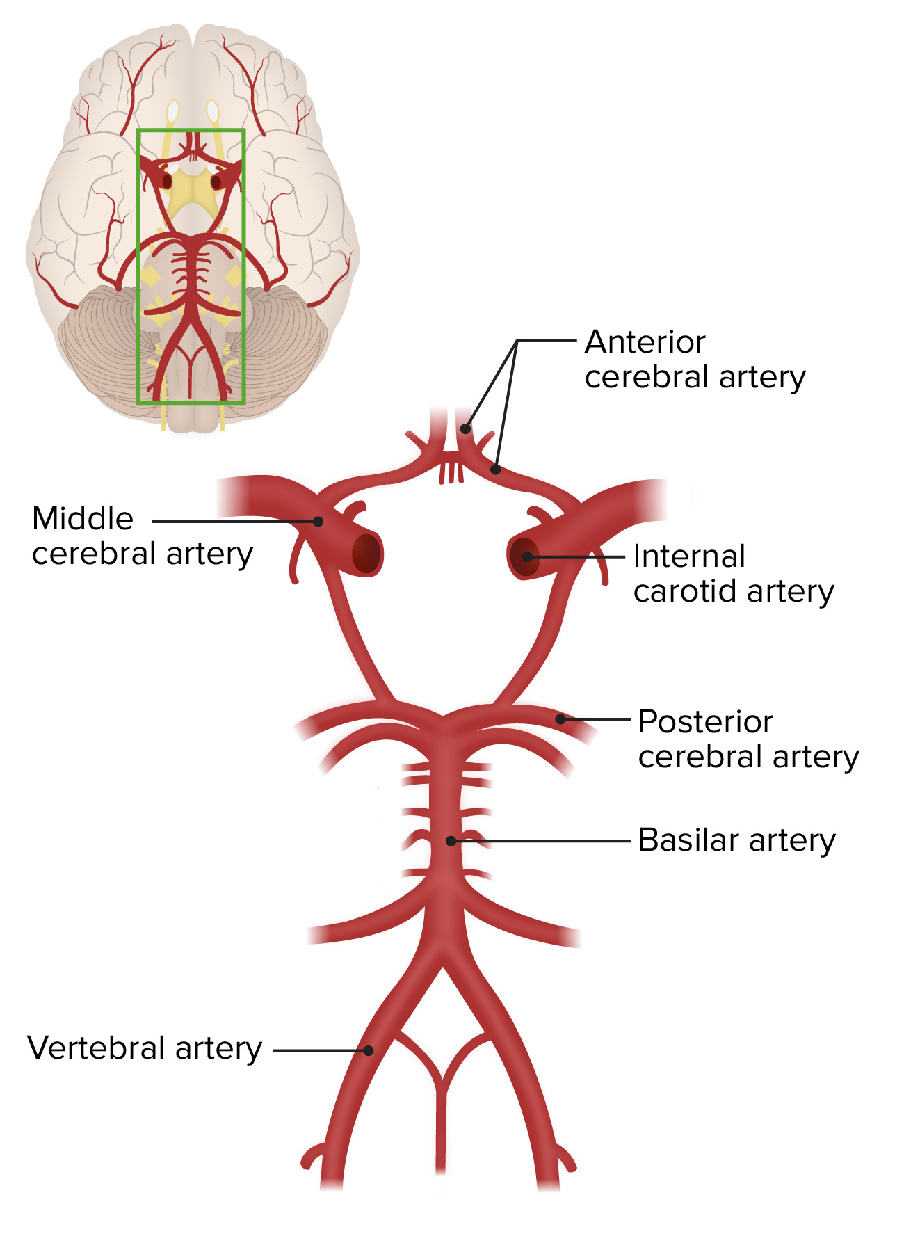
Internal carotid arteriesCarotid ArteriesEither of the two principal arteries on both sides of the neck that supply blood to the head and neck; each divides into two branches, the internal carotid artery and the external carotid artery.Carotid Arterial System: Anatomy (anterior circulationCirculationThe movement of the blood as it is pumped through the cardiovascular system.ABCDE Assessment):
Bifurcates from common carotid arteryCommon carotid arteryThe two principal arteries supplying the structures of the head and neck. They ascend in the neck, one on each side, and at the level of the upper border of the thyroid cartilage, each divides into two branches, the external and internal carotid arteries.Carotid Arterial System: Anatomy at the level of C4
Continues into brainBrainThe part of central nervous system that is contained within the skull (cranium). Arising from the neural tube, the embryonic brain is comprised of three major parts including prosencephalon (the forebrain); mesencephalon (the midbrain); and rhombencephalon (the hindbrain). The developed brain consists of cerebrum; cerebellum; and other structures in the brain stem.Nervous System: Anatomy, Structure, and Classification and becomes the middle cerebral artery
Anterior cerebral artery branches off the internal carotid arteryInternal carotid arteryBranch of the common carotid artery which supplies the anterior part of the brain, the eye and its appendages, the forehead and nose.Carotid Arterial System: Anatomy.
Vertebrobasilar system (posterior circulationCirculationThe movement of the blood as it is pumped through the cardiovascular system.ABCDE Assessment):
Vertebral arteriesArteriesArteries are tubular collections of cells that transport oxygenated blood and nutrients from the heart to the tissues of the body. The blood passes through the arteries in order of decreasing luminal diameter, starting in the largest artery (the aorta) and ending in the small arterioles. Arteries are classified into 3 types: large elastic arteries, medium muscular arteries, and small arteries and arterioles. Arteries: Histology arise from the subclavian arteriesArteriesArteries are tubular collections of cells that transport oxygenated blood and nutrients from the heart to the tissues of the body. The blood passes through the arteries in order of decreasing luminal diameter, starting in the largest artery (the aorta) and ending in the small arterioles. Arteries are classified into 3 types: large elastic arteries, medium muscular arteries, and small arteries and arterioles. Arteries: Histology.
Vertebral arteriesArteriesArteries are tubular collections of cells that transport oxygenated blood and nutrients from the heart to the tissues of the body. The blood passes through the arteries in order of decreasing luminal diameter, starting in the largest artery (the aorta) and ending in the small arterioles. Arteries are classified into 3 types: large elastic arteries, medium muscular arteries, and small arteries and arterioles. Arteries: Histology join to become the basilar artery.
Basilar artery bifurcates to become the posterior cerebral arteriesArteriesArteries are tubular collections of cells that transport oxygenated blood and nutrients from the heart to the tissues of the body. The blood passes through the arteries in order of decreasing luminal diameter, starting in the largest artery (the aorta) and ending in the small arterioles. Arteries are classified into 3 types: large elastic arteries, medium muscular arteries, and small arteries and arterioles. Arteries: Histology.
Cerebellar arteriesArteriesArteries are tubular collections of cells that transport oxygenated blood and nutrients from the heart to the tissues of the body. The blood passes through the arteries in order of decreasing luminal diameter, starting in the largest artery (the aorta) and ending in the small arterioles. Arteries are classified into 3 types: large elastic arteries, medium muscular arteries, and small arteries and arterioles. Arteries: Histology arise from the vertebral and basilar arteriesArteriesArteries are tubular collections of cells that transport oxygenated blood and nutrients from the heart to the tissues of the body. The blood passes through the arteries in order of decreasing luminal diameter, starting in the largest artery (the aorta) and ending in the small arterioles. Arteries are classified into 3 types: large elastic arteries, medium muscular arteries, and small arteries and arterioles. Arteries: Histology.
Blood supply to the brain is derived from 2 sources—the internal carotid arteries and the vertebrobasilar system.
Image by Lecturio.
Cerebral blood supply
Anterior cerebral artery:
Origin: smaller terminal branch of the internal carotid arteryInternal carotid arteryBranch of the common carotid artery which supplies the anterior part of the brain, the eye and its appendages, the forehead and nose.Carotid Arterial System: Anatomy
Course: occupies the longitudinal fissureFissureA crack or split that extends into the dermisGeneralized and Localized Rashes and travels posteriorly, ultimately anastomosing with the posterior cerebral arteriesArteriesArteries are tubular collections of cells that transport oxygenated blood and nutrients from the heart to the tissues of the body. The blood passes through the arteries in order of decreasing luminal diameter, starting in the largest artery (the aorta) and ending in the small arterioles. Arteries are classified into 3 types: large elastic arteries, medium muscular arteries, and small arteries and arterioles. Arteries: Histology
Divisions: distributes numerous anteromedial central branches
Supplies:
Medial frontalFrontalThe bone that forms the frontal aspect of the skull. Its flat part forms the forehead, articulating inferiorly with the nasal bone and the cheek bone on each side of the face.Skull: Anatomy and parietalParietalOne of a pair of irregularly shaped quadrilateral bones situated between the frontal bone and occipital bone, which together form the sides of the cranium.Skull: Anatomy lobes
Anterior limb of internal capsuleCapsuleAn envelope of loose gel surrounding a bacterial cell which is associated with the virulence of pathogenic bacteria. Some capsules have a well-defined border, whereas others form a slime layer that trails off into the medium. Most capsules consist of relatively simple polysaccharides but there are some bacteria whose capsules are made of polypeptides.Bacteroides
Most of corpus callosum
Middle cerebral artery:
Origin: larger terminal branch of the internal carotid arteryInternal carotid arteryBranch of the common carotid artery which supplies the anterior part of the brain, the eye and its appendages, the forehead and nose.Carotid Arterial System: Anatomy
Lateral frontalFrontalThe bone that forms the frontal aspect of the skull. Its flat part forms the forehead, articulating inferiorly with the nasal bone and the cheek bone on each side of the face.Skull: Anatomy and parietalParietalOne of a pair of irregularly shaped quadrilateral bones situated between the frontal bone and occipital bone, which together form the sides of the cranium.Skull: Anatomy lobes
Portion of temporal lobes
Genu and posterior limb of internal capsuleCapsuleAn envelope of loose gel surrounding a bacterial cell which is associated with the virulence of pathogenic bacteria. Some capsules have a well-defined border, whereas others form a slime layer that trails off into the medium. Most capsules consist of relatively simple polysaccharides but there are some bacteria whose capsules are made of polypeptides.Bacteroides
Most of basal gangliaBasal GangliaBasal ganglia are a group of subcortical nuclear agglomerations involved in movement, and are located deep to the cerebral hemispheres. Basal ganglia include the striatum (caudate nucleus and putamen), globus pallidus, substantia nigra, and subthalamic nucleus. Basal Ganglia: Anatomy
Posterior cerebral artery:
Origin: arises as terminal branches of basilar arteriesArteriesArteries are tubular collections of cells that transport oxygenated blood and nutrients from the heart to the tissues of the body. The blood passes through the arteries in order of decreasing luminal diameter, starting in the largest artery (the aorta) and ending in the small arterioles. Arteries are classified into 3 types: large elastic arteries, medium muscular arteries, and small arteries and arterioles. Arteries: Histology
Course:
Travels parallel to superior cerebellar artery
Receives the posterior communicating artery as it runs laterally
Supplies:
Occipital lobeOccipital lobePosterior portion of the cerebral hemispheres responsible for processing visual sensory information. It is located posterior to the parieto-occipital sulcus and extends to the preoccipital notch.Cerebral Cortex: Anatomy
Posteromedial temporal lobes
MidbrainMidbrainThe middle of the three primitive cerebral vesicles of the embryonic brain. Without further subdivision, midbrain develops into a short, constricted portion connecting the pons and the diencephalon. Midbrain contains two major parts, the dorsal tectum mesencephali and the ventral tegmentum mesencephali, housing components of auditory, visual, and other sensorimotor systems.Brain Stem: Anatomy
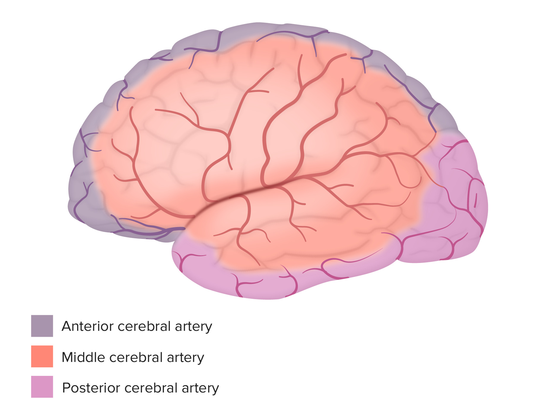
Lateral surface view shows the arterial supply of the brain: Note the distribution of the anterior cerebral artery supplying the anteromedial surface, the middle cerebral artery supplying the lateral surface, and the posterior cerebral artery supplying the posterior and inferior surfaces.
Image by Lecturio.
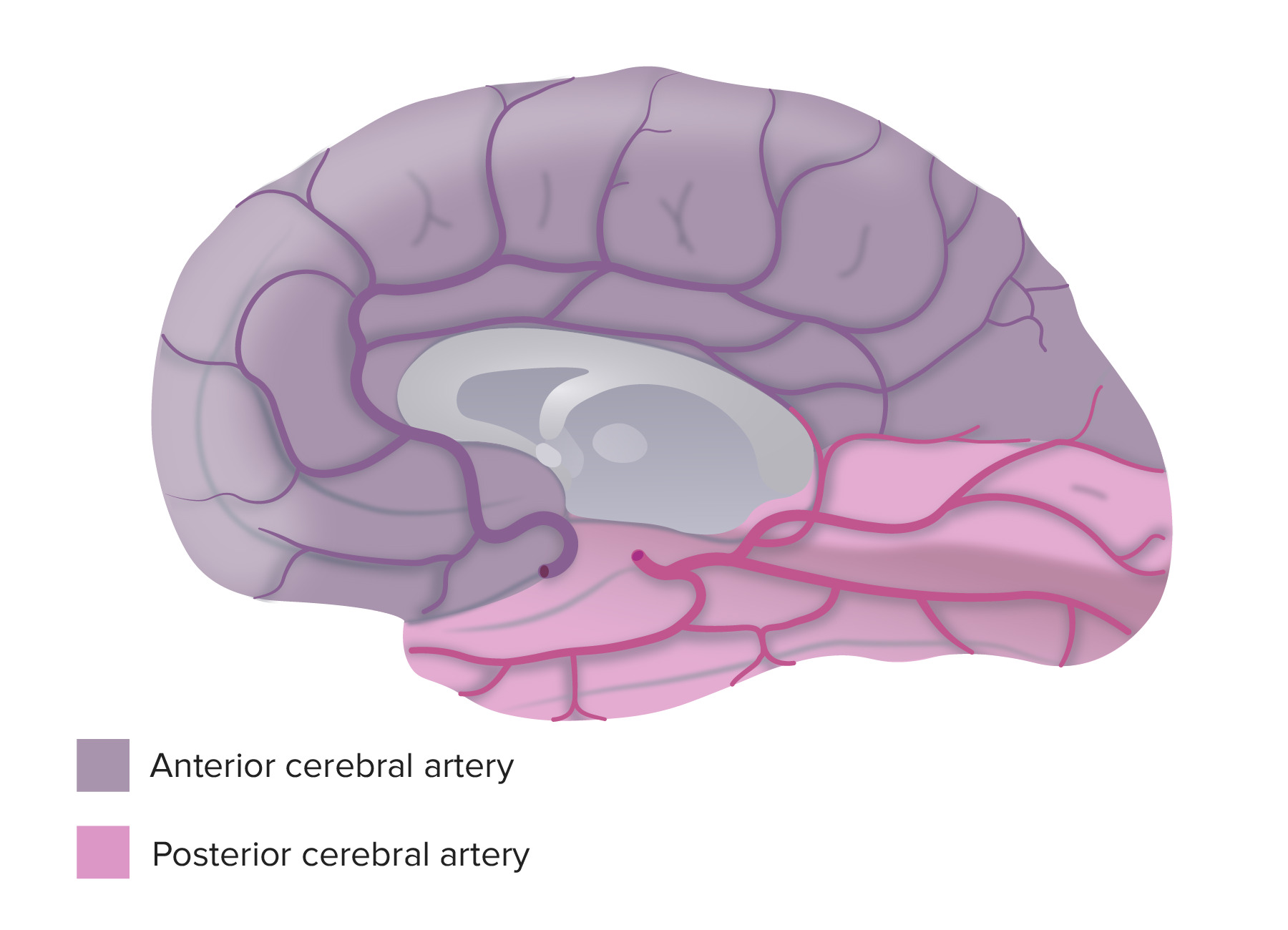
Medial view shows the arterial supply of the brain: Note the distribution of blood flow. The anterior cerebral artery is in purple and the posterior cerebral artery in pink.
Image by Lecturio.
Cerebellar blood supply
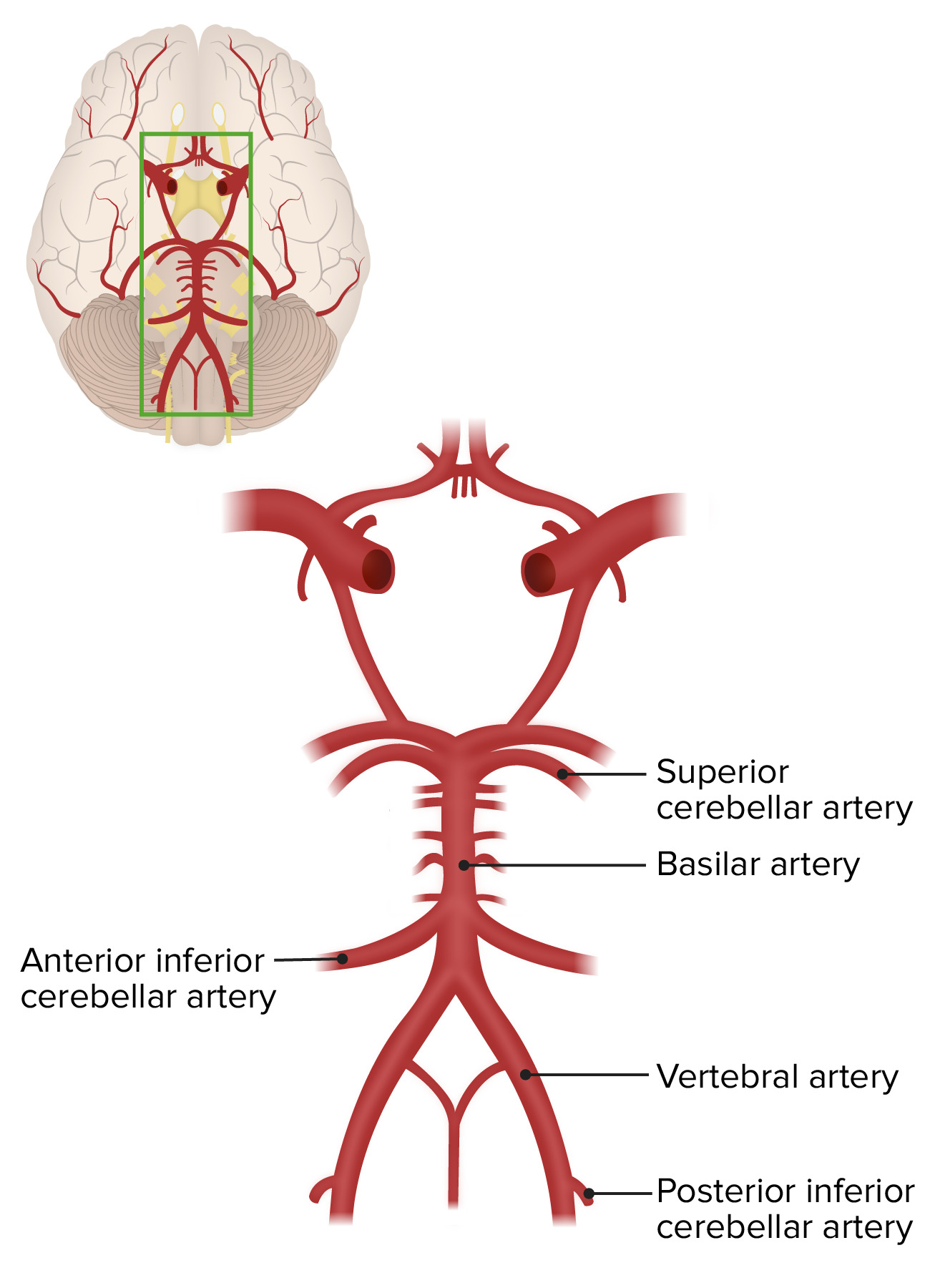
The cerebellumCerebellumThe cerebellum, Latin for “little brain,” is located in the posterior cranial fossa, dorsal to the pons and midbrain, and its principal role is in the coordination of movements. The cerebellum consists of 3 lobes on either side of its 2 hemispheres and is connected in the middle by the vermis. Cerebellum: Anatomy is supplied by branches of the vertebral and basilar arteriesArteriesArteries are tubular collections of cells that transport oxygenated blood and nutrients from the heart to the tissues of the body. The blood passes through the arteries in order of decreasing luminal diameter, starting in the largest artery (the aorta) and ending in the small arterioles. Arteries are classified into 3 types: large elastic arteries, medium muscular arteries, and small arteries and arterioles. Arteries: Histology.
Superior cerebellar artery:
Origin: arises from distal aspect of the basilar artery
Supplies:
Superior cerebellumCerebellumThe cerebellum, Latin for “little brain,” is located in the posterior cranial fossa, dorsal to the pons and midbrain, and its principal role is in the coordination of movements. The cerebellum consists of 3 lobes on either side of its 2 hemispheres and is connected in the middle by the vermis. Cerebellum: Anatomy
Pineal body
Anterior inferior cerebellar artery:
Origin: arises from proximal aspect of the basilar artery
Supplies:
Anterior inferior surface of cerebellumCerebellumThe cerebellum, Latin for “little brain,” is located in the posterior cranial fossa, dorsal to the pons and midbrain, and its principal role is in the coordination of movements. The cerebellum consists of 3 lobes on either side of its 2 hemispheres and is connected in the middle by the vermis. Cerebellum: Anatomy
Inferolateral aspect of the ponsPonsThe front part of the hindbrain (rhombencephalon) that lies between the medulla and the midbrain (mesencephalon) ventral to the cerebellum. It is composed of two parts, the dorsal and the ventral. The pons serves as a relay station for neural pathways between the cerebellum to the cerebrum.Brain Stem: Anatomy
Posterior inferior surface of cerebellumCerebellumThe cerebellum, Latin for “little brain,” is located in the posterior cranial fossa, dorsal to the pons and midbrain, and its principal role is in the coordination of movements. The cerebellum consists of 3 lobes on either side of its 2 hemispheres and is connected in the middle by the vermis. Cerebellum: Anatomy
ChoroidChoroidThe thin, highly vascular membrane covering most of the posterior of the eye between the retina and sclera.Eye: Anatomy plexus of 4th ventricle
The cerebellum is supplied by the superior cerebellar artery, anterior inferior cerebellar artery, and posterior inferior cerebellar artery.
Image by Lecturio.
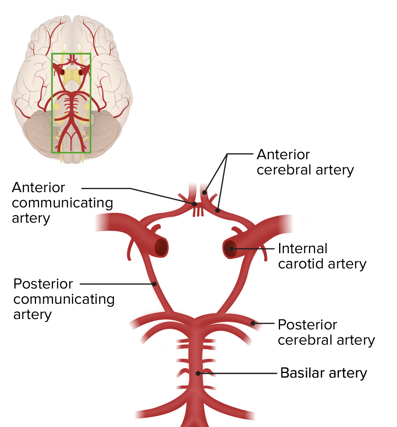
Circle of WillisCircle of WillisA polygonal anastomosis at the base of the brain formed by the internal carotid, proximal parts of the anterior, middle, and posterior cerebral arteries, the anterior communicating artery and the posterior communicating arteries.Subarachnoid Hemorrhage
The circle of WillisCircle of WillisA polygonal anastomosis at the base of the brain formed by the internal carotid, proximal parts of the anterior, middle, and posterior cerebral arteries, the anterior communicating artery and the posterior communicating arteries.Subarachnoid Hemorrhage is an interconnected network of arteriesArteriesArteries are tubular collections of cells that transport oxygenated blood and nutrients from the heart to the tissues of the body. The blood passes through the arteries in order of decreasing luminal diameter, starting in the largest artery (the aorta) and ending in the small arterioles. Arteries are classified into 3 types: large elastic arteries, medium muscular arteries, and small arteries and arterioles. Arteries: Histology within the brainBrainThe part of central nervous system that is contained within the skull (cranium). Arising from the neural tube, the embryonic brain is comprised of three major parts including prosencephalon (the forebrain); mesencephalon (the midbrain); and rhombencephalon (the hindbrain). The developed brain consists of cerebrum; cerebellum; and other structures in the brain stem.Nervous System: Anatomy, Structure, and Classification. The circle of WillisCircle of WillisA polygonal anastomosis at the base of the brain formed by the internal carotid, proximal parts of the anterior, middle, and posterior cerebral arteries, the anterior communicating artery and the posterior communicating arteries.Subarachnoid Hemorrhage represents the collateral pathways of arterial blood, and it has many anatomical variants. The circle is formed via anastomoses between the anterior and posterior arterial systems that supply blood to the brainBrainThe part of central nervous system that is contained within the skull (cranium). Arising from the neural tube, the embryonic brain is comprised of three major parts including prosencephalon (the forebrain); mesencephalon (the midbrain); and rhombencephalon (the hindbrain). The developed brain consists of cerebrum; cerebellum; and other structures in the brain stem.Nervous System: Anatomy, Structure, and Classification. This duality provides a safety net if 1 of the systems fails via occlusion, trauma, or a neoplastic process. Vessels comprising the circle of WillisCircle of WillisA polygonal anastomosis at the base of the brain formed by the internal carotid, proximal parts of the anterior, middle, and posterior cerebral arteries, the anterior communicating artery and the posterior communicating arteries.Subarachnoid Hemorrhage include:
Components of anterior circulationCirculationThe movement of the blood as it is pumped through the cardiovascular system.ABCDE Assessment:
Left and right internal carotid arteriesCarotid ArteriesEither of the two principal arteries on both sides of the neck that supply blood to the head and neck; each divides into two branches, the internal carotid artery and the external carotid artery.Carotid Arterial System: Anatomy
Horizontal segments of the left and right anterior cerebral arteriesArteriesArteries are tubular collections of cells that transport oxygenated blood and nutrients from the heart to the tissues of the body. The blood passes through the arteries in order of decreasing luminal diameter, starting in the largest artery (the aorta) and ending in the small arterioles. Arteries are classified into 3 types: large elastic arteries, medium muscular arteries, and small arteries and arterioles. Arteries: Histology
Single anterior communicating artery
Posterior circulationCirculationThe movement of the blood as it is pumped through the cardiovascular system.ABCDE Assessment:
Left and right posterior communicating arteriesArteriesArteries are tubular collections of cells that transport oxygenated blood and nutrients from the heart to the tissues of the body. The blood passes through the arteries in order of decreasing luminal diameter, starting in the largest artery (the aorta) and ending in the small arterioles. Arteries are classified into 3 types: large elastic arteries, medium muscular arteries, and small arteries and arterioles. Arteries: Histology
Horizontal segments of the left and right posterior cerebral arteriesArteriesArteries are tubular collections of cells that transport oxygenated blood and nutrients from the heart to the tissues of the body. The blood passes through the arteries in order of decreasing luminal diameter, starting in the largest artery (the aorta) and ending in the small arterioles. Arteries are classified into 3 types: large elastic arteries, medium muscular arteries, and small arteries and arterioles. Arteries: Histology, arising from the single basilar artery
The circle of Willis has 5 components, including the anterior communicating artery, the anterior cerebral arteries, the internal carotid artery, the posterior communicating artery, and the posterior cerebral arteries.
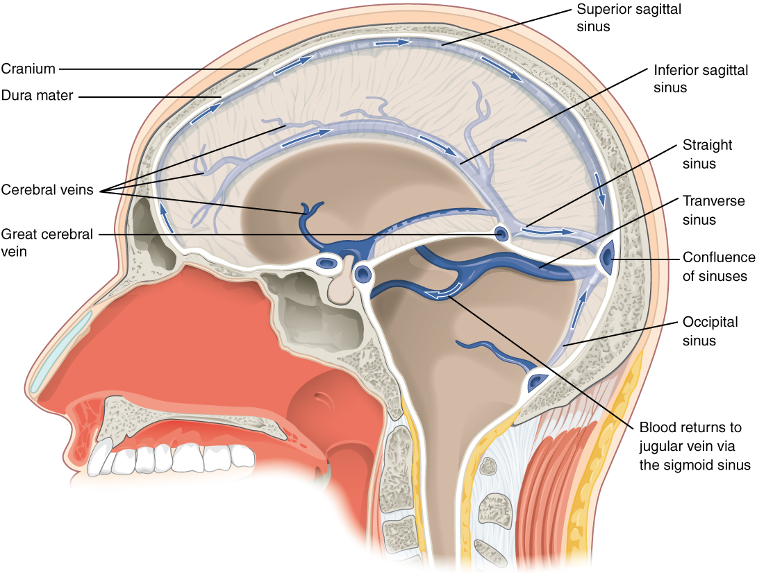
Venous drainage of the brainBrainThe part of central nervous system that is contained within the skull (cranium). Arising from the neural tube, the embryonic brain is comprised of three major parts including prosencephalon (the forebrain); mesencephalon (the midbrain); and rhombencephalon (the hindbrain). The developed brain consists of cerebrum; cerebellum; and other structures in the brain stem.Nervous System: Anatomy, Structure, and Classification occurs via the cerebral veinsVeinsVeins are tubular collections of cells, which transport deoxygenated blood and waste from the capillary beds back to the heart. Veins are classified into 3 types: small veins/venules, medium veins, and large veins. Each type contains 3 primary layers: tunica intima, tunica media, and tunica adventitia. Veins: Histology, which ultimately drain into the straight sinus, transverse sinus, and finally the sagittalSagittalComputed Tomography (CT) sinus before reaching the internal jugular veinInternal jugular veinParapharyngeal Abscess and traveling back to the heart.
Venous drainage of the brainBrainThe part of central nervous system that is contained within the skull (cranium). Arising from the neural tube, the embryonic brain is comprised of three major parts including prosencephalon (the forebrain); mesencephalon (the midbrain); and rhombencephalon (the hindbrain). The developed brain consists of cerebrum; cerebellum; and other structures in the brain stem.Nervous System: Anatomy, Structure, and Classification occurs via the superficial and deep venous systems:
Unlike all other veinsVeinsVeins are tubular collections of cells, which transport deoxygenated blood and waste from the capillary beds back to the heart. Veins are classified into 3 types: small veins/venules, medium veins, and large veins. Each type contains 3 primary layers: tunica intima, tunica media, and tunica adventitia. Veins: Histology of the body, cerebral veinsVeinsVeins are tubular collections of cells, which transport deoxygenated blood and waste from the capillary beds back to the heart. Veins are classified into 3 types: small veins/venules, medium veins, and large veins. Each type contains 3 primary layers: tunica intima, tunica media, and tunica adventitia. Veins: Histology do not have valves.
Superficial venous system:
Composed of cortical veinsVeinsVeins are tubular collections of cells, which transport deoxygenated blood and waste from the capillary beds back to the heart. Veins are classified into 3 types: small veins/venules, medium veins, and large veins. Each type contains 3 primary layers: tunica intima, tunica media, and tunica adventitia. Veins: Histology
Drains the cerebral cortexCerebral cortexThe cerebral cortex is the largest and most developed part of the human brain and CNS. Occupying the upper part of the cranial cavity, the cerebral cortex has 4 lobes and is divided into 2 hemispheres that are joined centrally by the corpus callosum. Cerebral Cortex: Anatomy
Deep venous system:
Divisions:
Deep cerebral veinsVeinsVeins are tubular collections of cells, which transport deoxygenated blood and waste from the capillary beds back to the heart. Veins are classified into 3 types: small veins/venules, medium veins, and large veins. Each type contains 3 primary layers: tunica intima, tunica media, and tunica adventitia. Veins: Histology
Drains the deep brainBrainThe part of central nervous system that is contained within the skull (cranium). Arising from the neural tube, the embryonic brain is comprised of three major parts including prosencephalon (the forebrain); mesencephalon (the midbrain); and rhombencephalon (the hindbrain). The developed brain consists of cerebrum; cerebellum; and other structures in the brain stem.Nervous System: Anatomy, Structure, and Classification structures
Drain blood from the cerebral veinsVeinsVeins are tubular collections of cells, which transport deoxygenated blood and waste from the capillary beds back to the heart. Veins are classified into 3 types: small veins/venules, medium veins, and large veins. Each type contains 3 primary layers: tunica intima, tunica media, and tunica adventitia. Veins: Histology, orbits, and skullSkullThe skull (cranium) is the skeletal structure of the head supporting the face and forming a protective cavity for the brain. The skull consists of 22 bones divided into the viscerocranium (facial skeleton) and the neurocranium.Skull: Anatomy
Empty into the internal jugular veinsVeinsVeins are tubular collections of cells, which transport deoxygenated blood and waste from the capillary beds back to the heart. Veins are classified into 3 types: small veins/venules, medium veins, and large veins. Each type contains 3 primary layers: tunica intima, tunica media, and tunica adventitia. Veins: Histology
Sagittal view through the skull illustrating the venous drainage system: The arrows demonstrate the flow of blood from the cerebral veins and sinuses to the confluence of sinuses, ultimately returning to the jugular vein via the sigmoid sinus.
Image: “Blood drains from the brain through a series of sinuses that connect to the jugular veins” by OpenStax College. License: CC BY 4.0
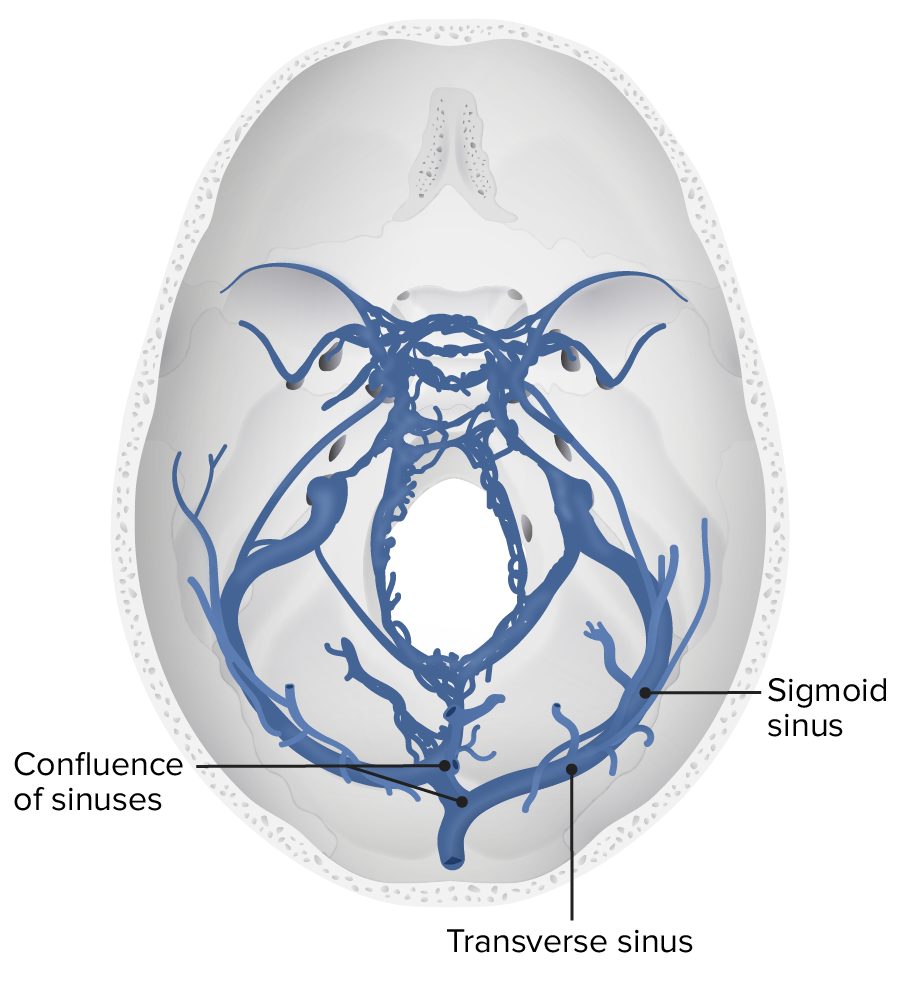
A transverse view of the cerebral deep venous system, specifically the sigmoid and transverse sinuses
Intracranial aneurysms: A cerebral, or intracranial, aneurysmAneurysmAn aneurysm is a bulging, weakened area of a blood vessel that causes an abnormal widening of its diameter > 1.5 times the size of the native vessel. Aneurysms occur more often in arteries than in veins and are at risk of dissection and rupture, which can be life-threatening. Thoracic Aortic Aneurysms is an abnormal dilation of a local area of the artery wall in the CNS. These aneurysms most often occur at junction points of the major arteriesArteriesArteries are tubular collections of cells that transport oxygenated blood and nutrients from the heart to the tissues of the body. The blood passes through the arteries in order of decreasing luminal diameter, starting in the largest artery (the aorta) and ending in the small arterioles. Arteries are classified into 3 types: large elastic arteries, medium muscular arteries, and small arteries and arterioles. Arteries: Histology of the brainBrainThe part of central nervous system that is contained within the skull (cranium). Arising from the neural tube, the embryonic brain is comprised of three major parts including prosencephalon (the forebrain); mesencephalon (the midbrain); and rhombencephalon (the hindbrain). The developed brain consists of cerebrum; cerebellum; and other structures in the brain stem.Nervous System: Anatomy, Structure, and Classification, usually around the circle of WillisCircle of WillisA polygonal anastomosis at the base of the brain formed by the internal carotid, proximal parts of the anterior, middle, and posterior cerebral arteries, the anterior communicating artery and the posterior communicating arteries.Subarachnoid Hemorrhage. They can either compress adjacent structures or rupture and cause a hemorrhagic strokeHemorrhagic strokeStroke due to rupture of a weakened blood vessel in the brain (e.g., cerebral hemispheres; cerebellum; subarachnoid space).Subarachnoid Hemorrhage.
Arterial dissections: The vertebral and carotid arteriesCarotid ArteriesEither of the two principal arteries on both sides of the neck that supply blood to the head and neck; each divides into two branches, the internal carotid artery and the external carotid artery.Carotid Arterial System: Anatomy are vital components of the blood supply to the brainBrainThe part of central nervous system that is contained within the skull (cranium). Arising from the neural tube, the embryonic brain is comprised of three major parts including prosencephalon (the forebrain); mesencephalon (the midbrain); and rhombencephalon (the hindbrain). The developed brain consists of cerebrum; cerebellum; and other structures in the brain stem.Nervous System: Anatomy, Structure, and Classification. Arterial dissections occur when the integrity of the arterial wall structure fails, usually abruptly, resulting in an intramural hematomaIntramural hematomaDissection of the Carotid and Vertebral Arteries formation and a false lumenFalse lumenAortic Dissection between the tunica mediaTunica mediaThe middle layer of blood vessel walls, composed principally of thin, cylindrical, smooth muscle cells and elastic tissue. It accounts for the bulk of the wall of most arteries. The smooth muscle cells are arranged in circular layers around the vessel, and the thickness of the coat varies with the size of the vessel.Arteries: Histology and adventitial layers. This process may result in aneurysmAneurysmAn aneurysm is a bulging, weakened area of a blood vessel that causes an abnormal widening of its diameter > 1.5 times the size of the native vessel. Aneurysms occur more often in arteries than in veins and are at risk of dissection and rupture, which can be life-threatening. Thoracic Aortic Aneurysms, stenosisStenosisHypoplastic Left Heart Syndrome (HLHS), or occlusion. Presentation is typically with unilateral head or neckNeckThe part of a human or animal body connecting the head to the rest of the body.Peritonsillar AbscesspainPainAn unpleasant sensation induced by noxious stimuli which are detected by nerve endings of nociceptive neurons.Pain: Types and Pathways and/or stroke-like symptoms.
Subclavian stealSubclavian stealA clinically significant reduction in blood supply to the brain stem and cerebellum (i.e., vertebrobasilar insufficiency) resulting from reversal of blood flow through the vertebral artery from occlusion or stenosis of the proximal subclavian or brachiocephalic artery. Common symptoms include vertigo, syncope, and intermittent claudication of the involved upper extremity. Subclavian steal may also occur in asymptomatic individuals.Subclavian Steal Syndrome syndrome: occurs when narrowing/occlusion of the subclavian artery proximal to the origin of the vertebral arteryVertebral arteryThe first branch of the subclavian artery with distribution to muscles of the neck; vertebrae; spinal cord; cerebellum; and interior of the cerebrum.Lateral Medullary Syndrome (Wallenberg Syndrome) causes a reversal of blood flowBlood flowBlood flow refers to the movement of a certain volume of blood through the vasculature over a given unit of time (e.g., mL per minute).Vascular Resistance, Flow, and Mean Arterial Pressure in the ipsilateral vertebral arteryVertebral arteryThe first branch of the subclavian artery with distribution to muscles of the neck; vertebrae; spinal cord; cerebellum; and interior of the cerebrum.Lateral Medullary Syndrome (Wallenberg Syndrome) to continue perfusing the ipsilateral armArmThe arm, or “upper arm” in common usage, is the region of the upper limb that extends from the shoulder to the elbow joint and connects inferiorly to the forearm through the cubital fossa. It is divided into 2 fascial compartments (anterior and posterior).Arm: Anatomy. The most common cause of subclavian stealSubclavian stealA clinically significant reduction in blood supply to the brain stem and cerebellum (i.e., vertebrobasilar insufficiency) resulting from reversal of blood flow through the vertebral artery from occlusion or stenosis of the proximal subclavian or brachiocephalic artery. Common symptoms include vertigo, syncope, and intermittent claudication of the involved upper extremity. Subclavian steal may also occur in asymptomatic individuals.Subclavian Steal Syndrome syndrome is atherosclerosisAtherosclerosisAtherosclerosis is a common form of arterial disease in which lipid deposition forms a plaque in the blood vessel walls. Atherosclerosis is an incurable disease, for which there are clearly defined risk factors that often can be reduced through a change in lifestyle and behavior of the patient. Atherosclerosis. Symptoms are rare, but when they occur, they are usually triggered by physical exertion of the armArmThe arm, or “upper arm” in common usage, is the region of the upper limb that extends from the shoulder to the elbow joint and connects inferiorly to the forearm through the cubital fossa. It is divided into 2 fascial compartments (anterior and posterior).Arm: Anatomy and subsequent hypoperfusion of the armArmThe arm, or “upper arm” in common usage, is the region of the upper limb that extends from the shoulder to the elbow joint and connects inferiorly to the forearm through the cubital fossa. It is divided into 2 fascial compartments (anterior and posterior).Arm: Anatomy or brainBrainThe part of central nervous system that is contained within the skull (cranium). Arising from the neural tube, the embryonic brain is comprised of three major parts including prosencephalon (the forebrain); mesencephalon (the midbrain); and rhombencephalon (the hindbrain). The developed brain consists of cerebrum; cerebellum; and other structures in the brain stem.Nervous System: Anatomy, Structure, and Classification.
Stroke: The 2 types of stroke are ischemic and hemorrhagic. Risk factors include hypertensionHypertensionHypertension, or high blood pressure, is a common disease that manifests as elevated systemic arterial pressures. Hypertension is most often asymptomatic and is found incidentally as part of a routine physical examination or during triage for an unrelated medical encounter. Hypertension, cerebral amyloid angiopathyAmyloid angiopathyA heterogeneous group of sporadic or familial disorders characterized by amyloid deposits in the walls of small and medium sized blood vessels of cerebral cortex and meninges. Clinical features include multiple, small lobar cerebral hemorrhage; cerebral ischemia; and cerebral infarction. Cerebral amyloid angiopathy is unrelated to generalized amyloidosis. Amyloidogenic peptides in this condition are nearly always the same ones found in alzheimer disease.Alzheimer Disease, neoplastic diseases, and cerebral aneurysms. Strokes can be diagnosed with brainBrainThe part of central nervous system that is contained within the skull (cranium). Arising from the neural tube, the embryonic brain is comprised of three major parts including prosencephalon (the forebrain); mesencephalon (the midbrain); and rhombencephalon (the hindbrain). The developed brain consists of cerebrum; cerebellum; and other structures in the brain stem.Nervous System: Anatomy, Structure, and Classification CT or MRI, and treatment is geared toward reperfusing the ischemic area or stopping the brainBrainThe part of central nervous system that is contained within the skull (cranium). Arising from the neural tube, the embryonic brain is comprised of three major parts including prosencephalon (the forebrain); mesencephalon (the midbrain); and rhombencephalon (the hindbrain). The developed brain consists of cerebrum; cerebellum; and other structures in the brain stem.Nervous System: Anatomy, Structure, and Classification hemorrhage.