Achieve Mastery of Medical Concepts

Study for medical school and boards with Lecturio

Subclavian Steal Syndrome

Subclavian steal syndrome occurs when narrowing/occlusion of the subclavian artery proximal to the origin of the vertebral artery Vertebral artery The first branch of the subclavian artery with distribution to muscles of the neck; vertebrae; spinal cord; cerebellum; and interior of the cerebrum. Lateral Medullary Syndrome (Wallenberg Syndrome) causes a reversal of blood flow Blood flow Blood flow refers to the movement of a certain volume of blood through the vasculature over a given unit of time (e.g., mL per minute). Vascular Resistance, Flow, and Mean Arterial Pressure in the ipsilateral vertebral artery Vertebral artery The first branch of the subclavian artery with distribution to muscles of the neck; vertebrae; spinal cord; cerebellum; and interior of the cerebrum. Lateral Medullary Syndrome (Wallenberg Syndrome) to continue perfusing the ipsilateral arm Arm The arm, or "upper arm" in common usage, is the region of the upper limb that extends from the shoulder to the elbow joint and connects inferiorly to the forearm through the cubital fossa. It is divided into 2 fascial compartments (anterior and posterior). Arm: Anatomy. The most common cause is atherosclerosis Atherosclerosis Atherosclerosis is a common form of arterial disease in which lipid deposition forms a plaque in the blood vessel walls. Atherosclerosis is an incurable disease, for which there are clearly defined risk factors that often can be reduced through a change in lifestyle and behavior of the patient. Atherosclerosis. Symptoms are rare, but when they occur are usually triggered by physical exertion of the arm Arm The arm, or "upper arm" in common usage, is the region of the upper limb that extends from the shoulder to the elbow joint and connects inferiorly to the forearm through the cubital fossa. It is divided into 2 fascial compartments (anterior and posterior). Arm: Anatomy and subsequent hypoperfusion of the arm Arm The arm, or "upper arm" in common usage, is the region of the upper limb that extends from the shoulder to the elbow joint and connects inferiorly to the forearm through the cubital fossa. It is divided into 2 fascial compartments (anterior and posterior). Arm: Anatomy or brain Brain The part of central nervous system that is contained within the skull (cranium). Arising from the neural tube, the embryonic brain is comprised of three major parts including prosencephalon (the forebrain); mesencephalon (the midbrain); and rhombencephalon (the hindbrain). The developed brain consists of cerebrum; cerebellum; and other structures in the brain stem. Nervous System: Anatomy, Structure, and Classification. Patients Patients Individuals participating in the health care system for the purpose of receiving therapeutic, diagnostic, or preventive procedures. Clinician–Patient Relationship may present with claudication, pain Pain An unpleasant sensation induced by noxious stimuli which are detected by nerve endings of nociceptive neurons. Pain: Types and Pathways, pallor, paresthesias Paresthesias Subjective cutaneous sensations (e.g., cold, warmth, tingling, pressure, etc.) that are experienced spontaneously in the absence of stimulation. Posterior Cord Syndrome, and weakened pulse in the affected extremity. Patients Patients Individuals participating in the health care system for the purpose of receiving therapeutic, diagnostic, or preventive procedures. Clinician–Patient Relationship may also present with transient neurologic disturbances concerning for a stroke. Diagnosis is made by clinical findings and imaging (ultrasound, CT, MRI). In addition to appropriate management of atherosclerosis Atherosclerosis Atherosclerosis is a common form of arterial disease in which lipid deposition forms a plaque in the blood vessel walls. Atherosclerosis is an incurable disease, for which there are clearly defined risk factors that often can be reduced through a change in lifestyle and behavior of the patient. Atherosclerosis, symptomatic patients Patients Individuals participating in the health care system for the purpose of receiving therapeutic, diagnostic, or preventive procedures. Clinician–Patient Relationship may need angioplasty Angioplasty Reconstruction or repair of a blood vessel, which includes the widening of a pathological narrowing of an artery or vein by the removal of atheromatous plaque material and/or the endothelial lining as well, or by dilatation (balloon angioplasty) to compress an atheroma. Except for endarterectomy, usually these procedures are performed via catheterization as minimally invasive endovascular procedures. Cardiac Surgery/stenting or surgical revascularization Revascularization Thromboangiitis Obliterans (Buerger’s Disease).

Last updated: Aug 18, 2022

Editorial responsibility: Stanley Oiseth, Lindsay Jones, Evelin Maza

Overview

Definition

Subclavian steal syndrome is the reversal of blood flow Blood flow Blood flow refers to the movement of a certain volume of blood through the vasculature over a given unit of time (e.g., mL per minute). Vascular Resistance, Flow, and Mean Arterial Pressure in the vertebral artery Vertebral artery The first branch of the subclavian artery with distribution to muscles of the neck; vertebrae; spinal cord; cerebellum; and interior of the cerebrum. Lateral Medullary Syndrome (Wallenberg Syndrome) on the side of a stenotic subclavian artery that produces symptoms of arterial insufficiency in the brain Brain The part of central nervous system that is contained within the skull (cranium). Arising from the neural tube, the embryonic brain is comprised of three major parts including prosencephalon (the forebrain); mesencephalon (the midbrain); and rhombencephalon (the hindbrain). The developed brain consists of cerebrum; cerebellum; and other structures in the brain stem. Nervous System: Anatomy, Structure, and Classification or upper extremity.
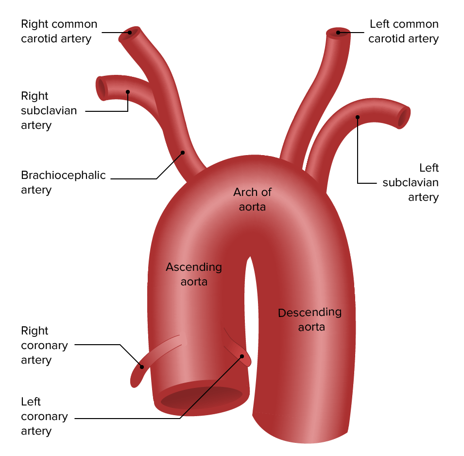
Epidemiology

  • 30% of patients Patients Individuals participating in the health care system for the purpose of receiving therapeutic, diagnostic, or preventive procedures. Clinician–Patient Relationship with peripheral artery disease Peripheral artery disease Peripheral artery disease (PAD) is obstruction of the arterial lumen resulting in decreased blood flow to the distal limbs. The disease can be a result of atherosclerosis or thrombosis. Patients may be asymptomatic or have progressive claudication, skin discoloration, ischemic ulcers, or gangrene. Peripheral Artery Disease have subclavian stenosis Stenosis Hypoplastic Left Heart Syndrome (HLHS).
  • Only a minority of those patients Patients Individuals participating in the health care system for the purpose of receiving therapeutic, diagnostic, or preventive procedures. Clinician–Patient Relationship develop clinical subclavian steal syndrome.
  • The left subclavian artery is more commonly affected (> 75% of cases).
  • Incidence Incidence The number of new cases of a given disease during a given period in a specified population. It also is used for the rate at which new events occur in a defined population. It is differentiated from prevalence, which refers to all cases in the population at a given time. Measures of Disease Frequency increases with age.
  • More common in men

Etiology

Pathophysiology and Clinical Presentation

Anatomy

  • The main blood supply to the upper extremities comes from axillary arteries Arteries Arteries are tubular collections of cells that transport oxygenated blood and nutrients from the heart to the tissues of the body. The blood passes through the arteries in order of decreasing luminal diameter, starting in the largest artery (the aorta) and ending in the small arterioles. Arteries are classified into 3 types: large elastic arteries, medium muscular arteries, and small arteries and arterioles. Arteries: Histology.
  • Axillary artery Axillary Artery The continuation of the subclavian artery; it distributes over the upper limb, axilla, chest and shoulder. Axilla and Brachial Plexus: Anatomy is a continuation of a subclavian artery once it exits the thoracic outlet Thoracic Outlet Thoracic Outlet Syndrome:
  • In most individuals, the vertebral arteries Arteries Arteries are tubular collections of cells that transport oxygenated blood and nutrients from the heart to the tissues of the body. The blood passes through the arteries in order of decreasing luminal diameter, starting in the largest artery (the aorta) and ending in the small arterioles. Arteries are classified into 3 types: large elastic arteries, medium muscular arteries, and small arteries and arterioles. Arteries: Histology originate from the subclavian arteries Arteries Arteries are tubular collections of cells that transport oxygenated blood and nutrients from the heart to the tissues of the body. The blood passes through the arteries in order of decreasing luminal diameter, starting in the largest artery (the aorta) and ending in the small arterioles. Arteries are classified into 3 types: large elastic arteries, medium muscular arteries, and small arteries and arterioles. Arteries: Histology.
  • Internal mammary artery (IMA), thyrocervical and costocervical trunk also originate from the subclavian artery.

Pathophysiology of subclavian steal

  • Stenosis Stenosis Hypoplastic Left Heart Syndrome (HLHS) or occlusion of the subclavian artery proximal to the origin of the vertebral artery Vertebral artery The first branch of the subclavian artery with distribution to muscles of the neck; vertebrae; spinal cord; cerebellum; and interior of the cerebrum. Lateral Medullary Syndrome (Wallenberg Syndrome) →  hypoperfusion of the ipsilateral subclavian and vertebral arteries Arteries Arteries are tubular collections of cells that transport oxygenated blood and nutrients from the heart to the tissues of the body. The blood passes through the arteries in order of decreasing luminal diameter, starting in the largest artery (the aorta) and ending in the small arterioles. Arteries are classified into 3 types: large elastic arteries, medium muscular arteries, and small arteries and arterioles. Arteries: Histology 
  • The reversal of blood flow Blood flow Blood flow refers to the movement of a certain volume of blood through the vasculature over a given unit of time (e.g., mL per minute). Vascular Resistance, Flow, and Mean Arterial Pressure occurs in the vertebral artery Vertebral artery The first branch of the subclavian artery with distribution to muscles of the neck; vertebrae; spinal cord; cerebellum; and interior of the cerebrum. Lateral Medullary Syndrome (Wallenberg Syndrome) from the basilar and contralateral vertebral artery Vertebral artery The first branch of the subclavian artery with distribution to muscles of the neck; vertebrae; spinal cord; cerebellum; and interior of the cerebrum. Lateral Medullary Syndrome (Wallenberg Syndrome).
  • The ipsilateral arm Arm The arm, or “upper arm” in common usage, is the region of the upper limb that extends from the shoulder to the elbow joint and connects inferiorly to the forearm through the cubital fossa. It is divided into 2 fascial compartments (anterior and posterior). Arm: Anatomy with the stenotic subclavian artery is usually supplied by collateral vessels around the shoulder.
  • Physical activity creates increased demands not met MET Preoperative Care by collateral circulation Circulation The movement of the blood as it is pumped through the cardiovascular system. ABCDE Assessment and claudication may develop.
  • The subclavian artery then gets a reverse flow Flow Blood flows through the heart, arteries, capillaries, and veins in a closed, continuous circuit. Flow is the movement of volume per unit of time. Flow is affected by the pressure gradient and the resistance fluid encounters between 2 points. Vascular resistance is the opposition to flow, which is caused primarily by blood friction against vessel walls. Vascular Resistance, Flow, and Mean Arterial Pressure from the ipsilateral vertebral artery Vertebral artery The first branch of the subclavian artery with distribution to muscles of the neck; vertebrae; spinal cord; cerebellum; and interior of the cerebrum. Lateral Medullary Syndrome (Wallenberg Syndrome) as a physiologic mechanism to increase perfusion to the affected arm Arm The arm, or “upper arm” in common usage, is the region of the upper limb that extends from the shoulder to the elbow joint and connects inferiorly to the forearm through the cubital fossa. It is divided into 2 fascial compartments (anterior and posterior). Arm: Anatomy.
  • Reversal of blood flow Blood flow Blood flow refers to the movement of a certain volume of blood through the vasculature over a given unit of time (e.g., mL per minute). Vascular Resistance, Flow, and Mean Arterial Pressure can result in hypoperfusion of the basilar artery going to the brainstem (usually triggered by exertion of the affected arm Arm The arm, or “upper arm” in common usage, is the region of the upper limb that extends from the shoulder to the elbow joint and connects inferiorly to the forearm through the cubital fossa. It is divided into 2 fascial compartments (anterior and posterior). Arm: Anatomy).
  • Patients Patients Individuals participating in the health care system for the purpose of receiving therapeutic, diagnostic, or preventive procedures. Clinician–Patient Relationship with symptoms of vertebrobasilar ischemia Ischemia A hypoperfusion of the blood through an organ or tissue caused by a pathologic constriction or obstruction of its blood vessels, or an absence of blood circulation. Ischemic Cell Damage tend to have:
    • Additional cerebrovascular atherosclerotic lesions 
    • Anomalies of the circle of Willis Circle of Willis A polygonal anastomosis at the base of the brain formed by the internal carotid, proximal parts of the anterior, middle, and posterior cerebral arteries, the anterior communicating artery and the posterior communicating arteries. Subarachnoid Hemorrhage 
  • Coronary-subclavian steal:
    • Described in patients Patients Individuals participating in the health care system for the purpose of receiving therapeutic, diagnostic, or preventive procedures. Clinician–Patient Relationship who underwent coronary artery Coronary Artery Truncus Arteriosus bypass surgery using IMA
    • If subclavian artery is stenotic proximal to the origin of IMA, a reversal of flow Flow Blood flows through the heart, arteries, capillaries, and veins in a closed, continuous circuit. Flow is the movement of volume per unit of time. Flow is affected by the pressure gradient and the resistance fluid encounters between 2 points. Vascular resistance is the opposition to flow, which is caused primarily by blood friction against vessel walls. Vascular Resistance, Flow, and Mean Arterial Pressure may occur in IMA, “stealing” from the coronary circulation Circulation The movement of the blood as it is pumped through the cardiovascular system. ABCDE Assessment.
    • Usually happens during exercise/exertion
    • Simultaneous coronary and cerebrovascular steal may occur.
Subclavian steal

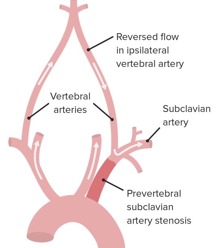
Pathophysiology of subclavian steal syndrome:
Note the reversed blood flow in the ipsilateral vertebral artery.

Image by Lecturio.

Clinical presentation

Most patients Patients Individuals participating in the health care system for the purpose of receiving therapeutic, diagnostic, or preventive procedures. Clinician–Patient Relationship with subclavian stenosis Stenosis Hypoplastic Left Heart Syndrome (HLHS)/steal are asymptomatic.

Symptoms of (ipsilateral) limb ischemia Ischemia A hypoperfusion of the blood through an organ or tissue caused by a pathologic constriction or obstruction of its blood vessels, or an absence of blood circulation. Ischemic Cell Damage/hypoperfusion:

  • Claudication ( pain Pain An unpleasant sensation induced by noxious stimuli which are detected by nerve endings of nociceptive neurons. Pain: Types and Pathways or discomfort in arm Arm The arm, or “upper arm” in common usage, is the region of the upper limb that extends from the shoulder to the elbow joint and connects inferiorly to the forearm through the cubital fossa. It is divided into 2 fascial compartments (anterior and posterior). Arm: Anatomy with activity that improves with rest)
  • Paleness
  • Cool skin Skin The skin, also referred to as the integumentary system, is the largest organ of the body. The skin is primarily composed of the epidermis (outer layer) and dermis (deep layer). The epidermis is primarily composed of keratinocytes that undergo rapid turnover, while the dermis contains dense layers of connective tissue. Skin: Structure and Functions
  • Paresthesias Paresthesias Subjective cutaneous sensations (e.g., cold, warmth, tingling, pressure, etc.) that are experienced spontaneously in the absence of stimulation. Posterior Cord Syndrome
  • Weakness
  • Weakened pulse in affected arm Arm The arm, or “upper arm” in common usage, is the region of the upper limb that extends from the shoulder to the elbow joint and connects inferiorly to the forearm through the cubital fossa. It is divided into 2 fascial compartments (anterior and posterior). Arm: Anatomy

Symptoms related to cerebral hypoperfusion/vertebrobasilar insufficiency:

  • Syncope Syncope Syncope is a short-term loss of consciousness and loss of postural stability followed by spontaneous return of consciousness to the previous neurologic baseline without the need for resuscitation. The condition is caused by transient interruption of cerebral blood flow that may be benign or related to a underlying life-threatening condition. Syncope/ presyncope Presyncope Syncope
  • Dizziness Dizziness An imprecise term which may refer to a sense of spatial disorientation, motion of the environment, or lightheadedness. Lateral Medullary Syndrome (Wallenberg Syndrome)
  • Vertigo Vertigo Vertigo is defined as the perceived sensation of rotational motion while remaining still. A very common complaint in primary care and the ER, vertigo is more frequently experienced by women and its prevalence increases with age. Vertigo is classified into peripheral or central based on its etiology. Vertigo
  • Visual disturbances (e.g., blurring, double vision Vision Ophthalmic Exam, hemianopia Hemianopia Transient Ischemic Attack (TIA), nystagmus Nystagmus Involuntary movements of the eye that are divided into two types, jerk and pendular. Jerk nystagmus has a slow phase in one direction followed by a corrective fast phase in the opposite direction, and is usually caused by central or peripheral vestibular dysfunction. Pendular nystagmus features oscillations that are of equal velocity in both directions and this condition is often associated with visual loss early in life. Albinism)
  • Transient hearing loss Hearing loss Hearing loss, also known as hearing impairment, is any degree of impairment in the ability to apprehend sound as determined by audiometry to be below normal hearing thresholds. Clinical presentation may occur at birth or as a gradual loss of hearing with age, including a short-term or sudden loss at any point. Hearing Loss
  • Tinnitus Tinnitus A nonspecific symptom of hearing disorder characterized by the sensation of buzzing, ringing, clicking, pulsations, and other noises in the ear. Objective tinnitus refers to noises generated from within the ear or adjacent structures that can be heard by other individuals. The term subjective tinnitus is used when the sound is audible only to the affected individual. Tinnitus may occur as a manifestation of cochlear diseases; vestibulocochlear nerve diseases; intracranial hypertension; craniocerebral trauma; and other conditions. Cranial Nerve Palsies
  • Disequilibrium
  • Ataxia Ataxia Impairment of the ability to perform smoothly coordinated voluntary movements. This condition may affect the limbs, trunk, eyes, pharynx, larynx, and other structures. Ataxia may result from impaired sensory or motor function. Sensory ataxia may result from posterior column injury or peripheral nerve diseases. Motor ataxia may be associated with cerebellar diseases; cerebral cortex diseases; thalamic diseases; basal ganglia diseases; injury to the red nucleus; and other conditions. Ataxia-telangiectasia

Diagnosis

History

  • History of atherosclerosis Atherosclerosis Atherosclerosis is a common form of arterial disease in which lipid deposition forms a plaque in the blood vessel walls. Atherosclerosis is an incurable disease, for which there are clearly defined risk factors that often can be reduced through a change in lifestyle and behavior of the patient. Atherosclerosis/peripheral vascular disease
  • Unilateral arm Arm The arm, or “upper arm” in common usage, is the region of the upper limb that extends from the shoulder to the elbow joint and connects inferiorly to the forearm through the cubital fossa. It is divided into 2 fascial compartments (anterior and posterior). Arm: Anatomy paresthesias Paresthesias Subjective cutaneous sensations (e.g., cold, warmth, tingling, pressure, etc.) that are experienced spontaneously in the absence of stimulation. Posterior Cord Syndrome/claudication
  • Syncopal episodes
  • Vertigo Vertigo Vertigo is defined as the perceived sensation of rotational motion while remaining still. A very common complaint in primary care and the ER, vertigo is more frequently experienced by women and its prevalence increases with age. Vertigo is classified into peripheral or central based on its etiology. Vertigo, transient visual/hearing disturbances

Physical exam

  • Blood pressure difference > 15 mm MM Multiple myeloma (MM) is a malignant condition of plasma cells (activated B lymphocytes) primarily seen in the elderly. Monoclonal proliferation of plasma cells results in cytokine-driven osteoclastic activity and excessive secretion of IgG antibodies. Multiple Myeloma Hg between arms indicates subclavian stenosis Stenosis Hypoplastic Left Heart Syndrome (HLHS).
  • Blood pressure difference > 40 mm MM Multiple myeloma (MM) is a malignant condition of plasma cells (activated B lymphocytes) primarily seen in the elderly. Monoclonal proliferation of plasma cells results in cytokine-driven osteoclastic activity and excessive secretion of IgG antibodies. Multiple Myeloma Hg typically seen in those who are symptomatic
  • Difference in radial pulses
  • Hand Hand The hand constitutes the distal part of the upper limb and provides the fine, precise movements needed in activities of daily living. It consists of 5 metacarpal bones and 14 phalanges, as well as numerous muscles innervated by the median and ulnar nerves. Hand: Anatomy skin Skin The skin, also referred to as the integumentary system, is the largest organ of the body. The skin is primarily composed of the epidermis (outer layer) and dermis (deep layer). The epidermis is primarily composed of keratinocytes that undergo rapid turnover, while the dermis contains dense layers of connective tissue. Skin: Structure and Functions and nail beds:
    • Look for blue discoloration, ulcerations, and splinter hemorrhages Splinter hemorrhages Endocarditis, which may indicate emboli from subclavian atherosclerotic lesions.

Imaging

  • Duplex ultrasound:
  • MRA MRA Imaging of the Heart and Great Vessels or CT angiography Angiography Radiography of blood vessels after injection of a contrast medium. Cardiac Surgery:
    • Used to confirm and grade the disease
    • Used for detailed anatomic evaluation of subclavian and vertebral arteries Arteries Arteries are tubular collections of cells that transport oxygenated blood and nutrients from the heart to the tissues of the body. The blood passes through the arteries in order of decreasing luminal diameter, starting in the largest artery (the aorta) and ending in the small arterioles. Arteries are classified into 3 types: large elastic arteries, medium muscular arteries, and small arteries and arterioles. Arteries: Histology
  • Transcranial doppler Doppler Ultrasonography applying the doppler effect, with frequency-shifted ultrasound reflections produced by moving targets (usually red blood cells) in the bloodstream along the ultrasound axis in direct proportion to the velocity of movement of the targets, to determine both direction and velocity of blood flow. Ultrasound (Sonography):
    • Can be performed if reversal of flow Flow Blood flows through the heart, arteries, capillaries, and veins in a closed, continuous circuit. Flow is the movement of volume per unit of time. Flow is affected by the pressure gradient and the resistance fluid encounters between 2 points. Vascular resistance is the opposition to flow, which is caused primarily by blood friction against vessel walls. Vascular Resistance, Flow, and Mean Arterial Pressure in vertebral artery Vertebral artery The first branch of the subclavian artery with distribution to muscles of the neck; vertebrae; spinal cord; cerebellum; and interior of the cerebrum. Lateral Medullary Syndrome (Wallenberg Syndrome) is observed
    • Evaluates for reversal of flow Flow Blood flows through the heart, arteries, capillaries, and veins in a closed, continuous circuit. Flow is the movement of volume per unit of time. Flow is affected by the pressure gradient and the resistance fluid encounters between 2 points. Vascular resistance is the opposition to flow, which is caused primarily by blood friction against vessel walls. Vascular Resistance, Flow, and Mean Arterial Pressure in the basilar artery, which is more predictive of symptoms than vertebral flow Flow Blood flows through the heart, arteries, capillaries, and veins in a closed, continuous circuit. Flow is the movement of volume per unit of time. Flow is affected by the pressure gradient and the resistance fluid encounters between 2 points. Vascular resistance is the opposition to flow, which is caused primarily by blood friction against vessel walls. Vascular Resistance, Flow, and Mean Arterial Pressure alone
  • Percutaneous angiography Angiography Radiography of blood vessels after injection of a contrast medium. Cardiac Surgery:
    • Invasive procedure
    • Can be diagnostic and therapeutic ( angioplasty Angioplasty Reconstruction or repair of a blood vessel, which includes the widening of a pathological narrowing of an artery or vein by the removal of atheromatous plaque material and/or the endothelial lining as well, or by dilatation (balloon angioplasty) to compress an atheroma. Except for endarterectomy, usually these procedures are performed via catheterization as minimally invasive endovascular procedures. Cardiac Surgery/stenting)
Critical ostial subclavian artery stenosis

Percutaneous transradial catheter angiogram showing critical ostial subclavian artery stenosis that caused coronary subclavian steal syndrome

Image: “Critical ostial subclavian artery stenosis” by Section of Cardiology, Christiana Care Health System, Newark, DE 19718, USA. License: CC BY 3.0

Grading Grading Methods which attempt to express in replicable terms the level of cell differentiation in neoplasms as increasing anaplasia correlates with the aggressiveness of the neoplasm. Grading, Staging, and Metastasis of subclavian steal syndrome

  • Grade I (pre-subclavian steal):
    •  Reduced antegrade vertebral flow Flow Blood flows through the heart, arteries, capillaries, and veins in a closed, continuous circuit. Flow is the movement of volume per unit of time. Flow is affected by the pressure gradient and the resistance fluid encounters between 2 points. Vascular resistance is the opposition to flow, which is caused primarily by blood friction against vessel walls. Vascular Resistance, Flow, and Mean Arterial Pressure
    • < 70% subclavian stenosis Stenosis Hypoplastic Left Heart Syndrome (HLHS)
  • Grade II (partial/latent): 
  • Grade III (permanent/advanced): 
    • Permanent retrograde vertebral flow Flow Blood flows through the heart, arteries, capillaries, and veins in a closed, continuous circuit. Flow is the movement of volume per unit of time. Flow is affected by the pressure gradient and the resistance fluid encounters between 2 points. Vascular resistance is the opposition to flow, which is caused primarily by blood friction against vessel walls. Vascular Resistance, Flow, and Mean Arterial Pressure
    • 95%–100% subclavian stenosis Stenosis Hypoplastic Left Heart Syndrome (HLHS)

Management

Prevention

  • Asymptomatic cases do not require invasive intervention.
  • However, subclavian stenosis Stenosis Hypoplastic Left Heart Syndrome (HLHS) is a marker for:
    • Atherosclerotic disease
    • Increased risk of cardiovascular events
  • Patients Patients Individuals participating in the health care system for the purpose of receiving therapeutic, diagnostic, or preventive procedures. Clinician–Patient Relationship may benefit from secondary prevention:
    • Control of hypertension Hypertension Hypertension, or high blood pressure, is a common disease that manifests as elevated systemic arterial pressures. Hypertension is most often asymptomatic and is found incidentally as part of a routine physical examination or during triage for an unrelated medical encounter. Hypertension, hyperlipidemia, and diabetes Diabetes Diabetes mellitus (DM) is a metabolic disease characterized by hyperglycemia and dysfunction of the regulation of glucose metabolism by insulin. Type 1 DM is diagnosed mostly in children and young adults as the result of autoimmune destruction of β cells in the pancreas and the resulting lack of insulin. Type 2 DM has a significant association with obesity and is characterized by insulin resistance. Diabetes Mellitus
    • Smoking Smoking Willful or deliberate act of inhaling and exhaling smoke from burning substances or agents held by hand. Interstitial Lung Diseases cessation
    • Lifestyle modifications ( weight loss Weight loss Decrease in existing body weight. Bariatric Surgery, exercise)
    • Antithrombotic therapy/ antiplatelet agents Antiplatelet agents Antiplatelet agents are medications that inhibit platelet aggregation, a critical step in the formation of the initial platelet plug. Abnormal, or inappropriate, platelet aggregation is a key step in the pathophysiology of arterial ischemic events. The primary categories of antiplatelet agents include aspirin, ADP inhibitors, phosphodiesterase/adenosine uptake inhibitors, and glycoprotein IIb/IIIa inhibitors. Antiplatelet Drugs

Invasive treatment

  • Indicated for symptomatic patients Patients Individuals participating in the health care system for the purpose of receiving therapeutic, diagnostic, or preventive procedures. Clinician–Patient Relationship
  • Endovascular treatment:
    • Angioplasty Angioplasty Reconstruction or repair of a blood vessel, which includes the widening of a pathological narrowing of an artery or vein by the removal of atheromatous plaque material and/or the endothelial lining as well, or by dilatation (balloon angioplasty) to compress an atheroma. Except for endarterectomy, usually these procedures are performed via catheterization as minimally invasive endovascular procedures. Cardiac Surgery +/- stenting
    • For short proximal stenosis Stenosis Hypoplastic Left Heart Syndrome (HLHS)
  • Surgical revascularization Revascularization Thromboangiitis Obliterans (Buerger’s Disease):
    • Indicated for evidence of distal embolization Embolization A method of hemostasis utilizing various agents such as gelfoam, silastic, metal, glass, or plastic pellets, autologous clot, fat, and muscle as emboli. It has been used in the treatment of spinal cord and intracranial arteriovenous malformations, renal arteriovenous fistulas, gastrointestinal bleeding, epistaxis, hypersplenism, certain highly vascular tumors, traumatic rupture of blood vessels, and control of operative hemorrhage. Gastrointestinal Bleeding from subclavian lesion
    • For anatomy unfavorable for endovascular intervention
    • Options include:

Differential Diagnosis

  • Vertebrobasilar stroke: a less common type of cerebrovascular accident Cerebrovascular accident An ischemic stroke (also known as cerebrovascular accident) is an acute neurologic injury that occurs as a result of brain ischemia; this condition may be due to cerebral blood vessel occlusion by thrombosis or embolism, or rarely due to systemic hypoperfusion. Ischemic Stroke with a high mortality Mortality All deaths reported in a given population. Measures of Health Status rate caused by occlusion, embolism, or dissection affecting the extracranial and intracranial vertebral arteries Arteries Arteries are tubular collections of cells that transport oxygenated blood and nutrients from the heart to the tissues of the body. The blood passes through the arteries in order of decreasing luminal diameter, starting in the largest artery (the aorta) and ending in the small arterioles. Arteries are classified into 3 types: large elastic arteries, medium muscular arteries, and small arteries and arterioles. Arteries: Histology, basilar artery, or posterior cerebral arteries Arteries Arteries are tubular collections of cells that transport oxygenated blood and nutrients from the heart to the tissues of the body. The blood passes through the arteries in order of decreasing luminal diameter, starting in the largest artery (the aorta) and ending in the small arterioles. Arteries are classified into 3 types: large elastic arteries, medium muscular arteries, and small arteries and arterioles. Arteries: Histology. Presentation varies depending on the site of infarction. Magnetic resonance and CT angiography Angiography Radiography of blood vessels after injection of a contrast medium. Cardiac Surgery are used to confirm the diagnosis. There are few effective treatments for this type of stroke but angioplasty Angioplasty Reconstruction or repair of a blood vessel, which includes the widening of a pathological narrowing of an artery or vein by the removal of atheromatous plaque material and/or the endothelial lining as well, or by dilatation (balloon angioplasty) to compress an atheroma. Except for endarterectomy, usually these procedures are performed via catheterization as minimally invasive endovascular procedures. Cardiac Surgery or open surgical repair may be attempted depending on the lesion site. 
  • Upper extremity deep vein thrombosis Upper extremity deep vein thrombosis Deep vein thrombosis of an upper extremity vein (e.g., axillary vein; subclavian vein; and jugular veins). It is associated with mechanical factors secondary to other anatomic factors. Symptoms may include sudden onset of pain, warmth, redness, blueness, and swelling in the arm. Deep Vein Thrombosis ( DVT DVT Deep vein thrombosis (DVT) usually occurs in the deep veins of the lower extremities. The affected veins include the femoral, popliteal, iliofemoral, and pelvic veins. Proximal DVT is more likely to cause a pulmonary embolism (PE) and is generally considered more serious. Deep Vein Thrombosis): a rare condition primarily triggered by vigorous upper extremity activity, typically in a young, healthy individual. The axillary and subclavian veins Veins Veins are tubular collections of cells, which transport deoxygenated blood and waste from the capillary beds back to the heart. Veins are classified into 3 types: small veins/venules, medium veins, and large veins. Each type contains 3 primary layers: tunica intima, tunica media, and tunica adventitia. Veins: Histology are most often affected. Secondary causes include central venous cannulation and prothrombotic states. Symptoms include sudden onset of pain Pain An unpleasant sensation induced by noxious stimuli which are detected by nerve endings of nociceptive neurons. Pain: Types and Pathways, swelling Swelling Inflammation, and cyanosis Cyanosis A bluish or purplish discoloration of the skin and mucous membranes due to an increase in the amount of deoxygenated hemoglobin in the blood or a structural defect in the hemoglobin molecule. Pulmonary Examination in the extremity. Diagnosis is made via ultrasound imaging. Treatment includes anticoagulation Anticoagulation Pulmonary Hypertension Drugs, thrombolysis, and surgical interventions when needed. 
  • Takayasu arteritis: a large-vessel vasculitis Vasculitis Inflammation of any one of the blood vessels, including the arteries; veins; and rest of the vasculature system in the body. Systemic Lupus Erythematosus that most commonly affects women. This condition is characterized by granulomatous inflammation Granulomatous Inflammation Chalazion of the aortic wall and its branches. Symptoms include fatigue Fatigue The state of weariness following a period of exertion, mental or physical, characterized by a decreased capacity for work and reduced efficiency to respond to stimuli. Fibromyalgia, weight loss Weight loss Decrease in existing body weight. Bariatric Surgery, fevers, chest pain Pain An unpleasant sensation induced by noxious stimuli which are detected by nerve endings of nociceptive neurons. Pain: Types and Pathways, and high blood pressure. Blood tests, ultrasound, and angiography Angiography Radiography of blood vessels after injection of a contrast medium. Cardiac Surgery are used to confirm the diagnosis. Management involves corticosteroids Corticosteroids Chorioretinitis, immunosuppressants Immunosuppressants Immunosuppressants are a class of drugs widely used in the management of autoimmune conditions and organ transplant rejection. The general effect is dampening of the immune response. Immunosuppressants, biologic agents Biologic Agents Immunosuppressants, and surgical interventions when needed. 
  • Thoracic outlet Thoracic Outlet Thoracic Outlet Syndrome syndrome ( TOS TOS Thoracic outlet syndrome (TOS) is a broad term used for a spectrum of syndromes related to the general region of the thoracic outlet, which involves the compression or irritation of elements of the brachial plexus, subclavian artery, or subclavian vein. Thoracic Outlet Syndrome): compression Compression Blunt Chest Trauma of nerves or blood vessels in an area between the lower neck Neck The part of a human or animal body connecting the head to the rest of the body. Peritonsillar Abscess and upper chest called the thoracic outlet Thoracic Outlet Thoracic Outlet Syndrome. The syndrome may be caused by anatomical defects such as a cervical 1st rib, trauma, poor posture, bodybuilding, or repetitive motions involving the arm Arm The arm, or “upper arm” in common usage, is the region of the upper limb that extends from the shoulder to the elbow joint and connects inferiorly to the forearm through the cubital fossa. It is divided into 2 fascial compartments (anterior and posterior). Arm: Anatomy and shoulder. Provocative maneuvers on exam and imaging studies help confirm the diagnosis. Management of vascular TOS TOS Thoracic outlet syndrome (TOS) is a broad term used for a spectrum of syndromes related to the general region of the thoracic outlet, which involves the compression or irritation of elements of the brachial plexus, subclavian artery, or subclavian vein. Thoracic Outlet Syndrome involves thrombolytics Thrombolytics Thrombolytics, also known as fibrinolytics, include recombinant tissue plasminogen activator (TPa) (i.e., alteplase, reteplase, and tenecteplase), urokinase, and streptokinase. The agents promote the breakdown of a blood clot by converting plasminogen to plasmin, which then degrades fibrin. Thrombolytics, anticoagulants Anticoagulants Anticoagulants are drugs that retard or interrupt the coagulation cascade. The primary classes of available anticoagulants include heparins, vitamin K-dependent antagonists (e.g., warfarin), direct thrombin inhibitors, and factor Xa inhibitors. Anticoagulants, and surgical interventions.

References

  1. Barshes N.R. (2019). Overview of upper extremity peripheral artery disease. UpToDate. Retrieved February 15, 2021, from https://www.uptodate.com/contents/overview-of-upper-extremity-peripheral-artery-disease
  2. Bayat I. (2019). Subclavian Steal Syndrome. Retrieved February 19, 2021, from https://emedicine.medscape.com/article/462036-overview
  3. Labropoulos N., Nandivada P., Bekelis K. (2010). Prevalence and impact of the subclavian steal syndrome. Ann Surg. 2010 Jul. 252 (1):166-70. 
  4. Potter B.J., Pinto D.S. (2014). Subclavian steal syndrome. Circulation. 2014 Jun 3. 129 (22):2320-3.
  5. Spittell P.C. (2019). Subclavian steal syndrome. UpToDate. Retrieved February 15, 2021, from https://www.uptodate.com/contents/subclavian-steal-syndrome

Create your free account or log in to continue reading!

Sign up now and get free access to Lecturio with concept pages, medical videos, and questions for your medical education.

User Reviews

Details