Cardiac surgery is the surgical management of cardiac abnormalities and of the great vessels of the thorax. In general terms, surgical intervention of the heart is performed to directly restore adequate pumpPumpACES and RUSH: Resuscitation Ultrasound Protocols function, correct inherent structural issues, and reestablish proper blood supply via the coronary circulationCirculationThe movement of the blood as it is pumped through the cardiovascular system.ABCDE Assessment. Common interventions treat ischemic and valvular heart disease as well as disorders of the great vessels.
The operator needs to be familiar with the anatomy of the heartAnatomy of the heartThe heart is a 4-chambered muscular pump made primarily of cardiac muscle tissue. The heart is divided into 4 chambers: 2 upper chambers for receiving blood from the great vessels, known as the right and left atria, and 2 stronger lower chambers, known as the right and left ventricles, which pump blood throughout the body. Heart: Anatomy and surrounding structures, as well as vascular territories, to avoid damaging vessels and nerves and to achieve successful reperfusion of the tissues.
General anatomy of the heartAnatomy of the heartThe heart is a 4-chambered muscular pump made primarily of cardiac muscle tissue. The heart is divided into 4 chambers: 2 upper chambers for receiving blood from the great vessels, known as the right and left atria, and 2 stronger lower chambers, known as the right and left ventricles, which pump blood throughout the body. Heart: Anatomy
Enclosed within the pericardiumPericardiumA conical fibroserous sac surrounding the heart and the roots of the great vessels (aorta; venae cavae; pulmonary artery). Pericardium consists of two sacs: the outer fibrous pericardium and the inner serous pericardium. The latter consists of an outer parietal layer facing the fibrous pericardium, and an inner visceral layer (epicardium) resting next to the heart, and a pericardial cavity between these two layers.Heart: Anatomy
Relations:
Anterior: sternumSternumA long, narrow, and flat bone commonly known as breastbone occurring in the midsection of the anterior thoracic segment or chest region, which stabilizes the rib cage and serves as the point of origin for several muscles that move the arms, head, and neck.Chest Wall: Anatomy and rib cartilageCartilageCartilage is a type of connective tissue derived from embryonic mesenchyme that is responsible for structural support, resilience, and the smoothness of physical actions. Perichondrium (connective tissue membrane surrounding cartilage) compensates for the absence of vasculature in cartilage by providing nutrition and support. Cartilage: Histology
Posterior: vertebral columnVertebral columnThe human spine, or vertebral column, is the most important anatomical and functional axis of the human body. It consists of 7 cervical vertebrae, 12 thoracic vertebrae, and 5 lumbar vertebrae and is limited cranially by the skull and caudally by the sacrum. Vertebral Column: Anatomy (T5–T8), esophagusEsophagusThe esophagus is a muscular tube-shaped organ of around 25 centimeters in length that connects the pharynx to the stomach. The organ extends from approximately the 6th cervical vertebra to the 11th thoracic vertebra and can be divided grossly into 3 parts: the cervical part, the thoracic part, and the abdominal part. Esophagus: Anatomy, primary bronchiBronchiThe larger air passages of the lungs arising from the terminal bifurcation of the trachea. They include the largest two primary bronchi which branch out into secondary bronchi, and tertiary bronchi which extend into bronchioles and pulmonary alveoli.Bronchial Tree: Anatomy, great vessels
Lateral: lungsLungsLungs are the main organs of the respiratory system. Lungs are paired viscera located in the thoracic cavity and are composed of spongy tissue. The primary function of the lungs is to oxygenate blood and eliminate CO2. Lungs: Anatomy and pleuraPleuraThe pleura is a serous membrane that lines the walls of the thoracic cavity and the surface of the lungs. This structure of mesodermal origin covers both lungs, the mediastinum, the thoracic surface of the diaphragm, and the inner part of the thoracic cage. The pleura is divided into a visceral pleura and parietal pleura. Pleura: Anatomy
Inferior: diaphragmDiaphragmThe diaphragm is a large, dome-shaped muscle that separates the thoracic cavity from the abdominal cavity. The diaphragm consists of muscle fibers and a large central tendon, which is divided into right and left parts. As the primary muscle of inspiration, the diaphragm contributes 75% of the total inspiratory muscle force.Diaphragm: Anatomy
Cone-shaped, with ⅔ of its massMassThree-dimensional lesion that occupies a space within the breastImaging of the Breast to the left of the midline
Coronary sinusCoronary SinusA short vein that collects about two thirds of the venous blood from the myocardium and drains into the right atrium. Coronary sinus, normally located between the left atrium and left ventricle on the posterior surface of the heart, can serve as an anatomical reference for cardiac procedures.Atrial Septal Defect (ASD)
Right atrial appendage (auricle): a muscular sac that increases atrial capacity
Left atrium: pulmonary veinsPulmonary veinsThe veins that return the oxygenated blood from the lungs to the left atrium of the heart.Lungs: Anatomy → left atrium → left ventricle
Receives oxygenated blood from the pulmonary veinsPulmonary veinsThe veins that return the oxygenated blood from the lungs to the left atrium of the heart.Lungs: Anatomy
Left atrial appendage (auricle): A muscular sac that increases atrial capacity, a common location for thrombi to lodge
Right ventricle: receives deoxygenated blood from right atrium through the tricuspid valveTricuspid valveThe valve consisting of three cusps situated between the right atrium and right ventricle of the heart.Heart: Anatomy, then transports it through the pulmonic valve and into the pulmonary arteryPulmonary arteryThe short wide vessel arising from the conus arteriosus of the right ventricle and conveying unaerated blood to the lungs.Lungs: Anatomy to the pulmonary circulationCirculationThe movement of the blood as it is pumped through the cardiovascular system.ABCDE Assessment for oxygenation
Left ventricle: receives oxygenated blood from the left atrium through the mitral valveMitral valveThe valve between the left atrium and left ventricle of the heart.Heart: Anatomy, then pumps it through the aortic valveAortic valveThe valve between the left ventricle and the ascending aorta which prevents backflow into the left ventricle.Heart: Anatomy and into the aortaAortaThe main trunk of the systemic arteries.Mediastinum and Great Vessels: Anatomy and the systemic circulationCirculationThe movement of the blood as it is pumped through the cardiovascular system.ABCDE Assessment
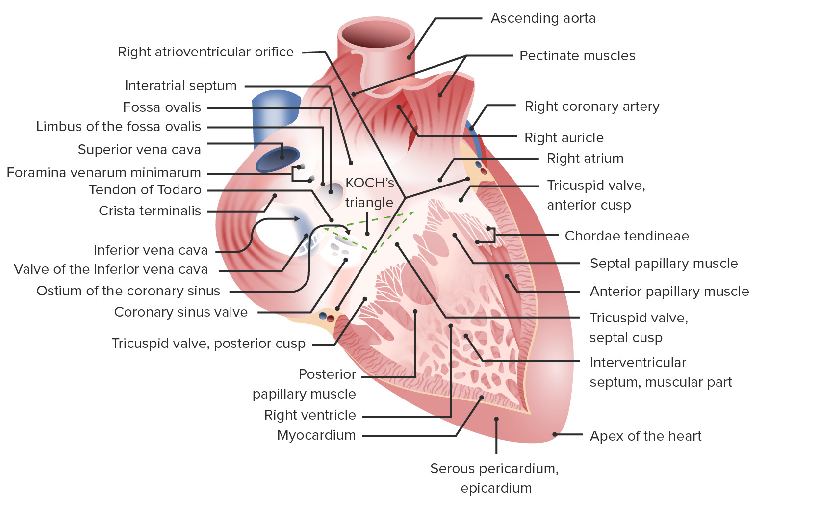
Right atrium and ventricle
Image by Lecturio.
Left atrium and ventricle
Image by Lecturio.
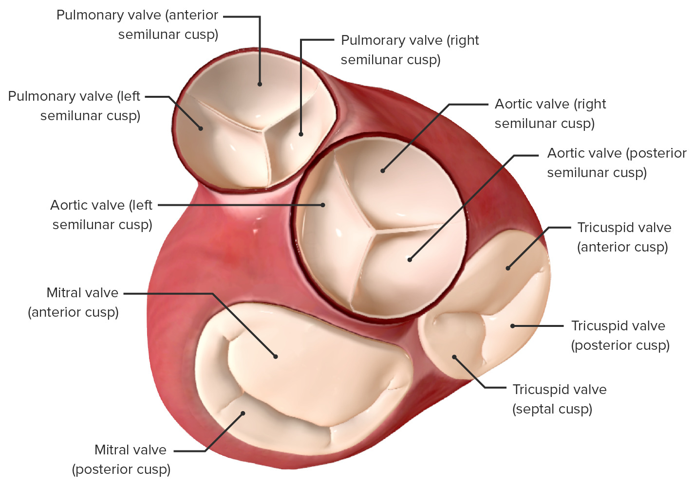
Valves
Prevent retrograde flowRetrograde flowVeins: Histology, their closure produces heart soundsHeart soundsHeart sounds are brief, transient sounds produced by valve opening and closure and by movement of blood in the heart. They are divided into systolic and diastolic sounds. In most cases, only the first (S1) and second (S2) heart sounds are heard. These are high-frequency sounds and arise from aortic and pulmonary valve closure (S1), as well as mitral and tricuspid valve closure (S2).Heart Sounds that can be heard on auscultation:
Mitral valveMitral valveThe valve between the left atrium and left ventricle of the heart.Heart: Anatomy:
2 cusps (bicuspid)
Between left atrium and left ventricle
Tricuspid valveTricuspid valveThe valve consisting of three cusps situated between the right atrium and right ventricle of the heart.Heart: Anatomy:
3 cusps (tricuspid)
Between right atrium and right ventricle
Pulmonary valvePulmonary valveA valve situated at the entrance to the pulmonary trunk from the right ventricle.Heart: Anatomy:
Subvalvular apparatus found under atrioventricular (AV) valves, consisting of chordae tendineaeChordae tendineaeThe tendinous cords that connect each cusp of the two atrioventricular heart valves to appropriate papillary muscles in the heart ventricles, preventing the valves from reversing themselves when the ventricles contract.Heart: Anatomy and papillary musclesPapillary musclesConical muscular projections from the walls of the cardiac ventricles, attached to the cusps of the atrioventricular valves by the chordae tendineae.Heart: Anatomy, act to prevent cusp prolapse into the atria
View of the valves of the heart from an atrial perspective: Atria removed
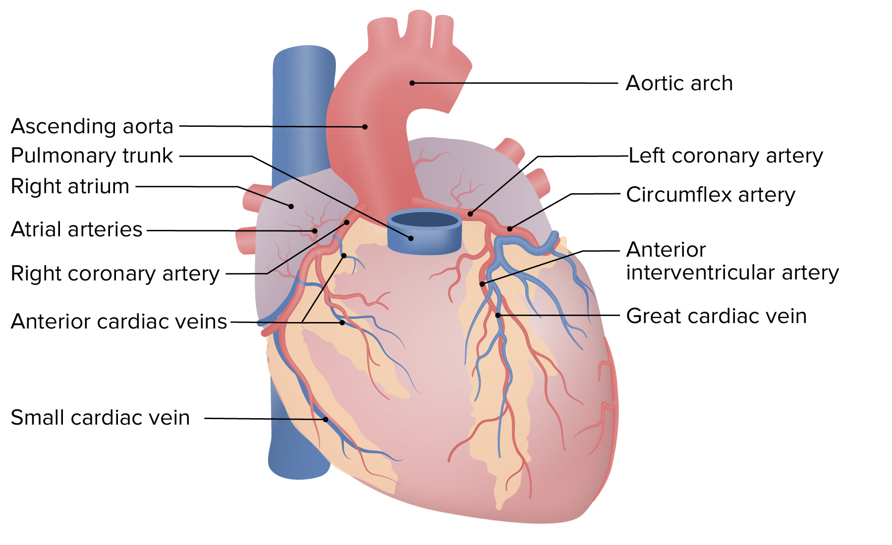
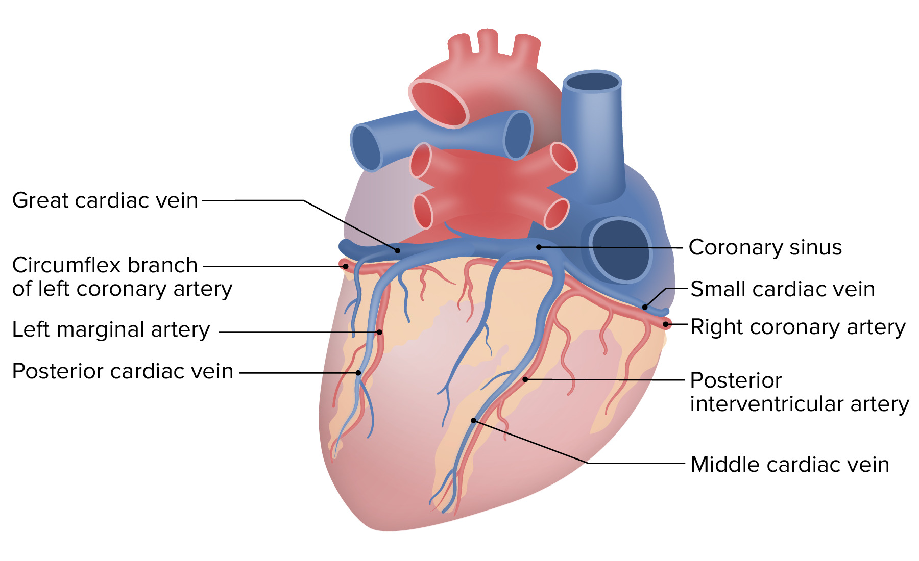
Coronary circulationCirculationThe movement of the blood as it is pumped through the cardiovascular system.ABCDE Assessment
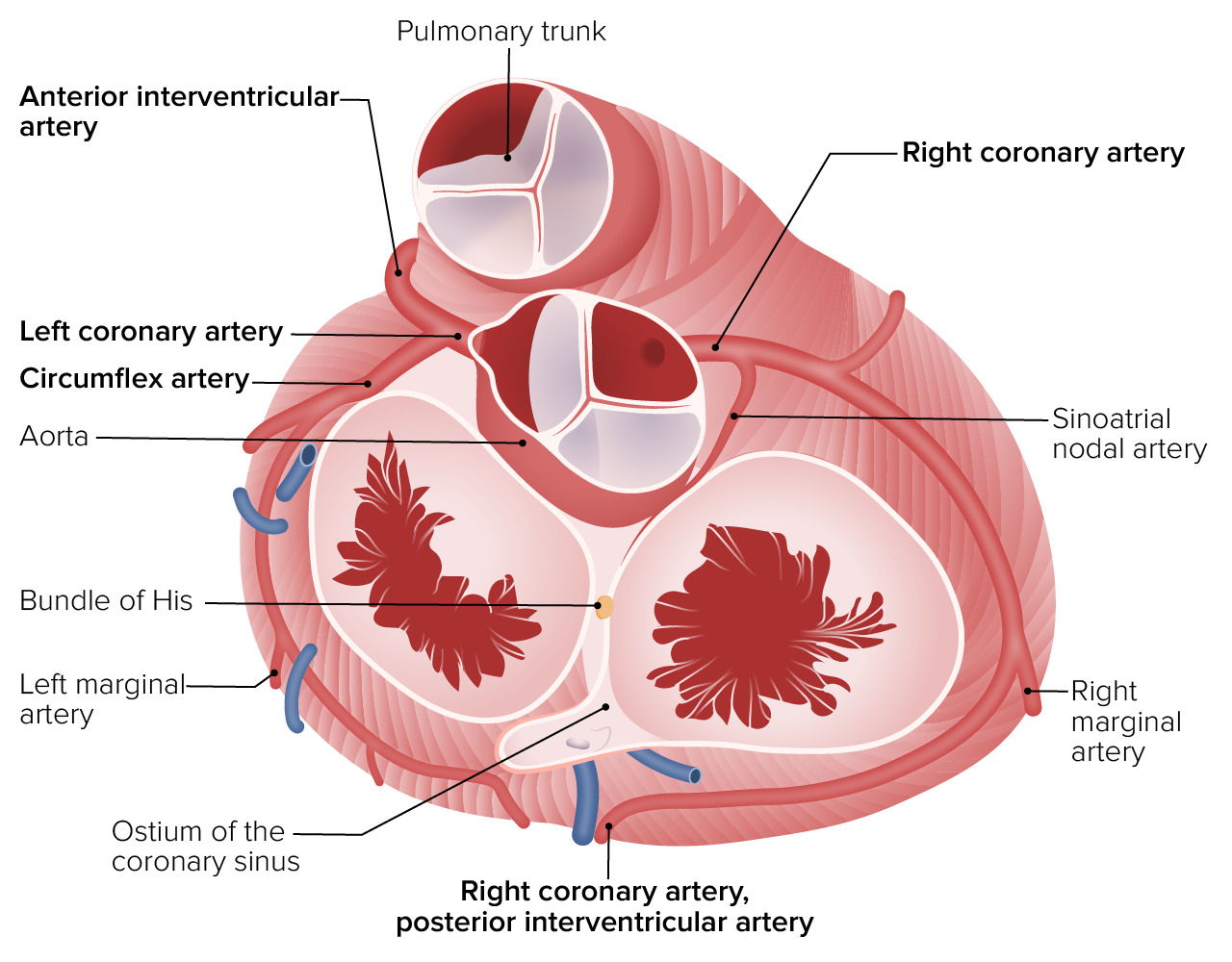
Assessment of the coronary arteriesArteriesArteries are tubular collections of cells that transport oxygenated blood and nutrients from the heart to the tissues of the body. The blood passes through the arteries in order of decreasing luminal diameter, starting in the largest artery (the aorta) and ending in the small arterioles. Arteries are classified into 3 types: large elastic arteries, medium muscular arteries, and small arteries and arterioles. Arteries: Histology starts with a review of their origin on an axialAxialComputed Tomography (CT) cut.
Roots (located just distal to the aortic valveAortic valveThe valve between the left ventricle and the ascending aorta which prevents backflow into the left ventricle.Heart: Anatomy):
ArteriesArteriesArteries are tubular collections of cells that transport oxygenated blood and nutrients from the heart to the tissues of the body. The blood passes through the arteries in order of decreasing luminal diameter, starting in the largest artery (the aorta) and ending in the small arterioles. Arteries are classified into 3 types: large elastic arteries, medium muscular arteries, and small arteries and arterioles. Arteries: Histology:
RCA:
Supplies blood to the right atrium, portion of both ventricles, and the cardiac conduction system
Gives rise to the posterior descending artery (PDAPDAThe ductus arteriosus (DA) allows blood to bypass pulmonary circulation. After birth, the DA remains open for up to 72 hours and then constricts and involutes, becoming the ligamentum arteriosum. Failure of this process to occur results in patent ductus arteriosus (PDA), a condition that causes up to 10% of congenital heart defects. Patent Ductus Arteriosus (PDA); most commonly, 70% of cases)
Gives rise to the left anterior descending artery and the circumflex artery
Patterns of circulationCirculationThe movement of the blood as it is pumped through the cardiovascular system.ABCDE Assessment:
Right dominant:
PDAPDAThe ductus arteriosus (DA) allows blood to bypass pulmonary circulation. After birth, the DA remains open for up to 72 hours and then constricts and involutes, becoming the ligamentum arteriosum. Failure of this process to occur results in patent ductus arteriosus (PDA), a condition that causes up to 10% of congenital heart defects. Patent Ductus Arteriosus (PDA) originates from the RCA
Majority (70%–80%) of the population
Left dominant:
PDAPDAThe ductus arteriosus (DA) allows blood to bypass pulmonary circulation. After birth, the DA remains open for up to 72 hours and then constricts and involutes, becoming the ligamentum arteriosum. Failure of this process to occur results in patent ductus arteriosus (PDA), a condition that causes up to 10% of congenital heart defects. Patent Ductus Arteriosus (PDA) originates from the LCA
Minority (10%) of the population
Codominant (also known as balanced):
PDAPDAThe ductus arteriosus (DA) allows blood to bypass pulmonary circulation. After birth, the DA remains open for up to 72 hours and then constricts and involutes, becoming the ligamentum arteriosum. Failure of this process to occur results in patent ductus arteriosus (PDA), a condition that causes up to 10% of congenital heart defects. Patent Ductus Arteriosus (PDA) made up from contributions of the LCA and RCA.
Minority (20%) of the population
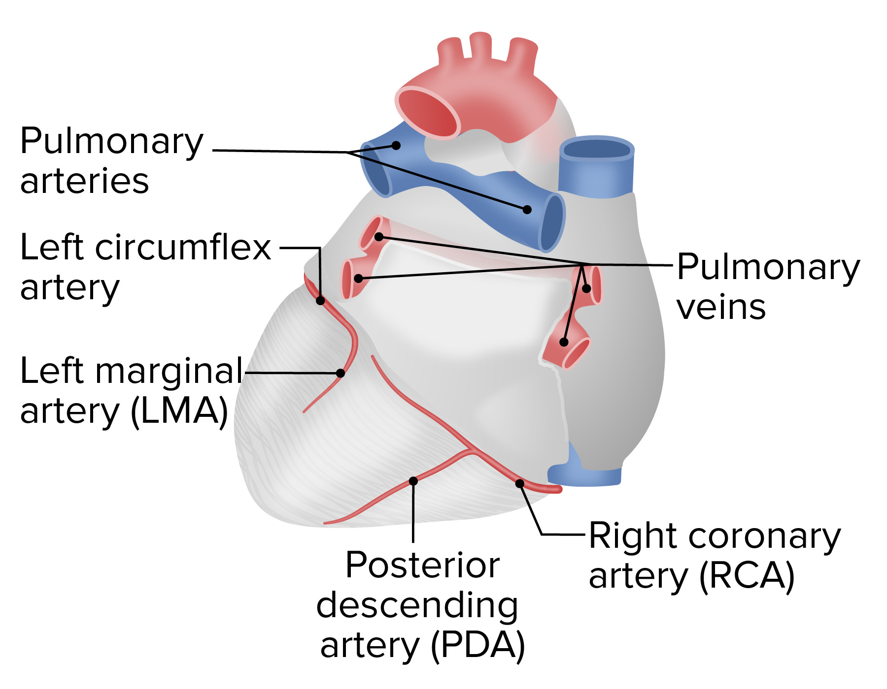
Coronary circulation, anterior view
Image by Lecturio.
Coronary circulation, posterior view
Image by Lecturio.
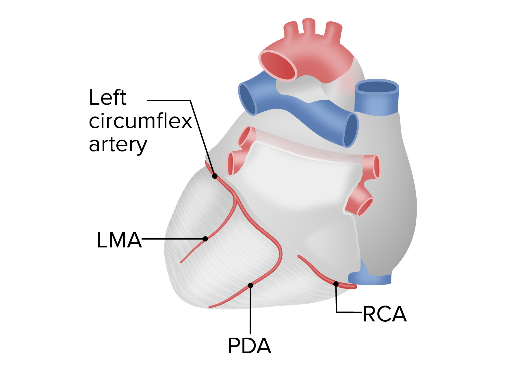
Right-dominant pattern of coronary circulation
Image by Lecturio.
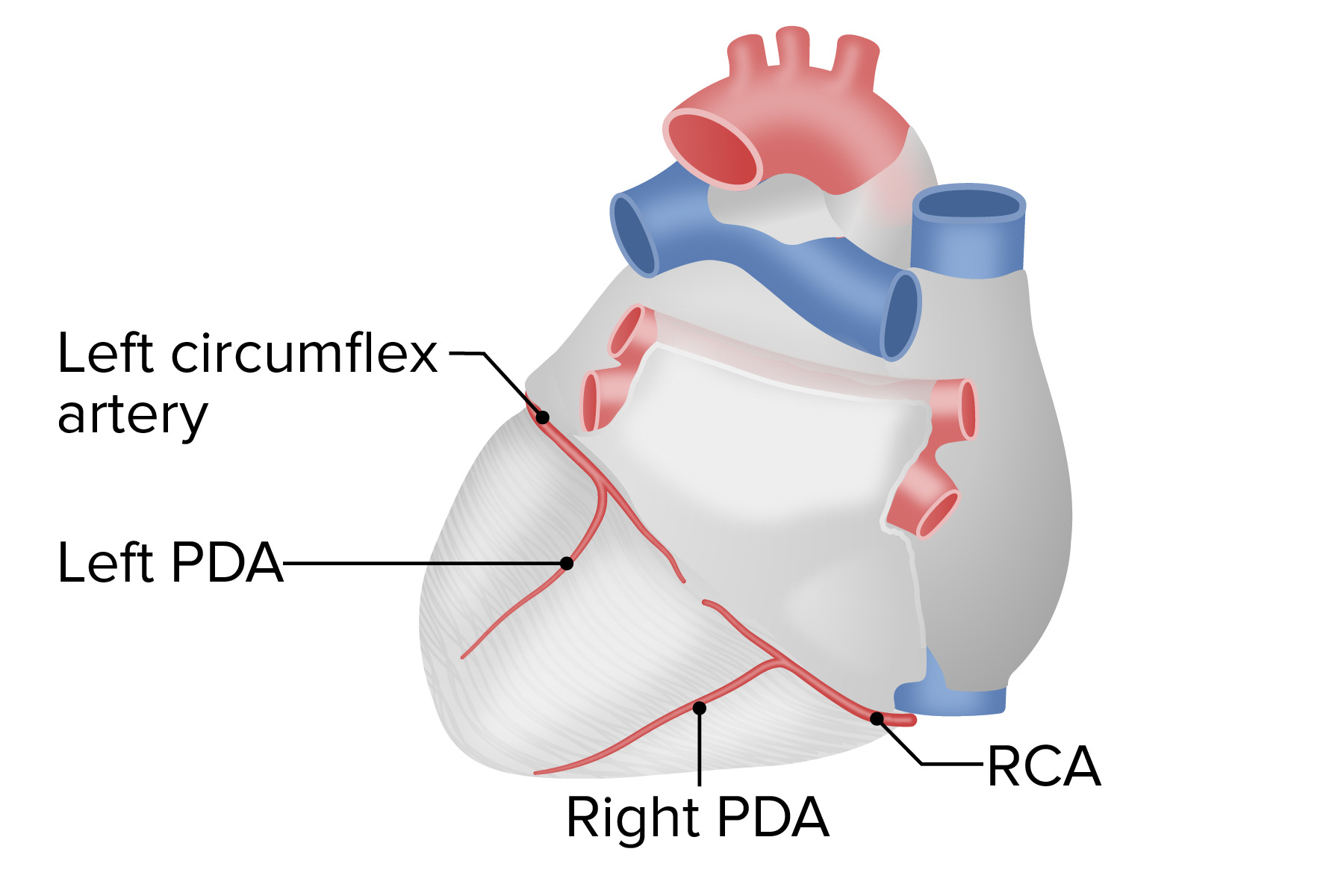
Left-dominant pattern of coronary circulation
Image by Lecturio.
Codominant pattern of coronary circulation
Image by Lecturio.
Ramification of the left and right coronary arteries from the ascending aorta
Image by Lecturio.
PericardiumPericardiumA conical fibroserous sac surrounding the heart and the roots of the great vessels (aorta; venae cavae; pulmonary artery). Pericardium consists of two sacs: the outer fibrous pericardium and the inner serous pericardium. The latter consists of an outer parietal layer facing the fibrous pericardium, and an inner visceral layer (epicardium) resting next to the heart, and a pericardial cavity between these two layers.Heart: Anatomy
FibrousFibrousFibrocystic ChangepericardiumPericardiumA conical fibroserous sac surrounding the heart and the roots of the great vessels (aorta; venae cavae; pulmonary artery). Pericardium consists of two sacs: the outer fibrous pericardium and the inner serous pericardium. The latter consists of an outer parietal layer facing the fibrous pericardium, and an inner visceral layer (epicardium) resting next to the heart, and a pericardial cavity between these two layers.Heart: Anatomy:
Tough outer layer composed of connective tissueConnective tissueConnective tissues originate from embryonic mesenchyme and are present throughout the body except inside the brain and spinal cord. The main function of connective tissues is to provide structural support to organs. Connective tissues consist of cells and an extracellular matrix.Connective Tissue: Histology
Continuous with the adventitia of the neighboring great vessels
Serous pericardiumPericardiumA conical fibroserous sac surrounding the heart and the roots of the great vessels (aorta; venae cavae; pulmonary artery). Pericardium consists of two sacs: the outer fibrous pericardium and the inner serous pericardium. The latter consists of an outer parietal layer facing the fibrous pericardium, and an inner visceral layer (epicardium) resting next to the heart, and a pericardial cavity between these two layers.Heart: Anatomy:
Inner serous membrane
2 layers:
ParietalParietalOne of a pair of irregularly shaped quadrilateral bones situated between the frontal bone and occipital bone, which together form the sides of the cranium.Skull: Anatomy: inner lining of the outer layer, continuous with and inseparable from the fibrousFibrousFibrocystic ChangepericardiumPericardiumA conical fibroserous sac surrounding the heart and the roots of the great vessels (aorta; venae cavae; pulmonary artery). Pericardium consists of two sacs: the outer fibrous pericardium and the inner serous pericardium. The latter consists of an outer parietal layer facing the fibrous pericardium, and an inner visceral layer (epicardium) resting next to the heart, and a pericardial cavity between these two layers.Heart: Anatomy
Visceral (also known as epicardiumEpicardiumHeart: Anatomy): inner layer covering the myocardial surface
Potential space between the parietalParietalOne of a pair of irregularly shaped quadrilateral bones situated between the frontal bone and occipital bone, which together form the sides of the cranium.Skull: Anatomy and visceral layer of the pericardiumPericardiumA conical fibroserous sac surrounding the heart and the roots of the great vessels (aorta; venae cavae; pulmonary artery). Pericardium consists of two sacs: the outer fibrous pericardium and the inner serous pericardium. The latter consists of an outer parietal layer facing the fibrous pericardium, and an inner visceral layer (epicardium) resting next to the heart, and a pericardial cavity between these two layers.Heart: Anatomy
Contains serous fluid that lubricates the movements of the heart within the pericardiumPericardiumA conical fibroserous sac surrounding the heart and the roots of the great vessels (aorta; venae cavae; pulmonary artery). Pericardium consists of two sacs: the outer fibrous pericardium and the inner serous pericardium. The latter consists of an outer parietal layer facing the fibrous pericardium, and an inner visceral layer (epicardium) resting next to the heart, and a pericardial cavity between these two layers.Heart: Anatomy and allows some degree of cushion to the heart
Diagnostic pericardiocentesis (obtaining pericardial fluidPericardial fluidWatery fluid produced in the serous and visceral pericardium surrounding the surface of the heart.Heart: Anatomy for diagnosis):
Suspicion of bacterial, mycobacterial, or fungal effusion
Suspicion of malignant effusion
Therapeutic pericardiocentesis:
Cardiac tamponadeTamponadePericardial effusion, usually of rapid onset, exceeding ventricular filling pressures and causing collapse of the heart with a markedly reduced cardiac output.Pericarditis: restriction in cardiac filling due to excessive accumulation of fluid within pericardial cavityPericardial cavityHeart: Anatomy; leads to decreased cardiac outputCardiac outputThe volume of blood passing through the heart per unit of time. It is usually expressed as liters (volume) per minute so as not to be confused with stroke volume (volume per beat).Cardiac Mechanics and potentially hemodynamic instability (hypotensionHypotensionHypotension is defined as low blood pressure, specifically < 90/60 mm Hg, and is most commonly a physiologic response. Hypotension may be mild, serious, or life threatening, depending on the cause. Hypotension and shockShockShock is a life-threatening condition associated with impaired circulation that results in tissue hypoxia. The different types of shock are based on the underlying cause: distributive (↑ cardiac output (CO), ↓ systemic vascular resistance (SVR)), cardiogenic (↓ CO, ↑ SVR), hypovolemic (↓ CO, ↑ SVR), obstructive (↓ CO), and mixed. Types of Shock)
Large pericardial effusionswith no known etiology
ContraindicationsContraindicationsA condition or factor associated with a recipient that makes the use of a drug, procedure, or physical agent improper or inadvisable. Contraindications may be absolute (life threatening) or relative (higher risk of complications in which benefits may outweigh risks).Noninvasive Ventilation
Aortic dissectionAortic dissectionAortic dissection occurs due to shearing stress from pulsatile pressure causing a tear in the tunica intima of the aortic wall. This tear allows blood to flow into the media, creating a “false lumen.” Aortic dissection is most commonly caused by uncontrolled hypertension.Aortic Dissection
Small or loculated pericardial effusionPericardial effusionFluid accumulation within the pericardium. Serous effusions are associated with pericardial diseases. Hemopericardium is associated with trauma. Lipid-containing effusion (chylopericardium) results from leakage of thoracic duct. Severe cases can lead to cardiac tamponade.Pericardial Effusion and Cardiac Tamponade
Nonviral infectious pericarditisPericarditisPericarditis is an inflammation of the pericardium, often with fluid accumulation. It can be caused by infection (often viral), myocardial infarction, drugs, malignancies, metabolic disorders, autoimmune disorders, or trauma. Acute, subacute, and chronic forms exist. Pericarditis
Procedure
Unless the individual is in cardiac arrestCardiac arrestCardiac arrest is the sudden, complete cessation of cardiac output with hemodynamic collapse. Patients present as pulseless, unresponsive, and apneic. Rhythms associated with cardiac arrest are ventricular fibrillation/tachycardia, asystole, or pulseless electrical activity. Cardiac Arrest or has an imminently life-threatening condition, a pericardiocentesis should be performed under ultrasound guidance.
EchocardiographyEchocardiographyUltrasonic recording of the size, motion, and composition of the heart and surrounding tissues. The standard approach is transthoracic.Tricuspid Valve Atresia (TVA) is performed (in emergency situations, this step is skipped).
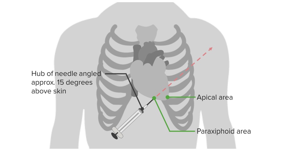
The left xiphochondral junction is located and marked with a pen.
The skinSkinThe skin, also referred to as the integumentary system, is the largest organ of the body. The skin is primarily composed of the epidermis (outer layer) and dermis (deep layer). The epidermis is primarily composed of keratinocytes that undergo rapid turnover, while the dermis contains dense layers of connective tissue.Skin: Structure and Functions is washed and draped, leaving a window for the left xiphochondral junction.
1 mL of lidocaineLidocaineA local anesthetic and cardiac depressant used as an antiarrhythmic agent. Its actions are more intense and its effects more prolonged than those of procaine but its duration of action is shorter than that of bupivacaine or prilocaine.Local Anesthetics is infiltrated into the puncture site, creating a skinSkinThe skin, also referred to as the integumentary system, is the largest organ of the body. The skin is primarily composed of the epidermis (outer layer) and dermis (deep layer). The epidermis is primarily composed of keratinocytes that undergo rapid turnover, while the dermis contains dense layers of connective tissue.Skin: Structure and FunctionswhealWhealUrticaria (Hives), and into the deeper tissues.
The skinSkinThe skin, also referred to as the integumentary system, is the largest organ of the body. The skin is primarily composed of the epidermis (outer layer) and dermis (deep layer). The epidermis is primarily composed of keratinocytes that undergo rapid turnover, while the dermis contains dense layers of connective tissue.Skin: Structure and Functions is punctured with a scalpel 1–2 cm inferior to the left xiphochondral junction.
The needle is introduced and advanced at a 45-degree angle through the small incision toward the left midscapula while maintaining negative pressure (pulling back on the plunger) on the syringe.
The needle is advanced until it reaches the pericardial space, confirmed by:
Aspiration of pericardial fluidPericardial fluidWatery fluid produced in the serous and visceral pericardium surrounding the surface of the heart.Heart: Anatomy or blood
Sensation of entrance to the cavity
Sensation of cardiac pulsations
Elevation of ST segmentST segmentIsoelectric segment between the s wave and the initial deflection of the t wave.Electrocardiogram (ECG) on ECGECGAn electrocardiogram (ECG) is a graphic representation of the electrical activity of the heart plotted against time. Adhesive electrodes are affixed to the skin surface allowing measurement of cardiac impulses from many angles. The ECG provides 3-dimensional information about the conduction system of the heart, the myocardium, and other cardiac structures. Electrocardiogram (ECG) (withdraw needle until normalized)
The pericardial content is aspirated.
The collected pericardial content is sent to the laboratory for:
GlucoseGlucoseA primary source of energy for living organisms. It is naturally occurring and is found in fruits and other parts of plants in its free state. It is used therapeutically in fluid and nutrient replacement.Lactose Intolerance
Biochemical examination (i.e., pHpHThe quantitative measurement of the acidity or basicity of a solution.Acid-Base Balance, LDHLDHOsteosarcoma)
The needle is removed once no more fluid can be aspirated.
Needle pericardiocentesis: To successfully reach the pericardial cavity, the needle has to be pointed at a 45-degree angle toward the left midscapula while maintaining negative pressure on the syringe.
Image by Lecturio.
Complications
Lack of clinical resolution: Improvement should be immediate after extraction of the pericardial fluidPericardial fluidWatery fluid produced in the serous and visceral pericardium surrounding the surface of the heart.Heart: Anatomy.
PenetrationPenetrationX-rays of the myocardial wall (right ventricle)
HemothoraxHemothoraxA hemothorax is a collection of blood in the pleural cavity. Hemothorax most commonly occurs due to damage to the intercostal arteries or from a lung laceration following chest trauma. Hemothorax can also occur as a complication of disease, or hemothorax may be spontaneous or iatrogenic. Hemothorax: accumulation of blood within the pleural spacePleural spaceThe thin serous membrane enveloping the lungs (lung) and lining the thoracic cavity. Pleura consist of two layers, the inner visceral pleura lying next to the pulmonary parenchyma and the outer parietal pleura. Between the two layers is the pleural cavity which contains a thin film of liquid.Pleuritis due to profuse bleeding, usually seen in the context of trauma to the chest
PneumothoraxPneumothoraxA pneumothorax is a life-threatening condition in which air collects in the pleural space, causing partial or full collapse of the lung. A pneumothorax can be traumatic or spontaneous. Patients present with a sudden onset of sharp chest pain, dyspnea, and diminished breath sounds on exam.Pneumothorax: accumulation of air within the pleural spacePleural spaceThe thin serous membrane enveloping the lungs (lung) and lining the thoracic cavity. Pleura consist of two layers, the inner visceral pleura lying next to the pulmonary parenchyma and the outer parietal pleura. Between the two layers is the pleural cavity which contains a thin film of liquid.Pleuritis (between the parietalParietalOne of a pair of irregularly shaped quadrilateral bones situated between the frontal bone and occipital bone, which together form the sides of the cranium.Skull: Anatomy and visceral pleuraVisceral pleuraPleura: Anatomy), which can be open (communicationCommunicationThe exchange or transmission of ideas, attitudes, or beliefs between individuals or groups.Decision-making Capacity and Legal Competence with the atmosphere) or under tension (without an opening in the chest wallChest wallThe chest wall consists of skin, fat, muscles, bones, and cartilage. The bony structure of the chest wall is composed of the ribs, sternum, and thoracic vertebrae. The chest wall serves as armor for the vital intrathoracic organs and provides the stability necessary for the movement of the shoulders and arms. Chest Wall: Anatomy)
MIMIMI is ischemia and death of an area of myocardial tissue due to insufficient blood flow and oxygenation, usually from thrombus formation on a ruptured atherosclerotic plaque in the epicardial arteries. Clinical presentation is most commonly with chest pain, but women and patients with diabetes may have atypical symptoms.Myocardial Infarction
Pneumopericardium
Hepatic injury
Bowel perforationBowel perforationPerforated viscus or GI perforation represents a condition in which the integrity of the GI wall is lost with subsequent leakage of enteric contents into the peritoneal cavity, resulting in peritonitis. The causes of perforated viscus include trauma, bowel ischemia, infections, or ulcerative conditions, all of which ultimately lead to a full-thickness disruption of the intestinal wall.Perforated Viscus
Pleuropericardial fistulaFistulaAbnormal communication most commonly seen between two internal organs, or between an internal organ and the surface of the body.Anal Fistula
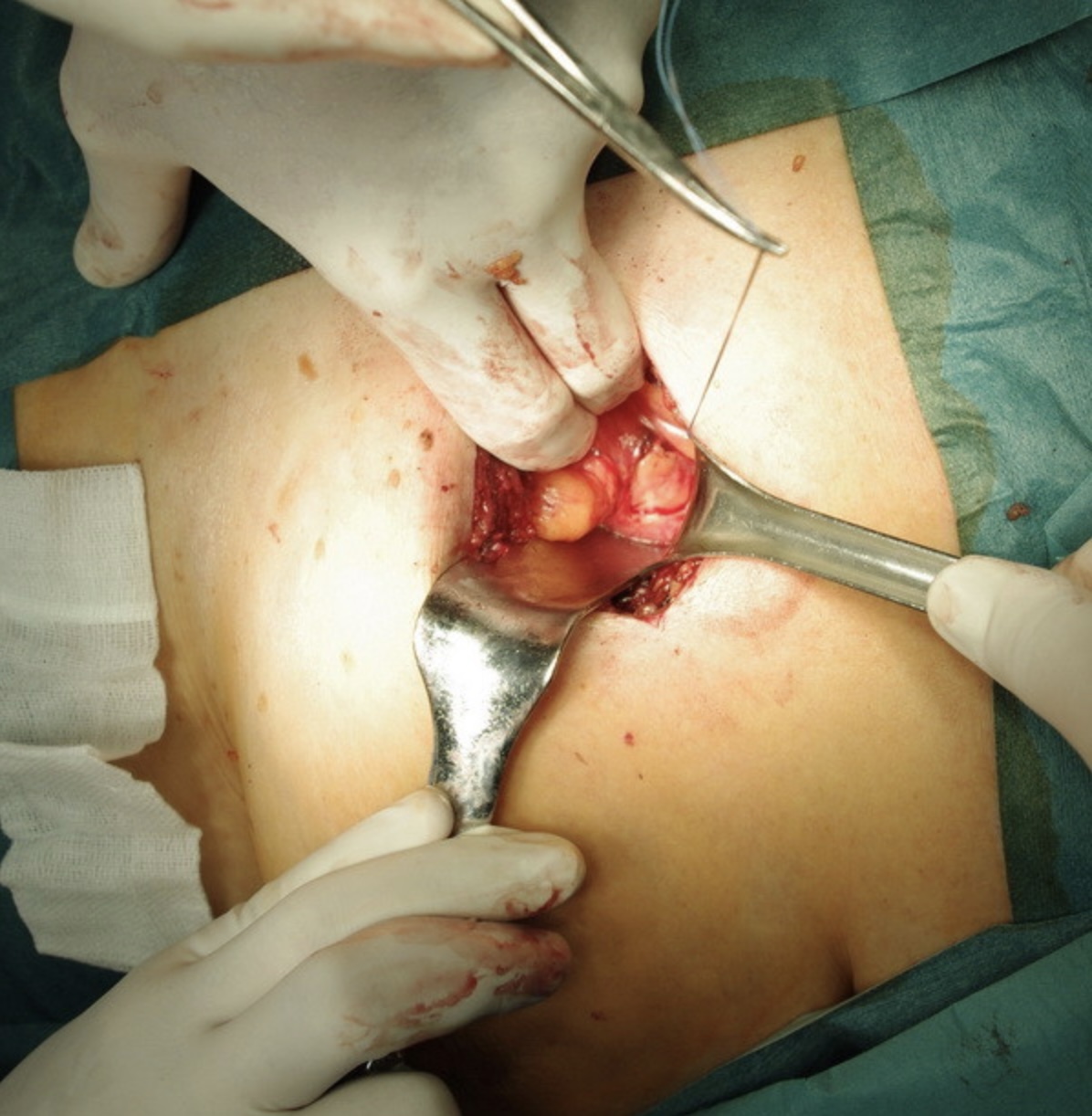
A pericardial window is a surgical procedure done on the parietalParietalOne of a pair of irregularly shaped quadrilateral bones situated between the frontal bone and occipital bone, which together form the sides of the cranium.Skull: AnatomypericardiumPericardiumA conical fibroserous sac surrounding the heart and the roots of the great vessels (aorta; venae cavae; pulmonary artery). Pericardium consists of two sacs: the outer fibrous pericardium and the inner serous pericardium. The latter consists of an outer parietal layer facing the fibrous pericardium, and an inner visceral layer (epicardium) resting next to the heart, and a pericardial cavity between these two layers.Heart: Anatomy to provide access to the pericardial spacePericardial SpacePericardial Effusion and Cardiac Tamponade and to evacuate occupying fluid and break loculations. The main goal of this procedure is to restore effective ventricular fillingVentricular fillingCardiac Cycle and cardiac outputCardiac outputThe volume of blood passing through the heart per unit of time. It is usually expressed as liters (volume) per minute so as not to be confused with stroke volume (volume per beat).Cardiac Mechanics.
Indications
Cardiac tamponadeTamponadePericardial effusion, usually of rapid onset, exceeding ventricular filling pressures and causing collapse of the heart with a markedly reduced cardiac output.Pericarditis
Symptomatic pericardial effusionPericardial effusionFluid accumulation within the pericardium. Serous effusions are associated with pericardial diseases. Hemopericardium is associated with trauma. Lipid-containing effusion (chylopericardium) results from leakage of thoracic duct. Severe cases can lead to cardiac tamponade.Pericardial Effusion and Cardiac Tamponade that persists despite 7–10 days of intensive medical treatment
ContraindicationsContraindicationsA condition or factor associated with a recipient that makes the use of a drug, procedure, or physical agent improper or inadvisable. Contraindications may be absolute (life threatening) or relative (higher risk of complications in which benefits may outweigh risks).Noninvasive Ventilation
There are no absolute contraindicationsContraindicationsA condition or factor associated with a recipient that makes the use of a drug, procedure, or physical agent improper or inadvisable. Contraindications may be absolute (life threatening) or relative (higher risk of complications in which benefits may outweigh risks).Noninvasive Ventilation. Relative contraindicationsContraindicationsA condition or factor associated with a recipient that makes the use of a drug, procedure, or physical agent improper or inadvisable. Contraindications may be absolute (life threatening) or relative (higher risk of complications in which benefits may outweigh risks).Noninvasive Ventilation include coagulopathy, thrombocytopeniaThrombocytopeniaThrombocytopenia occurs when the platelet count is < 150,000 per microliter. The normal range for platelets is usually 150,000-450,000/µL of whole blood. Thrombocytopenia can be a result of decreased production, increased destruction, or splenic sequestration of platelets. Patients are often asymptomatic until platelet counts are < 50,000/µL. Thrombocytopenia, and lack of operator experience.
Procedure
Subxiphoid approach (preferred because of its greater simplicity):
The diaphragmDiaphragmThe diaphragm is a large, dome-shaped muscle that separates the thoracic cavity from the abdominal cavity. The diaphragm consists of muscle fibers and a large central tendon, which is divided into right and left parts. As the primary muscle of inspiration, the diaphragm contributes 75% of the total inspiratory muscle force.Diaphragm: Anatomy is dissected away from the sternumSternumA long, narrow, and flat bone commonly known as breastbone occurring in the midsection of the anterior thoracic segment or chest region, which stabilizes the rib cage and serves as the point of origin for several muscles that move the arms, head, and neck.Chest Wall: Anatomy and xiphoid to access the thoracic cavity.
When the surgeon has direct visualization, the pericardiumPericardiumA conical fibroserous sac surrounding the heart and the roots of the great vessels (aorta; venae cavae; pulmonary artery). Pericardium consists of two sacs: the outer fibrous pericardium and the inner serous pericardium. The latter consists of an outer parietal layer facing the fibrous pericardium, and an inner visceral layer (epicardium) resting next to the heart, and a pericardial cavity between these two layers.Heart: Anatomy is opened.
Fluid is aspirated and loculations are gently broken.
Pericardial tissue and fluid are sent for bacteriologic and histologic study.
A pericardial window is made anterior to the phrenic nervePhrenic nerveThe motor nerve of the diaphragm. The phrenic nerve fibers originate in the cervical spinal column (mostly C4) and travel through the cervical plexus to the diaphragm.Diaphragm: Anatomy.
The skinSkinThe skin, also referred to as the integumentary system, is the largest organ of the body. The skin is primarily composed of the epidermis (outer layer) and dermis (deep layer). The epidermis is primarily composed of keratinocytes that undergo rapid turnover, while the dermis contains dense layers of connective tissue.Skin: Structure and Functions is loosely closed off using nonabsorbable suturesNonabsorbable SuturesSurgical Instruments and Sutures and cleansed of any residue (e.g., blood, adipose tissueAdipose tissueAdipose tissue is a specialized type of connective tissue that has both structural and highly complex metabolic functions, including energy storage, glucose homeostasis, and a multitude of endocrine capabilities. There are three types of adipose tissue, white adipose tissue, brown adipose tissue, and beige or “brite” adipose tissue, which is a transitional form.Adipose Tissue: Histology)
Image: “The place of pericardial window” by Toth et al. License: CC BY 2.0
Complications
Bleeding
Surgical site infectionSurgical site infectionInfection occurring at the site of a surgical incision.Surgical Complications (SSISSISurgical site infection (SSI) is a type of surgical infection that occurs at or near a surgical incision within 30 days of the procedure or within 90 days if prosthetic material is implanted. Surgical site infections are classified according to the depth of involvement as superficial, deep, or organ/space. Surgical Site Infections)
Arrhythmia
MIMIMI is ischemia and death of an area of myocardial tissue due to insufficient blood flow and oxygenation, usually from thrombus formation on a ruptured atherosclerotic plaque in the epicardial arteries. Clinical presentation is most commonly with chest pain, but women and patients with diabetes may have atypical symptoms.Myocardial Infarction
Hemodynamic collapse
Coronary Artery Bypass Graft (CABG)
Definition
Coronary arteryCoronary ArteryTruncus Arteriosus bypass graftGraftA piece of living tissue that is surgically transplantedOrgan Transplantation (CABG) surgery is an invasive revascularizationRevascularizationThromboangiitis Obliterans (Buerger Disease) procedure that consists of placing grafts between the arterial and coronary circulations in order to bypass obstructed segments of the coronary arteriesArteriesArteries are tubular collections of cells that transport oxygenated blood and nutrients from the heart to the tissues of the body. The blood passes through the arteries in order of decreasing luminal diameter, starting in the largest artery (the aorta) and ending in the small arterioles. Arteries are classified into 3 types: large elastic arteries, medium muscular arteries, and small arteries and arterioles. Arteries: Histology and supply the myocardiumMyocardiumThe muscle tissue of the heart. It is composed of striated, involuntary muscle cells connected to form the contractile pump to generate blood flow.Heart: Anatomy with oxygenated blood.
Indications
Individuals with activity-limiting angina despite maximum medical therapy
ContraindicationsContraindicationsA condition or factor associated with a recipient that makes the use of a drug, procedure, or physical agent improper or inadvisable. Contraindications may be absolute (life threatening) or relative (higher risk of complications in which benefits may outweigh risks).Noninvasive Ventilation
Coronary arteriesArteriesArteries are tubular collections of cells that transport oxygenated blood and nutrients from the heart to the tissues of the body. The blood passes through the arteries in order of decreasing luminal diameter, starting in the largest artery (the aorta) and ending in the small arterioles. Arteries are classified into 3 types: large elastic arteries, medium muscular arteries, and small arteries and arterioles. Arteries: Histology incompatible with grafting
Absence of viable myocardiumMyocardiumThe muscle tissue of the heart. It is composed of striated, involuntary muscle cells connected to form the contractile pump to generate blood flow.Heart: Anatomy to graftGraftA piece of living tissue that is surgically transplantedOrgan Transplantation
Procedure
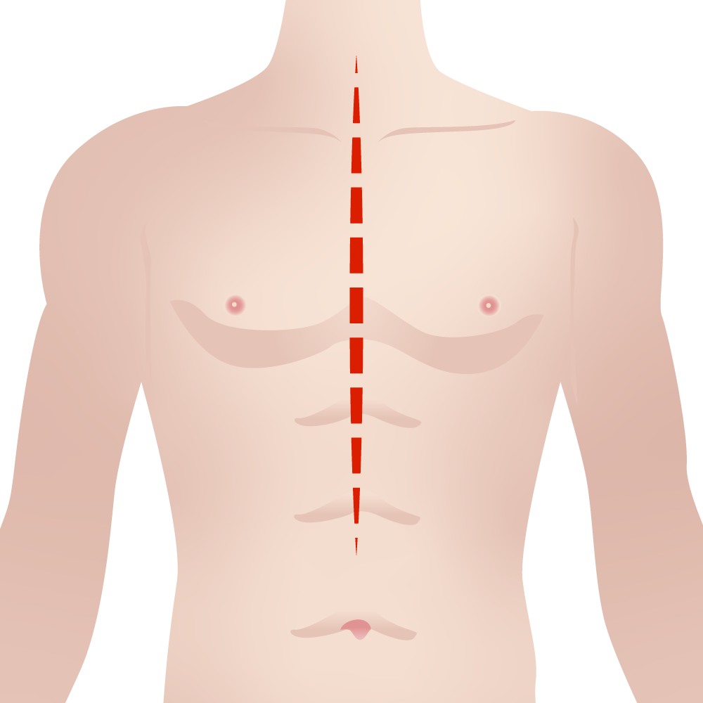
A median sternotomy is performed.
The saphenous vein is simultaneously removed from 1 or both legs using an open or video-assisted approach. The vein is used to make the grafts or conduits.
The heart is arrested using high-potassium cardioplegic solution.
The surgeon anastomoses the conduits to the coronary arteriesArteriesArteries are tubular collections of cells that transport oxygenated blood and nutrients from the heart to the tissues of the body. The blood passes through the arteries in order of decreasing luminal diameter, starting in the largest artery (the aorta) and ending in the small arterioles. Arteries are classified into 3 types: large elastic arteries, medium muscular arteries, and small arteries and arterioles. Arteries: Histology.
The conduits are later attached to new openings created in the proximal aortaAortaThe main trunk of the systemic arteries.Mediastinum and Great Vessels: Anatomy and/or other major vessels.
After the cardioplegic solution is washed out, the surgeon checks for conduit competence and bleeding from anastomosis sites.
If the procedure is considered satisfactory, the chest is closed using sternal wires.
A median sternotomy is performed during many cardiac operations to include a coronary artery bypass graft.
Image by Lecturio.
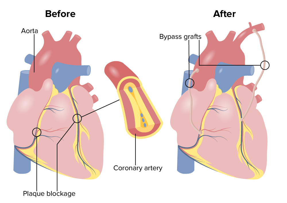
Illustration showing the heart before and after coronary artery bypass graft (CABG) surgery:
Vein grafts are used to bypass coronary obstructions.
Image by Lecturio.
Complications
GraftGraftA piece of living tissue that is surgically transplantedOrgan Transplantation failure: The inability of the graftGraftA piece of living tissue that is surgically transplantedOrgan Transplantation to adequately supply the myocardiumMyocardiumThe muscle tissue of the heart. It is composed of striated, involuntary muscle cells connected to form the contractile pump to generate blood flow.Heart: Anatomy with oxygenated blood.
Stroke: A serious complication of CABG in which injury to the brainBrainThe part of central nervous system that is contained within the skull (cranium). Arising from the neural tube, the embryonic brain is comprised of three major parts including prosencephalon (the forebrain); mesencephalon (the midbrain); and rhombencephalon (the hindbrain). The developed brain consists of cerebrum; cerebellum; and other structures in the brain stem.Nervous System: Anatomy, Structure, and Classification is caused by an interruption of blood flowBlood flowBlood flow refers to the movement of a certain volume of blood through the vasculature over a given unit of time (e.g., mL per minute).Vascular Resistance, Flow, and Mean Arterial Pressure (ischemic strokeIschemic StrokeAn ischemic stroke (also known as cerebrovascular accident) is an acute neurologic injury that occurs as a result of brain ischemia; this condition may be due to cerebral blood vessel occlusion by thrombosis or embolism, or rarely due to systemic hypoperfusion. Ischemic Stroke) or active hemorrhage (hemorrhagic strokeHemorrhagic strokeStroke due to rupture of a weakened blood vessel in the brain (e.g., cerebral hemispheres; cerebellum; subarachnoid space).Subarachnoid Hemorrhage), which has characteristic neurologic clinical features.
SSISSISurgical site infection (SSI) is a type of surgical infection that occurs at or near a surgical incision within 30 days of the procedure or within 90 days if prosthetic material is implanted. Surgical site infections are classified according to the depth of involvement as superficial, deep, or organ/space. Surgical Site Infections: A type of surgical infectionSurgical infectionAn infection is the proliferation of microorganisms within tissues, body cavities, or spaces, which induces an immune response and overwhelms the body’s natural defenses. In surgical patients, these infections are frequently caused by the translocation of commensal organisms into deeper tissues, combined with the impairment of host defenses due to surgical injury or stress. Surgical Infections that occurs at or near a surgical incisionSurgical IncisionSurgical Site Infections within 30 days after the procedure or within 90 days if prosthetic material is implanted. An SSISSISurgical site infection (SSI) is a type of surgical infection that occurs at or near a surgical incision within 30 days of the procedure or within 90 days if prosthetic material is implanted. Surgical site infections are classified according to the depth of involvement as superficial, deep, or organ/space. Surgical Site Infections is classified according to the depth of compromise as superficial, deep, or organ/space.
Atrial fibrillationAtrial fibrillationAtrial fibrillation (AF or Afib) is a supraventricular tachyarrhythmia and the most common kind of arrhythmia. It is caused by rapid, uncontrolled atrial contractions and uncoordinated ventricular responses. Atrial Fibrillation: A supraventricular tachyarrhythmiaTachyarrhythmiaA tachyarrhythmia is a rapid heart rhythm, regular or irregular, with a rate > 100 beats/min. Tachyarrhythmia may or may not be accompanied by symptoms of hemodynamic change.Tachyarrhythmias that is the most common kind of arrhythmia. Atrial fibrillationAtrial fibrillationAtrial fibrillation (AF or Afib) is a supraventricular tachyarrhythmia and the most common kind of arrhythmia. It is caused by rapid, uncontrolled atrial contractions and uncoordinated ventricular responses. Atrial Fibrillation is caused by rapid, uncontrolled atrial contractions and uncoordinated ventricular responses.
Cardiac catheterization is an invasive procedure that consists of inserting a catheter into the systemic arterial circulationCirculationThe movement of the blood as it is pumped through the cardiovascular system.ABCDE Assessment and advancing it toward the heart to inject contrast dye. This procedure allows for diagnostic vascular imaging and potential therapeutic intervention.
Indications
This procedure may be interventional or diagnostic.
Coronary arteryCoronary ArteryTruncus Arteriosus disease: Referred to as ischemic heart diseaseIschemic heart diseaseCoronary heart disease (CHD), or ischemic heart disease, describes a situation in which an inadequate supply of blood to the myocardium exists due to a stenosis of the coronary arteries, typically from atherosclerosis. Coronary Heart Disease, this condition is caused by an inadequate supply of blood to the myocardiumMyocardiumThe muscle tissue of the heart. It is composed of striated, involuntary muscle cells connected to form the contractile pump to generate blood flow.Heart: Anatomy due to a stenosisStenosisHypoplastic Left Heart Syndrome (HLHS) of the coronary arteriesArteriesArteries are tubular collections of cells that transport oxygenated blood and nutrients from the heart to the tissues of the body. The blood passes through the arteries in order of decreasing luminal diameter, starting in the largest artery (the aorta) and ending in the small arterioles. Arteries are classified into 3 types: large elastic arteries, medium muscular arteries, and small arteries and arterioles. Arteries: Histology, typically from atherosclerosisAtherosclerosisAtherosclerosis is a common form of arterial disease in which lipid deposition forms a plaque in the blood vessel walls. Atherosclerosis is an incurable disease, for which there are clearly defined risk factors that often can be reduced through a change in lifestyle and behavior of the patient. Atherosclerosis.
Heart failureHeart FailureA heterogeneous condition in which the heart is unable to pump out sufficient blood to meet the metabolic need of the body. Heart failure can be caused by structural defects, functional abnormalities (ventricular dysfunction), or a sudden overload beyond its capacity. Chronic heart failure is more common than acute heart failure which results from sudden insult to cardiac function, such as myocardial infarction.Total Anomalous Pulmonary Venous Return (TAPVR):This condition refers to the inability of the heart to supply the body with normal cardiac outputCardiac outputThe volume of blood passing through the heart per unit of time. It is usually expressed as liters (volume) per minute so as not to be confused with stroke volume (volume per beat).Cardiac Mechanics to meet metabolic needs.
ContraindicationsContraindicationsA condition or factor associated with a recipient that makes the use of a drug, procedure, or physical agent improper or inadvisable. Contraindications may be absolute (life threatening) or relative (higher risk of complications in which benefits may outweigh risks).Noninvasive Ventilation
There are no absolute contraindicationsContraindicationsA condition or factor associated with a recipient that makes the use of a drug, procedure, or physical agent improper or inadvisable. Contraindications may be absolute (life threatening) or relative (higher risk of complications in which benefits may outweigh risks).Noninvasive Ventilation for cardiac catheterization. However, if the likelihood of complications is reasonably high, the surgeon may consider other methods. Individuals with renal impairment should be treated cautiously, as contrast mediaContrast MediaSubstances used to allow enhanced visualization of tissues.Computed Tomography (CT) may be nephrotoxic.
Procedure
In the OR:
The individual is placed in the supine position.
A fluoroscope is placed over the individual.
IV access is obtained.
A Foley catheter is placed for quantification of urine output.
Continuous monitoring:
HR
Blood pressure
O2 saturation (pulse oximetry)
ECGECGAn electrocardiogram (ECG) is a graphic representation of the electrical activity of the heart plotted against time. Adhesive electrodes are affixed to the skin surface allowing measurement of cardiac impulses from many angles. The ECG provides 3-dimensional information about the conduction system of the heart, the myocardium, and other cardiac structures. Electrocardiogram (ECG) rhythm monitor
Moderate sedation and mild analgesiaAnalgesiaMethods of pain relief that may be used with or in place of analgesics.Anesthesiology: History and Basic Concepts coupled with local anesthesiaAnesthesiaA state characterized by loss of feeling or sensation. This depression of nerve function is usually the result of pharmacologic action and is induced to allow performance of surgery or other painful procedures.Anesthesiology: History and Basic Concepts are used.
Operative care:
Cardiac catheterization can be done through an arterial or venous access. A modified version of the Seldinger techniqueSeldinger TechniqueCentral Venous Catheter is used for percutaneous catheter insertion.
The vessel is punctured with a needle under ultrasound guidance.
A guidewire is advanced through the needle.
The needle is withdrawn and a dilator is inserted over the guidewire.
The guidewire is advanced through the systemic circulationCirculationThe movement of the blood as it is pumped through the cardiovascular system.ABCDE Assessment to the heart.
The operator inserts the catheter over the guidewire toward the heart.
Once the catheter reaches the heart, the surgeon performs the necessary assessments, according to the indications for the procedure.
The catheter and dilator are extracted while applying pressure to the puncture site to prevent bleeding.
Complications
HematomaHematomaA collection of blood outside the blood vessels. Hematoma can be localized in an organ, space, or tissue.Intussusception
PseudoaneurysmPseudoaneurysmNot an aneurysm but a well-defined collection of blood and connective tissue outside the wall of a blood vessel or the heart. It is the containment of a ruptured blood vessel or heart, such as sealing a rupture of the left ventricle. False aneurysm is formed by organized thrombus and hematoma in surrounding tissue.Thoracic Aortic Aneurysms
AV fistulaAV fistulaAn abnormal direct communication between an artery and a vein without passing through the capillaries. An a-v fistula usually leads to the formation of a dilated sac-like connection, arteriovenous aneurysm. The locations and size of the shunts determine the degree of effects on the cardiovascular functions such as blood pressure and heart rate.Vascular Surgery
Ventricular fibrillationVentricular fibrillationVentricular fibrillation (VF or V-fib) is a type of ventricular tachyarrhythmia (> 300/min) often preceded by ventricular tachycardia. In this arrhythmia, the ventricle beats rapidly and sporadically. The ventricular contraction is uncoordinated, leading to a decrease in cardiac output and immediate hemodynamic collapse. Ventricular Fibrillation (V-fib): a type of ventricular tachyarrhythmiaTachyarrhythmiaA tachyarrhythmia is a rapid heart rhythm, regular or irregular, with a rate > 100 beats/min. Tachyarrhythmia may or may not be accompanied by symptoms of hemodynamic change.Tachyarrhythmias characterized by uncoordinated ventricular contraction, which leads to a decrease in cardiac outputCardiac outputThe volume of blood passing through the heart per unit of time. It is usually expressed as liters (volume) per minute so as not to be confused with stroke volume (volume per beat).Cardiac Mechanics and immediate hemodynamic collapse
MIMIMI is ischemia and death of an area of myocardial tissue due to insufficient blood flow and oxygenation, usually from thrombus formation on a ruptured atherosclerotic plaque in the epicardial arteries. Clinical presentation is most commonly with chest pain, but women and patients with diabetes may have atypical symptoms.Myocardial Infarction: an injury to the myocardiumMyocardiumThe muscle tissue of the heart. It is composed of striated, involuntary muscle cells connected to form the contractile pump to generate blood flow.Heart: Anatomy due to ischemiaIschemiaA hypoperfusion of the blood through an organ or tissue caused by a pathologic constriction or obstruction of its blood vessels, or an absence of blood circulation.Ischemic Cell Damage, characterized by an increase in cardiac enzymesEnzymesEnzymes are complex protein biocatalysts that accelerate chemical reactions without being consumed by them. Due to the body’s constant metabolic needs, the absence of enzymes would make life unsustainable, as reactions would occur too slowly without these molecules. Basics of Enzymes (especially troponin T), ECGECGAn electrocardiogram (ECG) is a graphic representation of the electrical activity of the heart plotted against time. Adhesive electrodes are affixed to the skin surface allowing measurement of cardiac impulses from many angles. The ECG provides 3-dimensional information about the conduction system of the heart, the myocardium, and other cardiac structures. Electrocardiogram (ECG) changes suggestive of ischemiaIschemiaA hypoperfusion of the blood through an organ or tissue caused by a pathologic constriction or obstruction of its blood vessels, or an absence of blood circulation.Ischemic Cell Damage in 2 continuous leads, and chest painPainAn unpleasant sensation induced by noxious stimuli which are detected by nerve endings of nociceptive neurons.Pain: Types and Pathways
Stroke
Aortic dissectionAortic dissectionAortic dissection occurs due to shearing stress from pulsatile pressure causing a tear in the tunica intima of the aortic wall. This tear allows blood to flow into the media, creating a “false lumen.” Aortic dissection is most commonly caused by uncontrolled hypertension.Aortic Dissection: separation of the tunica intimaTunica intimaThe innermost layer of an artery or vein, made up of one layer of endothelial cells and supported by an internal elastic lamina.Arteries: Histology from the aortic wall due to shearing stress from pulsatile pressure that allows blood to flowFlowBlood flows through the heart, arteries, capillaries, and veins in a closed, continuous circuit. Flow is the movement of volume per unit of time. Flow is affected by the pressure gradient and the resistance fluid encounters between 2 points. Vascular resistance is the opposition to flow, which is caused primarily by blood friction against vessel walls.Vascular Resistance, Flow, and Mean Arterial Pressure into the tunica mediaTunica mediaThe middle layer of blood vessel walls, composed principally of thin, cylindrical, smooth muscle cells and elastic tissue. It accounts for the bulk of the wall of most arteries. The smooth muscle cells are arranged in circular layers around the vessel, and the thickness of the coat varies with the size of the vessel.Arteries: Histology, creating a “false lumenFalse lumenAortic Dissection”
Hypersensitivity reaction: There is growing evidence that some hypersensitivity reactions to contrast mediaContrast MediaSubstances used to allow enhanced visualization of tissues.Computed Tomography (CT), particularly those that are severe, may be IgE-mediated.
AKIAKIAcute kidney injury refers to sudden and often reversible loss of renal function, which develops over days or weeks. Azotemia refers to elevated levels of nitrogen-containing substances in the blood that accompany AKI, which include BUN and creatinine. Acute Kidney Injury: due to contrast required for the procedure
Ventriculography, the injection of contrast to visualize a ventricle, allows for measurement of ejection fraction.
Image: “Left ventriculography during systole showing apical ballooning akinesis with basal hyperkinesis in a characteristic takotsubo ventricle” by Tara C. Gangadhar. License: CC BY 2.0
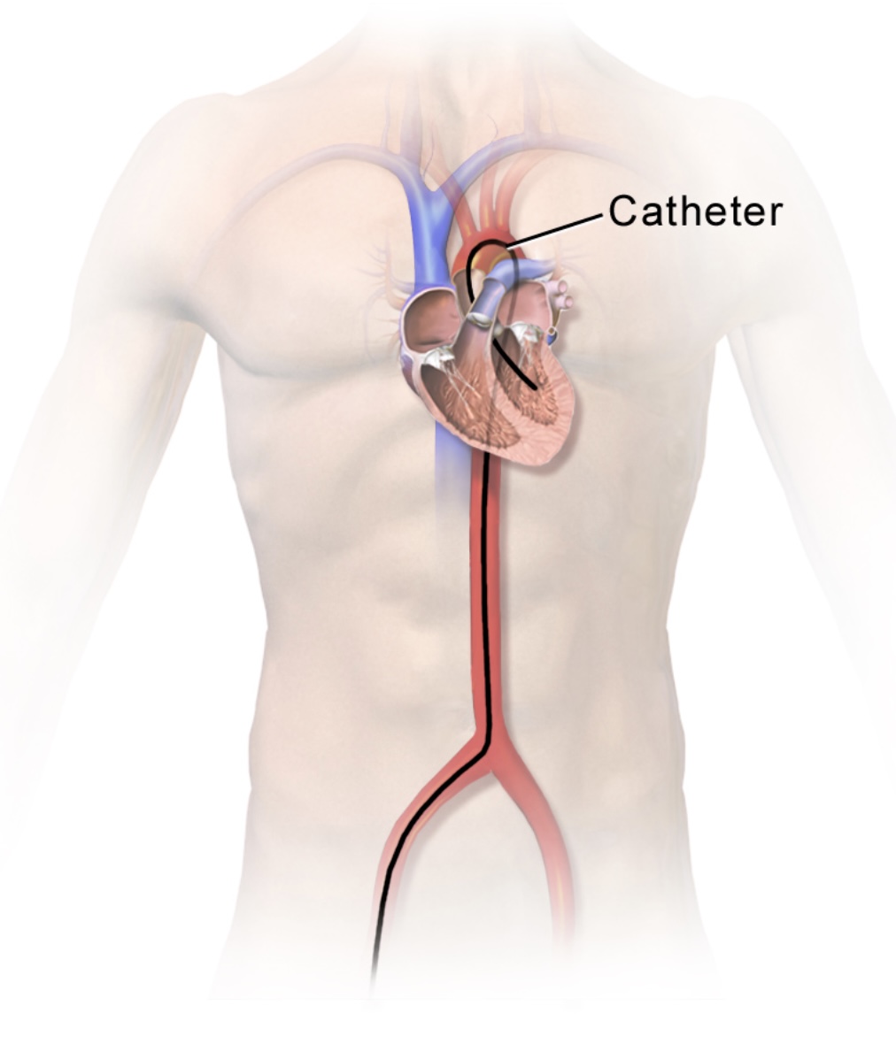
Left heart catheter inserted through a femoral approach
Image: “Left Heart Catheter” by BruceBlaus. License: CC BY-SA 4.0, cropped by Lecturio.
Formerly known as coronary angioplasty and stenting, PCI is an invasive, nonsurgical procedure that consists of introducing a catheter into the coronary arteriesArteriesArteries are tubular collections of cells that transport oxygenated blood and nutrients from the heart to the tissues of the body. The blood passes through the arteries in order of decreasing luminal diameter, starting in the largest artery (the aorta) and ending in the small arterioles. Arteries are classified into 3 types: large elastic arteries, medium muscular arteries, and small arteries and arterioles. Arteries: Histology and using a balloon and stent to relieve an occlusion within the vessel.
Indications
Critical coronary heart diseaseCoronary heart diseaseCoronary heart disease (CHD), or ischemic heart disease, describes a situation in which an inadequate supply of blood to the myocardium exists due to a stenosis of the coronary arteries, typically from atherosclerosis. Coronary Heart Disease (CHD):
The descriptive term denoting a situation in which an inadequate supply of blood to the myocardiumMyocardiumThe muscle tissue of the heart. It is composed of striated, involuntary muscle cells connected to form the contractile pump to generate blood flow.Heart: Anatomy exists because of stenosisStenosisHypoplastic Left Heart Syndrome (HLHS) of the coronary arteriesArteriesArteries are tubular collections of cells that transport oxygenated blood and nutrients from the heart to the tissues of the body. The blood passes through the arteries in order of decreasing luminal diameter, starting in the largest artery (the aorta) and ending in the small arterioles. Arteries are classified into 3 types: large elastic arteries, medium muscular arteries, and small arteries and arterioles. Arteries: Histology
Typically from atherosclerosisAtherosclerosisAtherosclerosis is a common form of arterial disease in which lipid deposition forms a plaque in the blood vessel walls. Atherosclerosis is an incurable disease, for which there are clearly defined risk factors that often can be reduced through a change in lifestyle and behavior of the patient. Atherosclerosis
PCI is indicated in individuals with CHD who do not qualify for CABG.
Acute ST-elevation MIMIMI is ischemia and death of an area of myocardial tissue due to insufficient blood flow and oxygenation, usually from thrombus formation on a ruptured atherosclerotic plaque in the epicardial arteries. Clinical presentation is most commonly with chest pain, but women and patients with diabetes may have atypical symptoms.Myocardial Infarction (STEMI):
Ischemic symptoms < 12 hours
Contraindicated in combination with fibrinolytic therapy
Non–ST-elevation acute coronary syndrome
Refractory angina
Recurrent angina
Symptoms of heart failureHeart FailureA heterogeneous condition in which the heart is unable to pump out sufficient blood to meet the metabolic need of the body. Heart failure can be caused by structural defects, functional abnormalities (ventricular dysfunction), or a sudden overload beyond its capacity. Chronic heart failure is more common than acute heart failure which results from sudden insult to cardiac function, such as myocardial infarction.Total Anomalous Pulmonary Venous Return (TAPVR)
Sustained ventricular tachycardiaTachycardiaAbnormally rapid heartbeat, usually with a heart rate above 100 beats per minute for adults. Tachycardia accompanied by disturbance in the cardiac depolarization (cardiac arrhythmia) is called tachyarrhythmia.Sepsis in Children/fibrillation
Angina (stable and unstable): substernal, pressure-like chest painPainAn unpleasant sensation induced by noxious stimuli which are detected by nerve endings of nociceptive neurons.Pain: Types and Pathways or discomfort, radiating to the neckNeckThe part of a human or animal body connecting the head to the rest of the body.Peritonsillar Abscess, jawJawThe jaw is made up of the mandible, which comprises the lower jaw, and the maxilla, which comprises the upper jaw. The mandible articulates with the temporal bone via the temporomandibular joint (TMJ). The 4 muscles of mastication produce the movements of the TMJ to ensure the efficient chewing of food. Jaw and Temporomandibular Joint: Anatomy, and/or left-upper limb that is associated with dyspneaDyspneaDyspnea is the subjective sensation of breathing discomfort. Dyspnea is a normal manifestation of heavy physical or psychological exertion, but also may be caused by underlying conditions (both pulmonary and extrapulmonary). Dyspnea, palpitationsPalpitationsEbstein’s Anomaly, anxietyAnxietyFeelings or emotions of dread, apprehension, and impending disaster but not disabling as with anxiety disorders.Generalized Anxiety Disorder, nauseaNauseaAn unpleasant sensation in the stomach usually accompanied by the urge to vomit. Common causes are early pregnancy, sea and motion sickness, emotional stress, intense pain, food poisoning, and various enteroviruses.Antiemetics, vomitingVomitingThe forcible expulsion of the contents of the stomach through the mouth.Hypokalemia, abdominal or epigastric painEpigastric painMallory-Weiss Syndrome (Mallory-Weiss Tear), and/or diaphoresis
Stable anginaStable anginaPersistent and reproducible chest discomfort usually precipitated by a physical exertion that dissipates upon cessation of such an activity. The symptoms are manifestations of myocardial ischemia.Stable and Unstable Angina (< 20 minutes)
ContraindicationsContraindicationsA condition or factor associated with a recipient that makes the use of a drug, procedure, or physical agent improper or inadvisable. Contraindications may be absolute (life threatening) or relative (higher risk of complications in which benefits may outweigh risks).Noninvasive Ventilation
Absolute:
Rejection of the procedure
Inability to take dual antiplatelet therapy.
Increased bleeding risk (i.e., thrombocytopeniaThrombocytopeniaThrombocytopenia occurs when the platelet count is < 150,000 per microliter. The normal range for platelets is usually 150,000-450,000/µL of whole blood. Thrombocytopenia can be a result of decreased production, increased destruction, or splenic sequestration of platelets. Patients are often asymptomatic until platelet counts are < 50,000/µL. Thrombocytopenia)
Restenosis due to multiple previous PCIs
Relative:
Long-term intolerance to antiplatelet therapy
HypercoagulableHypercoagulableHypercoagulable states (also referred to as thrombophilias) are a group of hematologic diseases defined by an increased risk of clot formation (i.e., thrombosis) due to either an increase in procoagulants, a decrease in anticoagulants, or a decrease in fibrinolysis. Hypercoagulable States state
Severe CKDCKDChronic kidney disease (CKD) is kidney impairment that lasts for ≥ 3 months, implying that it is irreversible. Hypertension and diabetes are the most common causes; however, there are a multitude of other etiologies. In the early to moderate stages, CKD is usually asymptomatic and is primarily diagnosed by laboratory abnormalities.Chronic Kidney Disease
Artery diameter < 1.5 mm
Critical left main stenosisStenosisHypoplastic Left Heart Syndrome (HLHS) with no collateral flowFlowBlood flows through the heart, arteries, capillaries, and veins in a closed, continuous circuit. Flow is the movement of volume per unit of time. Flow is affected by the pressure gradient and the resistance fluid encounters between 2 points. Vascular resistance is the opposition to flow, which is caused primarily by blood friction against vessel walls.Vascular Resistance, Flow, and Mean Arterial Pressure or patent bypass graftGraftA piece of living tissue that is surgically transplantedOrgan Transplantation
Preoperative carePreoperative CareThorough preoperative care is important for patients scheduled to undergo surgery so that they can have the best possible outcomes after their surgical procedure. The preoperative process begins once the decision has been made to proceed with a surgical procedure. Preoperative Care
Preparation for PCI is substantially similar to that for cardiac catheterization, however door-to-balloon time (D2B time) is an important factor to consider.
Percutaneous coronary intervention is often performed in the context of an emergency.
The D2B time is the time taken from arrival at the ED to balloon inflation of the culprit artery.
Reduction in D2B time is critical to increase the likelihood of in-hospital survival.
Antiplatelet therapy:
AspirinAspirinThe prototypical analgesic used in the treatment of mild to moderate pain. It has anti-inflammatory and antipyretic properties and acts as an inhibitor of cyclooxygenase which results in the inhibition of the biosynthesis of prostaglandins. Aspirin also inhibits platelet aggregation and is used in the prevention of arterial and venous thrombosis.Nonsteroidal Antiinflammatory Drugs (NSAIDs) 162–325 mg on the day of the procedure.
GPIIb/IIIa inhibitors (i.e., abciximab)
Dual antiplatelet therapy (DAPT): aspirinAspirinThe prototypical analgesic used in the treatment of mild to moderate pain. It has anti-inflammatory and antipyretic properties and acts as an inhibitor of cyclooxygenase which results in the inhibition of the biosynthesis of prostaglandins. Aspirin also inhibits platelet aggregation and is used in the prevention of arterial and venous thrombosis.Nonsteroidal Antiinflammatory Drugs (NSAIDs) and a P2Y12 inhibitor (e.g., clopidogrelClopidogrelA ticlopidine analog and platelet purinergic p2y receptor antagonist that inhibits adenosine diphosphate-mediated platelet aggregation. It is used to prevent thromboembolism in patients with arterial occlusive diseases; myocardial infarction; stroke; or atrial fibrillation.Antiplatelet Drugs, prasugrelPrasugrelA piperazine derivative and platelet aggregation inhibitor that is used to prevent thrombosis in patients with acute coronary syndrome; unstable angina and myocardial infarction, as well as in those undergoing percutaneous coronary interventions.Antiplatelet Drugs, ticagrelorTicagrelorAn adenosine triphosphate analogue and reversible p2y12 purinoceptor antagonist that inhibits adp-mediated platelet aggregation. It is used for the prevention of thromboembolism by patients with acute coronary syndrome or a history of myocardial infarction.Antiplatelet Drugs)
For anticoagulationAnticoagulationPulmonary Hypertension Drugs, unfractionated heparinUnfractionated heparinA highly acidic mucopolysaccharide formed of equal parts of sulfated d-glucosamine and d-glucuronic acid with sulfaminic bridges. The molecular weight ranges from six to twenty thousand. Heparin occurs in and is obtained from liver, lung, mast cells, etc. , of vertebrates. Its function is unknown, but it is used to prevent blood clotting in vivo and vitro, in the form of many different salts.Anticoagulants may be used at the time of PCI.
The coronary arteriesArteriesArteries are tubular collections of cells that transport oxygenated blood and nutrients from the heart to the tissues of the body. The blood passes through the arteries in order of decreasing luminal diameter, starting in the largest artery (the aorta) and ending in the small arterioles. Arteries are classified into 3 types: large elastic arteries, medium muscular arteries, and small arteries and arterioles. Arteries: Histology are reached via radial, axillary, or femoral approach. The radial arteryRadial ArteryThe direct continuation of the brachial trunk, originating at the bifurcation of the brachial artery opposite the neck of the radius. Its branches may be divided into three groups corresponding to the three regions in which the vessel is situated, the forearm, wrist, and hand.Forearm: Anatomy is preferred because of the decreased risk of bleeding. Stents are preferred over balloon angioplasty alone because of the decreased risk of restenosis.
Angiography:
Cardiac catheterization is performed to reach the coronary arteriesArteriesArteries are tubular collections of cells that transport oxygenated blood and nutrients from the heart to the tissues of the body. The blood passes through the arteries in order of decreasing luminal diameter, starting in the largest artery (the aorta) and ending in the small arterioles. Arteries are classified into 3 types: large elastic arteries, medium muscular arteries, and small arteries and arterioles. Arteries: Histology.
Contrast medium is introduced into the coronary arteryCoronary ArteryTruncus Arteriosus of interest and images are obtained with fluoroscopyFluoroscopyProduction of an image when x-rays strike a fluorescent screen.X-rays.
The guidewire is used to advance the deflated balloon and stent catheter to the stenotic segment.
Once it reaches the correct location, the balloon is expanded, stretching the stent over it, and opening the lumen of the vessel.
The balloon is deflated, the catheter is withdrawn, and adequate placement of the stent and resolution of stenosisStenosisHypoplastic Left Heart Syndrome (HLHS) is confirmed via imaging.
The catheter and dilator are extracted while applying pressure in the puncture site to prevent bleeding.
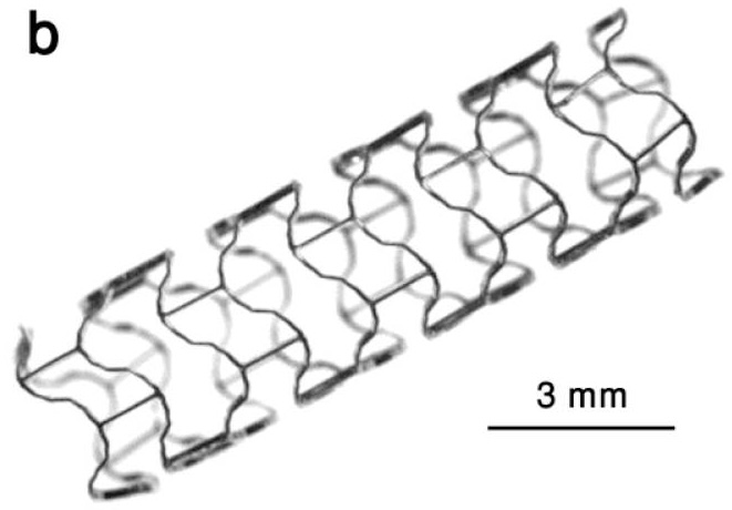
Example of a coronary stent, an expandable tubular metallic device
Image: “Lekton Magic coronary stent” by Maryam Moravej and Diego Mantovani. License: CC BY 3.0
Postoperative carePostoperative careAfter any procedure performed in the operating room, all patients must undergo close observation at least in the recovery room. After larger procedures and for patients who require hospitalization, observation must continue on the surgical ward. The primary intent of this practice is the early detection of postoperative complications. Postoperative Care
After the procedure, the individual is taken to the recovery roomRecovery roomHospital unit providing continuous monitoring of the patient following anesthesia.Postoperative Care for 6 hours, to be later transferred to the wards, if indicated.
Individuals who have undergone elective procedures and have low risk and no complications may be discharged 6–8 hours after the procedure.
SSISSISurgical site infection (SSI) is a type of surgical infection that occurs at or near a surgical incision within 30 days of the procedure or within 90 days if prosthetic material is implanted. Surgical site infections are classified according to the depth of involvement as superficial, deep, or organ/space. Surgical Site Infections
SepsisSepsisSystemic inflammatory response syndrome with a proven or suspected infectious etiology. When sepsis is associated with organ dysfunction distant from the site of infection, it is called severe sepsis. When sepsis is accompanied by hypotension despite adequate fluid infusion, it is called septic shock.Sepsis and Septic Shock
AKIAKIAcute kidney injury refers to sudden and often reversible loss of renal function, which develops over days or weeks. Azotemia refers to elevated levels of nitrogen-containing substances in the blood that accompany AKI, which include BUN and creatinine. Acute Kidney Injury: due to contrast from procedure
Stroke: due to thrombi generated during the procedure
MIMIMI is ischemia and death of an area of myocardial tissue due to insufficient blood flow and oxygenation, usually from thrombus formation on a ruptured atherosclerotic plaque in the epicardial arteries. Clinical presentation is most commonly with chest pain, but women and patients with diabetes may have atypical symptoms.Myocardial Infarction: due to dissection or thrombus within the stent
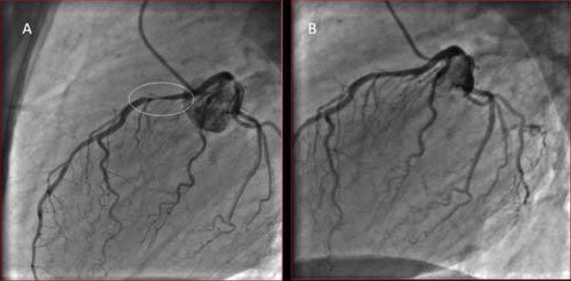
Coronary angiography showing severe stenosis of the left anterior descending (LAD) artery
Image: “Coronary angiography showing a severe proximal LAD stenosis” by V. Parisi et al. License: CC BY 2.0
Zipes, D., Libby, P., Bonow, R., Mann, D., Tomaselli, G., Braunwald, E. (2019). Braunwald’s Heart Disease: A Textbook of Cardiovascular Medicine. Elsevier/Saunders.
Toth, I., Szucs, G., Molnar, T. F. (2012). Mediastinoscope-controlled parasternal fenestration of the pericardium: definitive surgical palliation of malignant pericardial effusion. Journal of Cardiothoracic Surgery 7:56. https://doi.org/10.1186/1749-8090-7-56
Mueller, X. M., Tevaearai, H. T., Hurni, M., Ruchat, P., Fischer, A. P., Stumpe, F., von Segesser, L. K. (1997). Long-term results of surgical subxiphoid pericardial drainage. Thoracic and Cardiovascular Surgeon 45:65–69. https://doi.org/10.1055/s-2007-1013689