Vascular surgery is the specialized field of medicine that focuses on the surgical management of the pathologies of the peripheral circulationCirculationThe movement of the blood as it is pumped through the cardiovascular system.ABCDE Assessment. The main goal of most vascular procedures is to restore circulatory function to the affected vessels by relieving occlusions or by redirecting blood flowBlood flowBlood flow refers to the movement of a certain volume of blood through the vasculature over a given unit of time (e.g., mL per minute).Vascular Resistance, Flow, and Mean Arterial Pressure (e.g., bypass). Surgical intervention is either open or endovascular. Vascular interventions require a multidisciplinary approach, including vascular surgeons, interventional radiologists, anesthesiologistsAnesthesiologistsPhysicians specializing in anesthesiology.Anesthesiology: History and Basic Concepts (or anesthetists), nurses, physiotherapists, and occupational therapists.
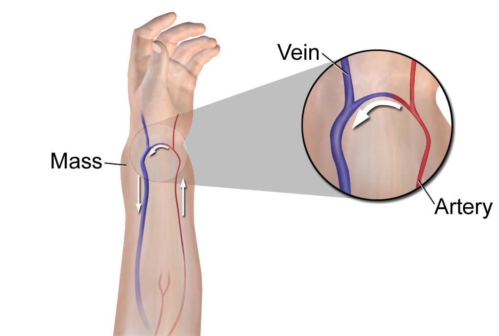
An AV fistulaFistulaAbnormal communication most commonly seen between two internal organs, or between an internal organ and the surface of the body.Anal Fistula is a surgically created anastomosis between an artery and a vein. This procedure is commonly performed for individuals with end-stage renal disease who require permanent vascular access for hemodialysisHemodialysisProcedures which temporarily or permanently remedy insufficient cleansing of body fluids by the kidneys.Crush Syndrome, although it may also be congenitalCongenitalChorioretinitis.
Indications
CKDCKDChronic kidney disease (CKD) is kidney impairment that lasts for ≥ 3 months, implying that it is irreversible. Hypertension and diabetes are the most common causes; however, there are a multitude of other etiologies. In the early to moderate stages, CKD is usually asymptomatic and is primarily diagnosed by laboratory abnormalities.Chronic Kidney Disease stage 5: defined by a GFRGFRThe volume of water filtered out of plasma through glomerular capillary walls into Bowman’s capsules per unit of time. It is considered to be equivalent to inulin clearance.Kidney Function Tests < 30 mL/min
Imminent need for dialysisDialysisRenal replacement therapy refers to dialysis and/or kidney transplantation. Dialysis is a procedure by which toxins and excess water are removed from the circulation. Hemodialysis and peritoneal dialysis (PD) are the two types of dialysis, and their primary difference is the location of the filtration process (external to the body in hemodialysis versus inside the body for PD).Peritoneal Dialysis and Hemodialysis
ContraindicationsContraindicationsA condition or factor associated with a recipient that makes the use of a drug, procedure, or physical agent improper or inadvisable. Contraindications may be absolute (life threatening) or relative (higher risk of complications in which benefits may outweigh risks).Noninvasive Ventilation
Venous occlusion
AmputationAmputationAn amputation is the separation of a portion of the limb or the entire limb from the body, along with the bone. Amputations are generally indicated for conditions that compromise the viability of the limb or promote the spread of a local process that could manifest systemically. Amputation
Advanced peripheral artery diseasePeripheral artery diseasePeripheral artery disease (PAD) is obstruction of the arterial lumen resulting in decreased blood flow to the distal limbs. The disease can be a result of atherosclerosis or thrombosis. Patients may be asymptomatic or have progressive claudication, skin discoloration, ischemic ulcers, or gangrene. Peripheral Artery Disease with necrosisNecrosisThe death of cells in an organ or tissue due to disease, injury or failure of the blood supply.Ischemic Cell Damage on the side of AV fistulaFistulaAbnormal communication most commonly seen between two internal organs, or between an internal organ and the surface of the body.Anal Fistula creation
Procedure
Preoperative carePreoperative CareThorough preoperative care is important for patients scheduled to undergo surgery so that they can have the best possible outcomes after their surgical procedure. The preoperative process begins once the decision has been made to proceed with a surgical procedure. Preoperative Care:
Previous fast (nil per os (NPO)) for 8 hours
Explain the procedure, benefits, risks, and alternatives to obtain informed consentInformed consentInformed consent is a medicolegal term describing the documented conversation between a patient and their physician wherein the physician discloses all relevant and necessary information to a patient who is competent to make an informed and voluntary decision regarding their care. Competency, disclosure, and voluntariness are the key elements upon which IC is based.Informed Consent.
Labs:
Platelet count > 50,000
PTT and PT within acceptable ranges
Required imaging: duplex ultrasonographyDuplex ultrasonographyUltrasonography applying the doppler effect combined with real-time imaging. The real-time image is created by rapid movement of the ultrasound beam. A powerful advantage of this technique is the ability to estimate the velocity of flow from the doppler shift frequency.Hypercoagulable States
AnticoagulantsAnticoagulantsAnticoagulants are drugs that retard or interrupt the coagulation cascade. The primary classes of available anticoagulants include heparins, vitamin K-dependent antagonists (e.g., warfarin), direct thrombin inhibitors, and factor Xa inhibitors. Anticoagulants are held before the procedure.
ECGECGAn electrocardiogram (ECG) is a graphic representation of the electrical activity of the heart plotted against time. Adhesive electrodes are affixed to the skin surface allowing measurement of cardiac impulses from many angles. The ECG provides 3-dimensional information about the conduction system of the heart, the myocardium, and other cardiac structures. Electrocardiogram (ECG) rhythm monitor
Operative care:
The most common AV fistulaFistulaAbnormal communication most commonly seen between two internal organs, or between an internal organ and the surface of the body.Anal Fistula techniques include:
Radiocephalic fistulaFistulaAbnormal communication most commonly seen between two internal organs, or between an internal organ and the surface of the body.Anal Fistula
Brachiocephalic fistulaFistulaAbnormal communication most commonly seen between two internal organs, or between an internal organ and the surface of the body.Anal Fistula
Transposed brachiobasilic fistulaFistulaAbnormal communication most commonly seen between two internal organs, or between an internal organ and the surface of the body.Anal Fistula
Recommended to perform this on the nondominant armArmThe arm, or “upper arm” in common usage, is the region of the upper limb that extends from the shoulder to the elbow joint and connects inferiorly to the forearm through the cubital fossa. It is divided into 2 fascial compartments (anterior and posterior).Arm: Anatomy
Access placed as distally as possible
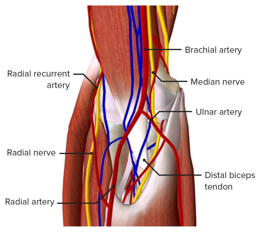
Radiocephalic fistulaFistulaAbnormal communication most commonly seen between two internal organs, or between an internal organ and the surface of the body.Anal Fistula (Brescia-Cimino fistulaFistulaAbnormal communication most commonly seen between two internal organs, or between an internal organ and the surface of the body.Anal Fistula):
A transverse incision is made in the wrist.
The radial arteryRadial ArteryThe direct continuation of the brachial trunk, originating at the bifurcation of the brachial artery opposite the neck of the radius. Its branches may be divided into three groups corresponding to the three regions in which the vessel is situated, the forearm, wrist, and hand.Forearm: Anatomy and cephalic vein are dissected.
An anterolateral arteriotomy is made on the radial arteryRadial ArteryThe direct continuation of the brachial trunk, originating at the bifurcation of the brachial artery opposite the neck of the radius. Its branches may be divided into three groups corresponding to the three regions in which the vessel is situated, the forearm, wrist, and hand.Forearm: Anatomy, and a corresponding venotomy is made on the cephalic vein.
The 2 vessels are anastomosed using a nonabsorbable suture.
Brachiocephalic fistulaFistulaAbnormal communication most commonly seen between two internal organs, or between an internal organ and the surface of the body.Anal Fistula (Kaufmann fistulaFistulaAbnormal communication most commonly seen between two internal organs, or between an internal organ and the surface of the body.Anal Fistula):
The brachial arteryBrachial ArteryThe continuation of the axillary artery; it branches into the radial and ulnar arteries.Cubital Fossa: Anatomy and cephalic vein are dissected.
Arteriotomy and venotomy are performed on each vessel.
The vessels are anastomosed using a nonabsorbable suture.
Transposed brachiobasilic fistulaFistulaAbnormal communication most commonly seen between two internal organs, or between an internal organ and the surface of the body.Anal Fistula (2-stage approach):
An arteriotomy is made in the distal brachial arteryBrachial ArteryThe continuation of the axillary artery; it branches into the radial and ulnar arteries.Cubital Fossa: Anatomy.
An end of the basilic veinBasilic veinArm: Anatomy is anastomosed to the arteriotomy on the brachial arteryBrachial ArteryThe continuation of the axillary artery; it branches into the radial and ulnar arteries.Cubital Fossa: Anatomy using nonabsorbable suture.
Final steps:
Vascular DopplerDopplerUltrasonography applying the doppler effect, with frequency-shifted ultrasound reflections produced by moving targets (usually red blood cells) in the bloodstream along the ultrasound axis in direct proportion to the velocity of movement of the targets, to determine both direction and velocity of blood flow.Ultrasound (Sonography) imaging is used to confirm patency.
The skinSkinThe skin, also referred to as the integumentary system, is the largest organ of the body. The skin is primarily composed of the epidermis (outer layer) and dermis (deep layer). The epidermis is primarily composed of keratinocytes that undergo rapid turnover, while the dermis contains dense layers of connective tissue.Skin: Structure and Functions is closed by layers using nonabsorbable suturesNonabsorbable SuturesSurgical Instruments and Sutures and cleansed of any residue (e.g., blood, adipose tissueAdipose tissueAdipose tissue is a specialized type of connective tissue that has both structural and highly complex metabolic functions, including energy storage, glucose homeostasis, and a multitude of endocrine capabilities. There are three types of adipose tissue, white adipose tissue, brown adipose tissue, and beige or “brite” adipose tissue, which is a transitional form.Adipose Tissue: Histology).
Postoperative carePostoperative careAfter any procedure performed in the operating room, all patients must undergo close observation at least in the recovery room. After larger procedures and for patients who require hospitalization, observation must continue on the surgical ward. The primary intent of this practice is the early detection of postoperative complications. Postoperative Care:
Observation in the recovery roomRecovery roomHospital unit providing continuous monitoring of the patient following anesthesia.Postoperative Care for 6 hours if necessary, or moved directly to the wards, according to each individual case.
FistulaFistulaAbnormal communication most commonly seen between two internal organs, or between an internal organ and the surface of the body.Anal Fistula maintenance:
Monitoring during dialysisDialysisRenal replacement therapy refers to dialysis and/or kidney transplantation. Dialysis is a procedure by which toxins and excess water are removed from the circulation. Hemodialysis and peritoneal dialysis (PD) are the two types of dialysis, and their primary difference is the location of the filtration process (external to the body in hemodialysis versus inside the body for PD).Peritoneal Dialysis and Hemodialysis sessions.
Individuals can be taught to examine the fistulaFistulaAbnormal communication most commonly seen between two internal organs, or between an internal organ and the surface of the body.Anal Fistula for a thrill (indicates patency). The fistulaFistulaAbnormal communication most commonly seen between two internal organs, or between an internal organ and the surface of the body.Anal Fistula should also be evaluated by a physician with routine appointments.
Ensure proper cleanliness and avoidance of clothing and jewelry over the access point to prevent flowFlowBlood flows through the heart, arteries, capillaries, and veins in a closed, continuous circuit. Flow is the movement of volume per unit of time. Flow is affected by the pressure gradient and the resistance fluid encounters between 2 points. Vascular resistance is the opposition to flow, which is caused primarily by blood friction against vessel walls.Vascular Resistance, Flow, and Mean Arterial Pressure restriction.
Bathing and everyday activities can be resumed as tolerated.
Complications
Steal syndrome:
Blood entering the limb moves through the fistulaFistulaAbnormal communication most commonly seen between two internal organs, or between an internal organ and the surface of the body.Anal Fistula without entering capillariesCapillariesCapillaries are the primary structures in the circulatory system that allow the exchange of gas, nutrients, and other materials between the blood and the extracellular fluid (ECF). Capillaries are the smallest of the blood vessels. Because a capillary diameter is so small, only 1 RBC may pass through at a time.Capillaries: Histology.
Clinical presentation: handHandThe hand constitutes the distal part of the upper limb and provides the fine, precise movements needed in activities of daily living. It consists of 5 metacarpal bones and 14 phalanges, as well as numerous muscles innervated by the median and ulnar nerves. Hand: AnatomypainPainAn unpleasant sensation induced by noxious stimuli which are detected by nerve endings of nociceptive neurons.Pain: Types and Pathways, coldness, sensorySensoryNeurons which conduct nerve impulses to the central nervous system.Nervous System: Histology and/or motorMotorNeurons which send impulses peripherally to activate muscles or secretory cells.Nervous System: Histology dysfunction, cyanosisCyanosisA bluish or purplish discoloration of the skin and mucous membranes due to an increase in the amount of deoxygenated hemoglobin in the blood or a structural defect in the hemoglobin molecule.Pulmonary Examination or pallor of the digits, and reduced or absent pulses,
Secondary to decreased blood flowBlood flowBlood flow refers to the movement of a certain volume of blood through the vasculature over a given unit of time (e.g., mL per minute).Vascular Resistance, Flow, and Mean Arterial Pressure to the distal extremity
ThrombosisThrombosisFormation and development of a thrombus or blood clot in the blood vessel.Epidemic Typhus
HematomaHematomaA collection of blood outside the blood vessels. Hematoma can be localized in an organ, space, or tissue.Intussusception
Hemorrhage
EdemaEdemaEdema is a condition in which excess serous fluid accumulates in the body cavity or interstitial space of connective tissues. Edema is a symptom observed in several medical conditions. It can be categorized into 2 types, namely, peripheral (in the extremities) and internal (in an organ or body cavity). Edema
AneurysmAneurysmAn aneurysm is a bulging, weakened area of a blood vessel that causes an abnormal widening of its diameter > 1.5 times the size of the native vessel. Aneurysms occur more often in arteries than in veins and are at risk of dissection and rupture, which can be life-threatening. Thoracic Aortic Aneurysms/pseudoaneurysmPseudoaneurysmNot an aneurysm but a well-defined collection of blood and connective tissue outside the wall of a blood vessel or the heart. It is the containment of a ruptured blood vessel or heart, such as sealing a rupture of the left ventricle. False aneurysm is formed by organized thrombus and hematoma in surrounding tissue.Thoracic Aortic Aneurysms:
Common complication because of repetitive needle sticks during dialysisDialysisRenal replacement therapy refers to dialysis and/or kidney transplantation. Dialysis is a procedure by which toxins and excess water are removed from the circulation. Hemodialysis and peritoneal dialysis (PD) are the two types of dialysis, and their primary difference is the location of the filtration process (external to the body in hemodialysis versus inside the body for PD).Peritoneal Dialysis and Hemodialysis
To avoid this complication, the needle should be inserted at different points in a rotating pattern.
FistulaFistulaAbnormal communication most commonly seen between two internal organs, or between an internal organ and the surface of the body.Anal Fistula infection
Image: “Blausen 0049 ArteriovenousFistula” by BruceBlaus. License: CC BY 3.0
Thrombectomy/Embolectomy
Definition
Thrombectomy is an interventional procedure by which a blood clot or thrombus is surgically removed from a vessel via endovascular devices under imaging guidance.
Recirculation thrombectomy: pulverizes the thrombus into microscopic fragments
Nonrecirculation thrombectomy: macerates the thrombus into macroscopic fragments
Energy-assisted thrombectomy: uses ultrasound, laser, or radiofrequency to lyse the thrombus
Indications
Stroke: injury undergone by brainBrainThe part of central nervous system that is contained within the skull (cranium). Arising from the neural tube, the embryonic brain is comprised of three major parts including prosencephalon (the forebrain); mesencephalon (the midbrain); and rhombencephalon (the hindbrain). The developed brain consists of cerebrum; cerebellum; and other structures in the brain stem.Nervous System: Anatomy, Structure, and Classification tissue after interruption of blood flowBlood flowBlood flow refers to the movement of a certain volume of blood through the vasculature over a given unit of time (e.g., mL per minute).Vascular Resistance, Flow, and Mean Arterial Pressure (ischemic strokeIschemic StrokeAn ischemic stroke (also known as cerebrovascular accident) is an acute neurologic injury that occurs as a result of brain ischemia; this condition may be due to cerebral blood vessel occlusion by thrombosis or embolism, or rarely due to systemic hypoperfusion. Ischemic Stroke) or active hemorrhage (hemorrhagic strokeHemorrhagic strokeStroke due to rupture of a weakened blood vessel in the brain (e.g., cerebral hemispheres; cerebellum; subarachnoid space).Subarachnoid Hemorrhage) that has characteristic neurologic deficitsNeurologic DeficitsHigh-Risk Headaches
MIMIMI is ischemia and death of an area of myocardial tissue due to insufficient blood flow and oxygenation, usually from thrombus formation on a ruptured atherosclerotic plaque in the epicardial arteries. Clinical presentation is most commonly with chest pain, but women and patients with diabetes may have atypical symptoms.Myocardial Infarction: injury to the myocardiumMyocardiumThe muscle tissue of the heart. It is composed of striated, involuntary muscle cells connected to form the contractile pump to generate blood flow.Heart: Anatomy due to ischemiaIschemiaA hypoperfusion of the blood through an organ or tissue caused by a pathologic constriction or obstruction of its blood vessels, or an absence of blood circulation.Ischemic Cell Damage, characterized by an increase in cardiac enzymesEnzymesEnzymes are complex protein biocatalysts that accelerate chemical reactions without being consumed by them. Due to the body’s constant metabolic needs, the absence of enzymes would make life unsustainable, as reactions would occur too slowly without these molecules. Basics of Enzymes (especially troponin T), ECGECGAn electrocardiogram (ECG) is a graphic representation of the electrical activity of the heart plotted against time. Adhesive electrodes are affixed to the skin surface allowing measurement of cardiac impulses from many angles. The ECG provides 3-dimensional information about the conduction system of the heart, the myocardium, and other cardiac structures. Electrocardiogram (ECG) changes suggestive of ischemiaIschemiaA hypoperfusion of the blood through an organ or tissue caused by a pathologic constriction or obstruction of its blood vessels, or an absence of blood circulation.Ischemic Cell Damage in 2 contiguous leads, and chest painPainAn unpleasant sensation induced by noxious stimuli which are detected by nerve endings of nociceptive neurons.Pain: Types and Pathways.
Pulmonary embolismPulmonary EmbolismPulmonary embolism (PE) is a potentially fatal condition that occurs as a result of intraluminal obstruction of the main pulmonary artery or its branches. The causative factors include thrombi, air, amniotic fluid, and fat. In PE, gas exchange is impaired due to the decreased return of deoxygenated blood to the lungs. Pulmonary Embolism (PE): potentially fatal condition that occurs as a result of intraluminal obstruction of the main pulmonary arteryPulmonary arteryThe short wide vessel arising from the conus arteriosus of the right ventricle and conveying unaerated blood to the lungs.Lungs: Anatomy or its branches by material (e.g., thrombus, air, amniotic fluidAmniotic fluidA clear, yellowish liquid that envelopes the fetus inside the sac of amnion. In the first trimester, it is likely a transudate of maternal or fetal plasma. In the second trimester, amniotic fluid derives primarily from fetal lung and kidney. Cells or substances in this fluid can be removed for prenatal diagnostic tests (amniocentesis).Placenta, Umbilical Cord, and Amniotic Cavity, or fat)
ContraindicationsContraindicationsA condition or factor associated with a recipient that makes the use of a drug, procedure, or physical agent improper or inadvisable. Contraindications may be absolute (life threatening) or relative (higher risk of complications in which benefits may outweigh risks).Noninvasive Ventilation
Intracranial hemorrhageIntracranial hemorrhageSubarachnoid hemorrhage (SAH) is a type of cerebrovascular accident (stroke) resulting from intracranial hemorrhage into the subarachnoid space between the arachnoid and the pia mater layers of the meninges surrounding the brain. Most sahs originate from a saccular aneurysm in the circle of willis but may also occur as a result of trauma, uncontrolled hypertension, vasculitis, anticoagulant use, or stimulant use.Subarachnoid Hemorrhage
Large infarctInfarctArea of necrotic cells in an organ, arising mainly from hypoxia and ischemiaIschemic Cell Damage core with minimal penumbra
Uncontrolled hypertensionUncontrolled hypertensionAlthough hypertension is defined as a blood pressure of > 130/80 mm Hg, individuals can present with comorbidities of severe asymptomatic or “uncontrolled” hypertension (≥ 180 mm Hg systolic and/or ≥ 120 mm Hg diastolic) that carries with it a significant risk of morbidity and mortality. Uncontrolled Hypertension
Procedure
Preoperative carePreoperative CareThorough preoperative care is important for patients scheduled to undergo surgery so that they can have the best possible outcomes after their surgical procedure. The preoperative process begins once the decision has been made to proceed with a surgical procedure. Preoperative Care:
Dietary fast (nil per os (NPO)) for 8 hours.
Explain the procedure, benefits, risks, and alternatives to obtain informed consentInformed consentInformed consent is a medicolegal term describing the documented conversation between a patient and their physician wherein the physician discloses all relevant and necessary information to a patient who is competent to make an informed and voluntary decision regarding their care. Competency, disclosure, and voluntariness are the key elements upon which IC is based.Informed Consent.
Labs:
Platelet count > 50,000
PTT and PT within acceptable ranges
Renal function: serum creatinine and blood ureaUreaA compound formed in the liver from ammonia produced by the deamination of amino acids. It is the principal end product of protein catabolism and constitutes about one half of the total urinary solids.Urea CyclenitrogenNitrogenAn element with the atomic symbol n, atomic number 7, and atomic weight [14. 00643; 14. 00728]. Nitrogen exists as a diatomic gas and makes up about 78% of the earth’s atmosphere by volume. It is a constituent of proteins and nucleic acids and found in all living cells.Urea Cycle within normal ranges.
HbA1c
Diagnostic images: ultrasound, head CT scan
AnticoagulantsAnticoagulantsAnticoagulants are drugs that retard or interrupt the coagulation cascade. The primary classes of available anticoagulants include heparins, vitamin K-dependent antagonists (e.g., warfarin), direct thrombin inhibitors, and factor Xa inhibitors. Anticoagulants are withheld before the procedure.
In the OR:
The individual is placed in the supine position.
IV access is obtained.
Continuous monitoring:
HR
Blood pressure
O2 saturation (pulse oximetry)
ECGECGAn electrocardiogram (ECG) is a graphic representation of the electrical activity of the heart plotted against time. Adhesive electrodes are affixed to the skin surface allowing measurement of cardiac impulses from many angles. The ECG provides 3-dimensional information about the conduction system of the heart, the myocardium, and other cardiac structures. Electrocardiogram (ECG) rhythm monitor
Operative care:
There are several thrombectomy techniques. The following uses a balloon catheter and stent for thrombus removal.
A balloon catheter is inserted through a groinGroinThe external junctural region between the lower part of the abdomen and the thigh.Male Genitourinary Examination puncture.
The catheter is advanced until it reaches the thrombus.
A guidewire is advanced through the thrombus.
A microcatheter is passed over the guidewire and through the thrombus.
A stent is deployed, and once it reaches the thrombus, it is deployed within the vessel.
The projections of the stent secure the thrombus to its surface.
Contrast is injected through the balloon catheter to ensure patency of the vessel.
The balloon is inflated to temporarily restrict flowFlowBlood flows through the heart, arteries, capillaries, and veins in a closed, continuous circuit. Flow is the movement of volume per unit of time. Flow is affected by the pressure gradient and the resistance fluid encounters between 2 points. Vascular resistance is the opposition to flow, which is caused primarily by blood friction against vessel walls.Vascular Resistance, Flow, and Mean Arterial Pressure and allow removal of the stent with the thrombus and microcatheter.
AngiographyAngiographyRadiography of blood vessels after injection of a contrast medium.Cardiac Surgery is performed to check if the thrombus was completely removed.
Postoperative carePostoperative careAfter any procedure performed in the operating room, all patients must undergo close observation at least in the recovery room. After larger procedures and for patients who require hospitalization, observation must continue on the surgical ward. The primary intent of this practice is the early detection of postoperative complications. Postoperative Care:
Observation in the recovery roomRecovery roomHospital unit providing continuous monitoring of the patient following anesthesia.Postoperative Care for 6 hours; later moved to the wards. Individuals with neurologic conditions may need to be admitted to the neurosurgeryNeurosurgeryNeurosurgery is a specialized field focused on the surgical management of pathologies of the brain, spine, spinal cord, and peripheral nerves. General neurosurgery includes cases of trauma and emergencies. There are a number of specialized neurosurgical practices, including oncologic neurosurgery, spinal neurosurgery, and pediatric neurosurgery. NeurosurgeryICUICUHospital units providing continuous surveillance and care to acutely ill patients.West Nile Virus.
Close monitoring of:
Blood pressure (hypertensionHypertensionHypertension, or high blood pressure, is a common disease that manifests as elevated systemic arterial pressures. Hypertension is most often asymptomatic and is found incidentally as part of a routine physical examination or during triage for an unrelated medical encounter. Hypertension → reperfusion injuryReperfusion injuryAdverse functional, metabolic, or structural changes in ischemic tissues resulting from the restoration of blood flow to the tissue (reperfusion), including swelling; hemorrhage; necrosis; and damage from free radicals. The most common instance is myocardial reperfusion injury.Ischemic Cell Damage)
Temperature (feverFeverFever is defined as a measured body temperature of at least 38°C (100.4°F). Fever is caused by circulating endogenous and/or exogenous pyrogens that increase levels of prostaglandin E2 in the hypothalamus. Fever is commonly associated with chills, rigors, sweating, and flushing of the skin. Fever)
Vessel perforationPerforationA pathological hole in an organ, blood vessel or other soft part of the body, occurring in the absence of external force.Esophagitis or dissection
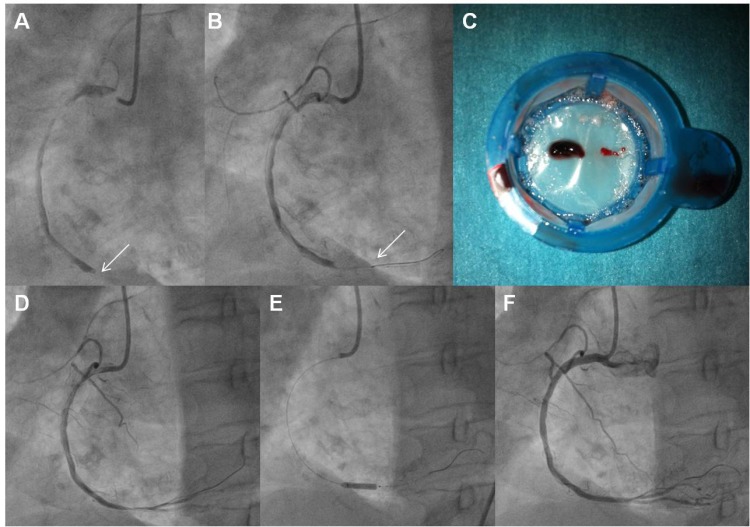
Image: “CCR-8-202_F2” by Dimitrios Alexopoulos* and Periklis A Davlouros. License: CC BY 2.5
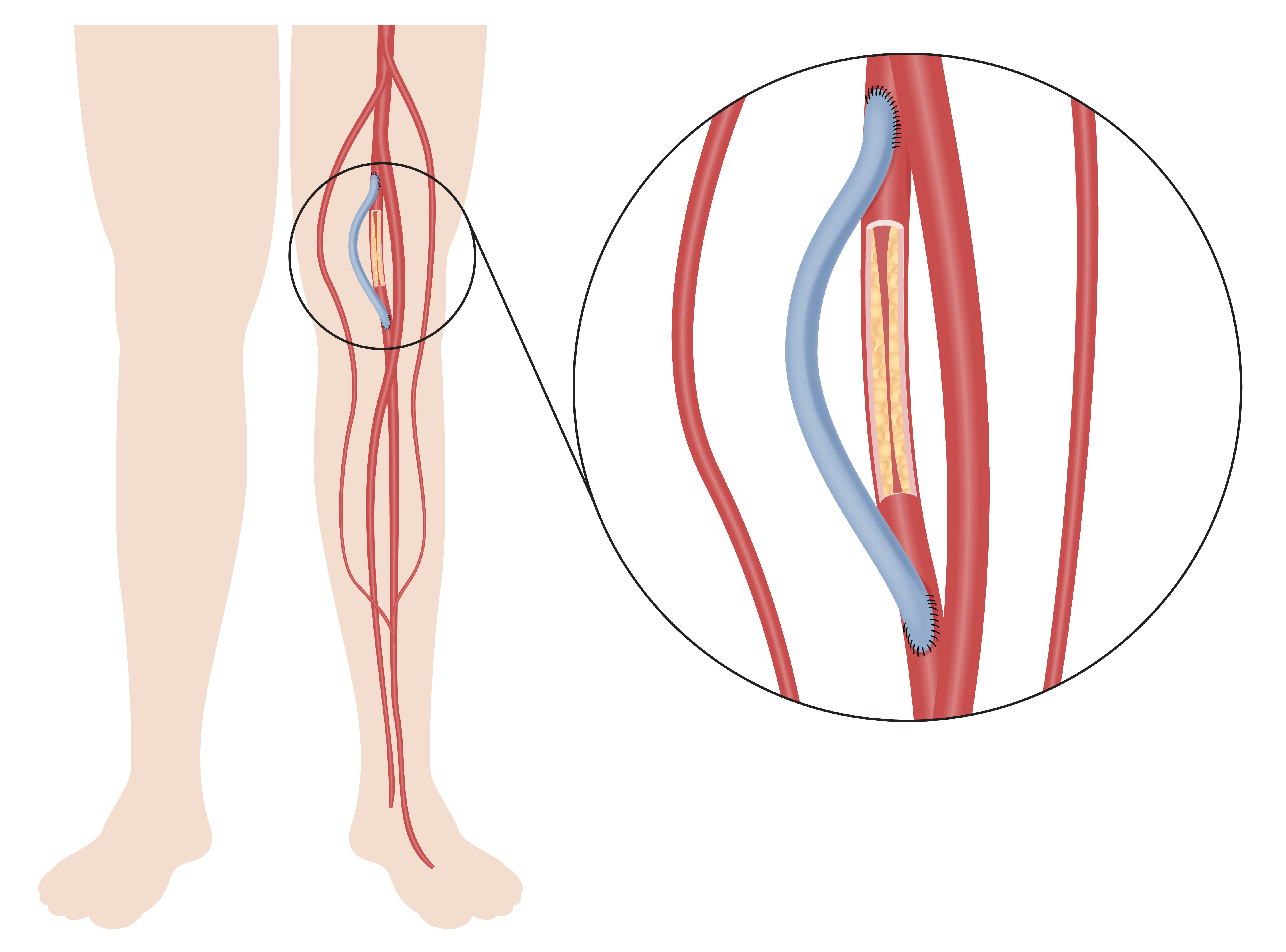
Peripheral Artery Bypass (PAB)
Definition
A PAB (also known as a peripheral vascular bypass) is a surgical procedure that uses a graftGraftA piece of living tissue that is surgically transplantedOrgan Transplantation to restore perfusion to a segment of the arterial circulationCirculationThe movement of the blood as it is pumped through the cardiovascular system.ABCDE Assessment distal to an occlusion. This procedure can be performed in potentially any segment of circulationCirculationThe movement of the blood as it is pumped through the cardiovascular system.ABCDE Assessment.
Indications
Peripheral artery diseasePeripheral artery diseasePeripheral artery disease (PAD) is obstruction of the arterial lumen resulting in decreased blood flow to the distal limbs. The disease can be a result of atherosclerosis or thrombosis. Patients may be asymptomatic or have progressive claudication, skin discoloration, ischemic ulcers, or gangrene. Peripheral Artery Disease (PAD): Obstruction of the arterial lumen, commonly secondary to atherosclerosisAtherosclerosisAtherosclerosis is a common form of arterial disease in which lipid deposition forms a plaque in the blood vessel walls. Atherosclerosis is an incurable disease, for which there are clearly defined risk factors that often can be reduced through a change in lifestyle and behavior of the patient. Atherosclerosis or thrombosisThrombosisFormation and development of a thrombus or blood clot in the blood vessel.Epidemic Typhus, that results in decreased blood flowBlood flowBlood flow refers to the movement of a certain volume of blood through the vasculature over a given unit of time (e.g., mL per minute).Vascular Resistance, Flow, and Mean Arterial Pressure to visceral organs or the distal limbs. Individuals may initially be asymptomatic and later develop organ dysfunction, claudication, skinSkinThe skin, also referred to as the integumentary system, is the largest organ of the body. The skin is primarily composed of the epidermis (outer layer) and dermis (deep layer). The epidermis is primarily composed of keratinocytes that undergo rapid turnover, while the dermis contains dense layers of connective tissue.Skin: Structure and Functions discoloration, ischemic ulcers, or gangreneGangreneDeath and putrefaction of tissue usually due to a loss of blood supply.Small Bowel Obstruction.
Narrowing in the brainBrainThe part of central nervous system that is contained within the skull (cranium). Arising from the neural tube, the embryonic brain is comprised of three major parts including prosencephalon (the forebrain); mesencephalon (the midbrain); and rhombencephalon (the hindbrain). The developed brain consists of cerebrum; cerebellum; and other structures in the brain stem.Nervous System: Anatomy, Structure, and Classification is termed cerebrovascular disease.
Narrowing in the kidneysKidneysThe kidneys are a pair of bean-shaped organs located retroperitoneally against the posterior wall of the abdomen on either side of the spine. As part of the urinary tract, the kidneys are responsible for blood filtration and excretion of water-soluble waste in the urine.Kidneys: Anatomy is termed renovascular disease.
Narrowing in the legs may cause legLegThe lower leg, or just “leg” in anatomical terms, is the part of the lower limb between the knee and the ankle joint. The bony structure is composed of the tibia and fibula bones, and the muscles of the leg are grouped into the anterior, lateral, and posterior compartments by extensions of fascia.Leg: AnatomypainPainAn unpleasant sensation induced by noxious stimuli which are detected by nerve endings of nociceptive neurons.Pain: Types and Pathways with walking (intermittent claudicationIntermittent claudicationA symptom complex characterized by pain and weakness in skeletal muscle group associated with exercise, such as leg pain and weakness brought on by walking. Such muscle limpness disappears after a brief rest and is often relates to arterial stenosis; muscle ischemia; and accumulation of lactate.Thromboangiitis Obliterans (Buerger Disease))
SmokingSmokingWillful or deliberate act of inhaling and exhaling smoke from burning substances or agents held by hand.Interstitial Lung Diseases is the strongest risk factor for PAD
Traumatic arterial injuries
Aneurysms
ContraindicationsContraindicationsA condition or factor associated with a recipient that makes the use of a drug, procedure, or physical agent improper or inadvisable. Contraindications may be absolute (life threatening) or relative (higher risk of complications in which benefits may outweigh risks).Noninvasive Ventilation
History of cardiac surgeryCardiac surgeryCardiac surgery is the surgical management of cardiac abnormalities and of the great vessels of the thorax. In general terms, surgical intervention of the heart is performed to directly restore adequate pump function, correct inherent structural issues, and reestablish proper blood supply via the coronary circulation.Cardiac Surgery (e.g., stenting, angioplastyAngioplastyReconstruction or repair of a blood vessel, which includes the widening of a pathological narrowing of an artery or vein by the removal of atheromatous plaque material and/or the endothelial lining as well, or by dilatation (balloon angioplasty) to compress an atheroma. Except for endarterectomy, usually these procedures are performed via catheterization as minimally invasive endovascular procedures.Cardiac Surgery or coronary arteryCoronary ArteryTruncus Arteriosus bypass)
Preoperative carePreoperative CareThorough preoperative care is important for patients scheduled to undergo surgery so that they can have the best possible outcomes after their surgical procedure. The preoperative process begins once the decision has been made to proceed with a surgical procedure. Preoperative Care:
Dietary fast (nil per os (NPO)) for 8 hours
Explain the procedure and obtain informed consentInformed consentInformed consent is a medicolegal term describing the documented conversation between a patient and their physician wherein the physician discloses all relevant and necessary information to a patient who is competent to make an informed and voluntary decision regarding their care. Competency, disclosure, and voluntariness are the key elements upon which IC is based.Informed Consent.
Labs:
Platelet count > 50,000
Adequate hemoglobin and hematocritHematocritThe volume of packed red blood cells in a blood specimen. The volume is measured by centrifugation in a tube with graduated markings, or with automated blood cell counters. It is an indicator of erythrocyte status in disease. For example, anemia shows a low value; polycythemia, a high value.Neonatal Polycythemia
PTT and PT within acceptable ranges
Blood type, cross, and match
Renal function: serum creatinine and BUN within normal ranges (unless performing renovascular intervention).
HbA1c
Required imaging: ultrasonography
These procedures may involve significant blood loss requiring transfusion. Blood products are made available for transfusion after blood type is obtained.
AnticoagulantsAnticoagulantsAnticoagulants are drugs that retard or interrupt the coagulation cascade. The primary classes of available anticoagulants include heparins, vitamin K-dependent antagonists (e.g., warfarin), direct thrombin inhibitors, and factor Xa inhibitors. Anticoagulants are withheld before the procedure.
ECGECGAn electrocardiogram (ECG) is a graphic representation of the electrical activity of the heart plotted against time. Adhesive electrodes are affixed to the skin surface allowing measurement of cardiac impulses from many angles. The ECG provides 3-dimensional information about the conduction system of the heart, the myocardium, and other cardiac structures. Electrocardiogram (ECG) rhythm monitor
Operative care:
The procedure itself will vary widely depending on the surgical anatomy and location of the obstruction. However, the underlying principle of PAB is establishing proximal and distal anastomoses in disease-free segments. Some examples of PAB include:
Iliofemoral bypass: ipsilateral or contralateral iliac artery is communicated with the common femoral arteryFemoral ArteryThe main artery of the thigh, a continuation of the external iliac artery.Femoral Region and Hernias: Anatomy (CFA)
Aortobifemoral bypass: the abdominal aortaAbdominal AortaThe aorta from the diaphragm to the bifurcation into the right and left common iliac arteries.Posterior Abdominal Wall: Anatomy is communicated with both CFAs
Femoropopliteal bypass: the popliteal arteryPopliteal ArteryThe continuation of the femoral artery coursing through the popliteal fossa; it divides into the anterior and posterior tibial arteries.Popliteal Fossa: Anatomy is communicated with a single femoral arteryFemoral ArteryThe main artery of the thigh, a continuation of the external iliac artery.Femoral Region and Hernias: Anatomy
Grafts are created with organic (e.g., harvested saphenous vein) or artificial (e.g., polytetrafluoroethylene) materials.
Final steps:
The skinSkinThe skin, also referred to as the integumentary system, is the largest organ of the body. The skin is primarily composed of the epidermis (outer layer) and dermis (deep layer). The epidermis is primarily composed of keratinocytes that undergo rapid turnover, while the dermis contains dense layers of connective tissue.Skin: Structure and Functions is closed by layers using nonabsorbable suturesNonabsorbable SuturesSurgical Instruments and Sutures and cleansed of any residue (e.g., blood, adipose tissueAdipose tissueAdipose tissue is a specialized type of connective tissue that has both structural and highly complex metabolic functions, including energy storage, glucose homeostasis, and a multitude of endocrine capabilities. There are three types of adipose tissue, white adipose tissue, brown adipose tissue, and beige or “brite” adipose tissue, which is a transitional form.Adipose Tissue: Histology).
Postoperative carePostoperative careAfter any procedure performed in the operating room, all patients must undergo close observation at least in the recovery room. After larger procedures and for patients who require hospitalization, observation must continue on the surgical ward. The primary intent of this practice is the early detection of postoperative complications. Postoperative Care:
Observation in recovery roomRecovery roomHospital unit providing continuous monitoring of the patient following anesthesia.Postoperative Care for 6 hours; later moved to the wards
Physical examination:
Vascular: reduced pulses, prolonged capillary filling time, hematomas
Neurologic: paresthesiasParesthesiasSubjective cutaneous sensations (e.g., cold, warmth, tingling, pressure, etc.) that are experienced spontaneously in the absence of stimulation.Posterior Cord Syndrome, paralysis
Complications
Surgical site infectionSurgical site infectionInfection occurring at the site of a surgical incision.Surgical Complications (SSISSISurgical site infection (SSI) is a type of surgical infection that occurs at or near a surgical incision within 30 days of the procedure or within 90 days if prosthetic material is implanted. Surgical site infections are classified according to the depth of involvement as superficial, deep, or organ/space. Surgical Site Infections): A type of infection that occurs at or near a surgical incisionSurgical IncisionSurgical Site Infections within 30 days after the procedure or within 90 days if prosthetic material is implanted. An SSISSISurgical site infection (SSI) is a type of surgical infection that occurs at or near a surgical incision within 30 days of the procedure or within 90 days if prosthetic material is implanted. Surgical site infections are classified according to the depth of involvement as superficial, deep, or organ/space. Surgical Site Infections is classified according to the depth of compromise—superficial, deep, or organ/space.
Hemorrhage/hematomaHematomaA collection of blood outside the blood vessels. Hematoma can be localized in an organ, space, or tissue.Intussusception
AneurysmAneurysmAn aneurysm is a bulging, weakened area of a blood vessel that causes an abnormal widening of its diameter > 1.5 times the size of the native vessel. Aneurysms occur more often in arteries than in veins and are at risk of dissection and rupture, which can be life-threatening. Thoracic Aortic Aneurysms/pseudoaneurysmPseudoaneurysmNot an aneurysm but a well-defined collection of blood and connective tissue outside the wall of a blood vessel or the heart. It is the containment of a ruptured blood vessel or heart, such as sealing a rupture of the left ventricle. False aneurysm is formed by organized thrombus and hematoma in surrounding tissue.Thoracic Aortic Aneurysms
Failure of anastomoses
PneumoniaPneumoniaPneumonia or pulmonary inflammation is an acute or chronic inflammation of lung tissue. Causes include infection with bacteria, viruses, or fungi. In more rare cases, pneumonia can also be caused through toxic triggers through inhalation of toxic substances, immunological processes, or in the course of radiotherapy.Pneumonia
A CEACEAA glycoprotein that is secreted into the luminal surface of the epithelia in the gastrointestinal tract. It is found in the feces and pancreaticobiliary secretions and is used to monitor the response to colon cancer treatment.Serum Tumor Markers is a surgical procedure in which atherosclerotic plaquePlaquePrimary Skin Lesions is manually removed from the common and/or internal carotid arteryInternal carotid arteryBranch of the common carotid artery which supplies the anterior part of the brain, the eye and its appendages, the forehead and nose.Carotid Arterial System: Anatomy. The therapeutic goals are to restore normal blood flowBlood flowBlood flow refers to the movement of a certain volume of blood through the vasculature over a given unit of time (e.g., mL per minute).Vascular Resistance, Flow, and Mean Arterial Pressure and reduce the likelihood of embolizationEmbolizationA method of hemostasis utilizing various agents such as gelfoam, silastic, metal, glass, or plastic pellets, autologous clot, fat, and muscle as emboli. It has been used in the treatment of spinal cord and intracranial arteriovenous malformations, renal arteriovenous fistulas, gastrointestinal bleeding, epistaxis, hypersplenism, certain highly vascular tumors, traumatic rupture of blood vessels, and control of operative hemorrhage.Gastrointestinal Bleeding.
History of ipsilateral stroke or transient ischemic attackTransient ischemic attackTransient ischemic attack (TIA) is a temporary episode of neurologic dysfunction caused by ischemia without infarction that resolves completely when blood supply is restored. Transient ischemic attack is a neurologic emergency that warrants urgent medical attention. Transient Ischemic Attack (TIA) (TIATIATransient ischemic attack (TIA) is a temporary episode of neurologic dysfunction caused by ischemia without infarction that resolves completely when blood supply is restored. Transient ischemic attack is a neurologic emergency that warrants urgent medical attention. Transient Ischemic Attack (TIA))
ContraindicationsContraindicationsA condition or factor associated with a recipient that makes the use of a drug, procedure, or physical agent improper or inadvisable. Contraindications may be absolute (life threatening) or relative (higher risk of complications in which benefits may outweigh risks).Noninvasive Ventilation
Very ill and symptomatic individuals who are unable to undergo an open surgical procedure
History of radiationRadiationEmission or propagation of acoustic waves (sound), electromagnetic energy waves (such as light; radio waves; gamma rays; or x-rays), or a stream of subatomic particles (such as electrons; neutrons; protons; or alpha particles).Osteosarcoma therapy to the neckNeckThe part of a human or animal body connecting the head to the rest of the body.Peritonsillar Abscess
Individuals with high-likelihood of poor outcomes must be carefully selected.
Older age (> 70 years)
Severe heart disease
Severe pulmonary dysfunction
Renal insufficiency or failure
Stroke as the indication for endarterectomyEndarterectomySurgical excision, performed under general anesthesia, of the atheromatous tunica intima of an artery. When reconstruction of an artery is performed as an endovascular procedure through a catheter, it is called atherectomy.Intestinal Ischemia
Procedure
Preoperative carePreoperative CareThorough preoperative care is important for patients scheduled to undergo surgery so that they can have the best possible outcomes after their surgical procedure. The preoperative process begins once the decision has been made to proceed with a surgical procedure. Preoperative Care:
Dietary fast (nil per os (NPO)) of 8 hours
Explain the procedure and obtain informed consentInformed consentInformed consent is a medicolegal term describing the documented conversation between a patient and their physician wherein the physician discloses all relevant and necessary information to a patient who is competent to make an informed and voluntary decision regarding their care. Competency, disclosure, and voluntariness are the key elements upon which IC is based.Informed Consent.
Labs:
Platelet count > 50,000
PTT and PT within acceptable ranges
Renal function: serum creatinine and blood ureaUreaA compound formed in the liver from ammonia produced by the deamination of amino acids. It is the principal end product of protein catabolism and constitutes about one half of the total urinary solids.Urea CyclenitrogenNitrogenAn element with the atomic symbol n, atomic number 7, and atomic weight [14. 00643; 14. 00728]. Nitrogen exists as a diatomic gas and makes up about 78% of the earth’s atmosphere by volume. It is a constituent of proteins and nucleic acids and found in all living cells.Urea Cycle within normal ranges
HbA1c
Required images: carotid duplex ultrasonogram; head CT scan
Antiplatelet therapy with aspirinAspirinThe prototypical analgesic used in the treatment of mild to moderate pain. It has anti-inflammatory and antipyretic properties and acts as an inhibitor of cyclooxygenase which results in the inhibition of the biosynthesis of prostaglandins. Aspirin also inhibits platelet aggregation and is used in the prevention of arterial and venous thrombosis.Nonsteroidal Antiinflammatory Drugs (NSAIDs) (81–325 mg/day) prior to the procedure
AnticoagulantsAnticoagulantsAnticoagulants are drugs that retard or interrupt the coagulation cascade. The primary classes of available anticoagulants include heparins, vitamin K-dependent antagonists (e.g., warfarin), direct thrombin inhibitors, and factor Xa inhibitors. Anticoagulants are withheld if so indicated by the surgeon.
ECGECGAn electrocardiogram (ECG) is a graphic representation of the electrical activity of the heart plotted against time. Adhesive electrodes are affixed to the skin surface allowing measurement of cardiac impulses from many angles. The ECG provides 3-dimensional information about the conduction system of the heart, the myocardium, and other cardiac structures. Electrocardiogram (ECG) rhythm monitor
The individual can be placed under general or local anesthesiaAnesthesiaA state characterized by loss of feeling or sensation. This depression of nerve function is usually the result of pharmacologic action and is induced to allow performance of surgery or other painful procedures.Anesthesiology: History and Basic Concepts with sedation.
The skinSkinThe skin, also referred to as the integumentary system, is the largest organ of the body. The skin is primarily composed of the epidermis (outer layer) and dermis (deep layer). The epidermis is primarily composed of keratinocytes that undergo rapid turnover, while the dermis contains dense layers of connective tissue.Skin: Structure and Functions is closed by layers using nonabsorbable suturesNonabsorbable SuturesSurgical Instruments and Sutures and cleansed of any residue (e.g., blood, adipose tissueAdipose tissueAdipose tissue is a specialized type of connective tissue that has both structural and highly complex metabolic functions, including energy storage, glucose homeostasis, and a multitude of endocrine capabilities. There are three types of adipose tissue, white adipose tissue, brown adipose tissue, and beige or “brite” adipose tissue, which is a transitional form.Adipose Tissue: Histology).
Postoperative carePostoperative careAfter any procedure performed in the operating room, all patients must undergo close observation at least in the recovery room. After larger procedures and for patients who require hospitalization, observation must continue on the surgical ward. The primary intent of this practice is the early detection of postoperative complications. Postoperative Care:
Observation in recovery roomRecovery roomHospital unit providing continuous monitoring of the patient following anesthesia.Postoperative Care for 6 hours; later moved to the wards
Neurologic examination every hour.
Blood pressure control (hypertensionHypertensionHypertension, or high blood pressure, is a common disease that manifests as elevated systemic arterial pressures. Hypertension is most often asymptomatic and is found incidentally as part of a routine physical examination or during triage for an unrelated medical encounter. Hypertension → neckNeckThe part of a human or animal body connecting the head to the rest of the body.Peritonsillar AbscesshematomaHematomaA collection of blood outside the blood vessels. Hematoma can be localized in an organ, space, or tissue.Intussusception)
Complications
Hemorrhage
NeckNeckThe part of a human or animal body connecting the head to the rest of the body.Peritonsillar AbscesshematomaHematomaA collection of blood outside the blood vessels. Hematoma can be localized in an organ, space, or tissue.Intussusception
DysphagiaDysphagiaDysphagia is the subjective sensation of difficulty swallowing. Symptoms can range from a complete inability to swallow, to the sensation of solids or liquids becoming “stuck.” Dysphagia is classified as either oropharyngeal or esophageal, with esophageal dysphagia having 2 sub-types: functional and mechanical. Dysphagia
MIMIMI is ischemia and death of an area of myocardial tissue due to insufficient blood flow and oxygenation, usually from thrombus formation on a ruptured atherosclerotic plaque in the epicardial arteries. Clinical presentation is most commonly with chest pain, but women and patients with diabetes may have atypical symptoms.Myocardial Infarction
Cerebral hyperperfusion syndrome: A clinical presentation secondary to increased cerebral perfusionCerebral PerfusionSyncope after carotid endarterectomyEndarterectomySurgical excision, performed under general anesthesia, of the atheromatous tunica intima of an artery. When reconstruction of an artery is performed as an endovascular procedure through a catheter, it is called atherectomy.Intestinal Ischemia that is characterized by ipsilateral headacheHeadacheThe symptom of pain in the cranial region. It may be an isolated benign occurrence or manifestation of a wide variety of headache disorders.Brain Abscess, hypertensionHypertensionHypertension, or high blood pressure, is a common disease that manifests as elevated systemic arterial pressures. Hypertension is most often asymptomatic and is found incidentally as part of a routine physical examination or during triage for an unrelated medical encounter. Hypertension, seizuresSeizuresA seizure is abnormal electrical activity of the neurons in the cerebral cortex that can manifest in numerous ways depending on the region of the brain affected. Seizures consist of a sudden imbalance that occurs between the excitatory and inhibitory signals in cortical neurons, creating a net excitation. The 2 major classes of seizures are focal and generalized. Seizures, and focal neurological deficitsFocal Neurological DeficitsBrain Abscess. If not treated, cerebral hyperperfusion can result in severe cerebral edemaCerebral edemaIncreased intracellular or extracellular fluid in brain tissue. Cytotoxic brain edema (swelling due to increased intracellular fluid) is indicative of a disturbance in cell metabolism, and is commonly associated with hypoxic or ischemic injuries. An increase in extracellular fluid may be caused by increased brain capillary permeability (vasogenic edema), an osmotic gradient, local blockages in interstitial fluid pathways, or by obstruction of CSF flow (e.g., obstructive hydrocephalus).Increased Intracranial Pressure (ICP) or intracerebral or subarachnoid hemorrhageSubarachnoid HemorrhageSubarachnoid hemorrhage (SAH) is a type of cerebrovascular accident (stroke) resulting from intracranial hemorrhage into the subarachnoid space between the arachnoid and the pia mater layers of the meninges surrounding the brain. Most SAHs originate from a saccular aneurysm in the circle of Willis but may also occur as a result of trauma, uncontrolled hypertension, vasculitis, anticoagulant use, or stimulant use. Subarachnoid Hemorrhage.
Perioperative stroke and TIATIATransient ischemic attack (TIA) is a temporary episode of neurologic dysfunction caused by ischemia without infarction that resolves completely when blood supply is restored. Transient ischemic attack is a neurologic emergency that warrants urgent medical attention. Transient Ischemic Attack (TIA)
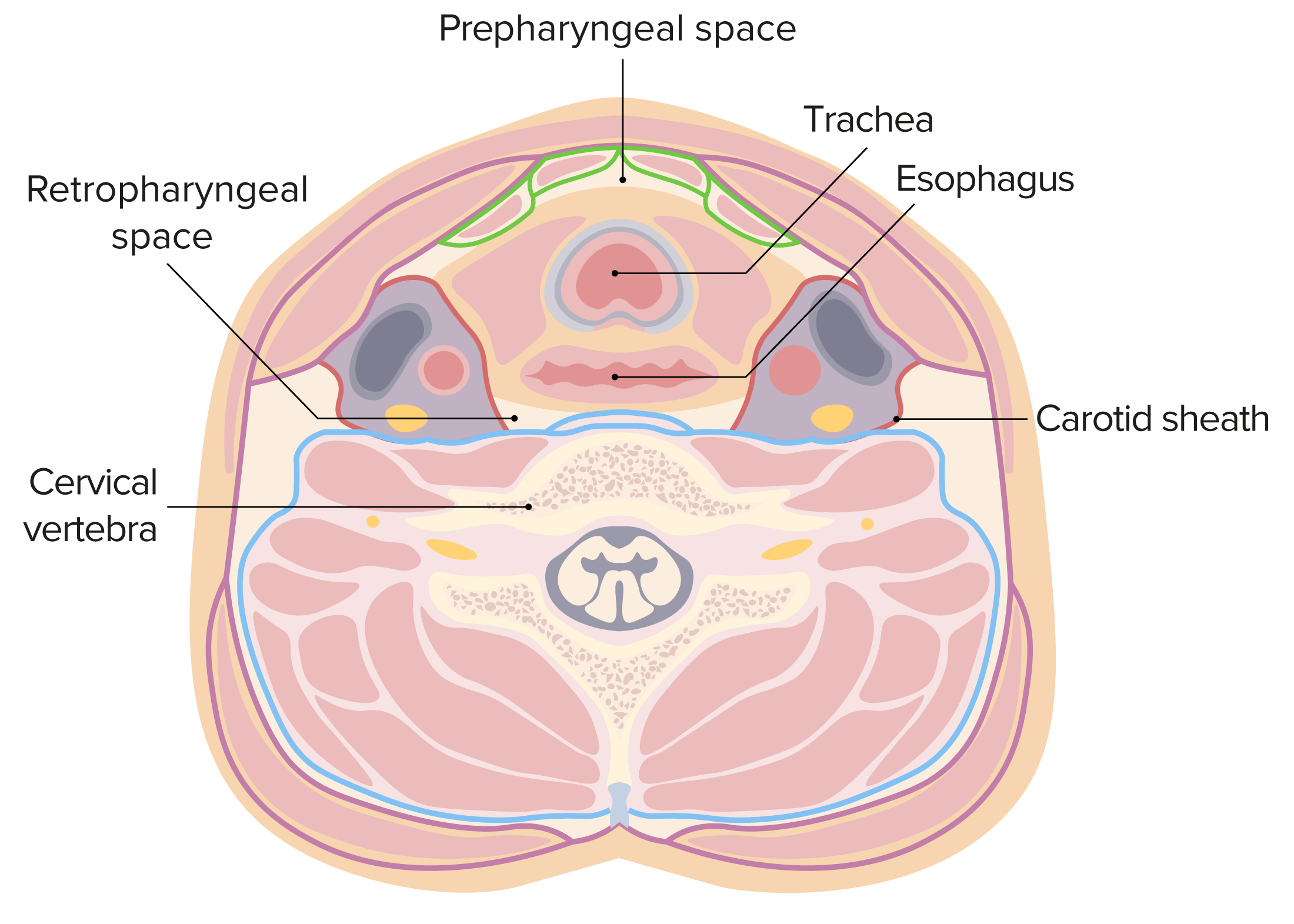
Cross section of the structures of the neck. Note the location of the carotid sheaths deep to the platysma and sternocleidomastoid muscles, as well as the orientation of the contents within the carotid sheath.
Elias, N., Stapleton, S. (2020). Creation of a radial-cephalic arteriovenous fistula. Journal of Medical Insight. 2020(110). https://doi.org/10.24296/jomi/110
Alexopoulos, D., Davlouros, P. A. (2012). Thrombus extraction catheters vs. angiojet rheolytic thrombectomy in thrombotic lesions/SV grafts. Current Cardiology Reviews. 8, 202–208. https://doi.org/10.2174/157340312803217265
Moore, W., Lawrence, P., Oderich, G. (2019). Moore’s Vascular and Endovascular Surgery: A Comprehensive Review. Elsevier.
Samaniego, E., Hasan, D. (2019). Acute stroke management in the era of thrombectomy. Cham: Springer.