Dysphagia is the subjective sensation of difficulty swallowingSwallowingThe act of taking solids and liquids into the gastrointestinal tract through the mouth and throat.Gastrointestinal Motility. Symptoms can range from a complete inability to swallow, to the sensation of solids or liquids becoming “stuck.” Dysphagia is classified as either oropharyngeal or esophageal, with esophageal dysphagia having 2 sub-types: functional and mechanical. Common causes of functional dysphagia include achalasiaAchalasiaAchalasia is a primary esophageal motility disorder that develops from the degeneration of the myenteric plexus. This condition results in impaired lower esophageal sphincter relaxation and absence of normal esophageal peristalsis. Patients typically present with dysphagia to solids and liquids along with regurgitation. Achalasia, sclerodermaSclerodermaScleroderma (systemic sclerosis) is an autoimmune condition characterized by diffuse collagen deposition and fibrosis. The clinical presentation varies from limited skin involvement to diffuse involvement of internal organs. Scleroderma, and diffuse esophageal spasm (DES). Mechanical causes of dysphagia include esophageal rings, webs, strictures, and cancer. Oropharyngeal dysphagia may be due to structural abnormalities or abnormal neuromuscular function and coordinationCoordinationCerebellar Disorders. The diagnostic workup depends on the patient’s presenting symptoms, but may include manometryManometryMeasurement of the pressure or tension of liquids or gases with a manometer.Achalasia, barium esophagram, or direct visualization with nasopharyngeal laryngoscopy or esophageal endoscopyEndoscopyProcedures of applying endoscopes for disease diagnosis and treatment. Endoscopy involves passing an optical instrument through a small incision in the skin i.e., percutaneous; or through a natural orifice and along natural body pathways such as the digestive tract; and/or through an incision in the wall of a tubular structure or organ, i.e. Transluminal, to examine or perform surgery on the interior parts of the body.Gastroesophageal Reflux Disease (GERD). Treatment varies depending on the underlying cause.
Dysphagia is a condition in which there is a disruption of the swallowingSwallowingThe act of taking solids and liquids into the gastrointestinal tract through the mouth and throat.Gastrointestinal Motility process, typically interfering with the ability to eat and drink.
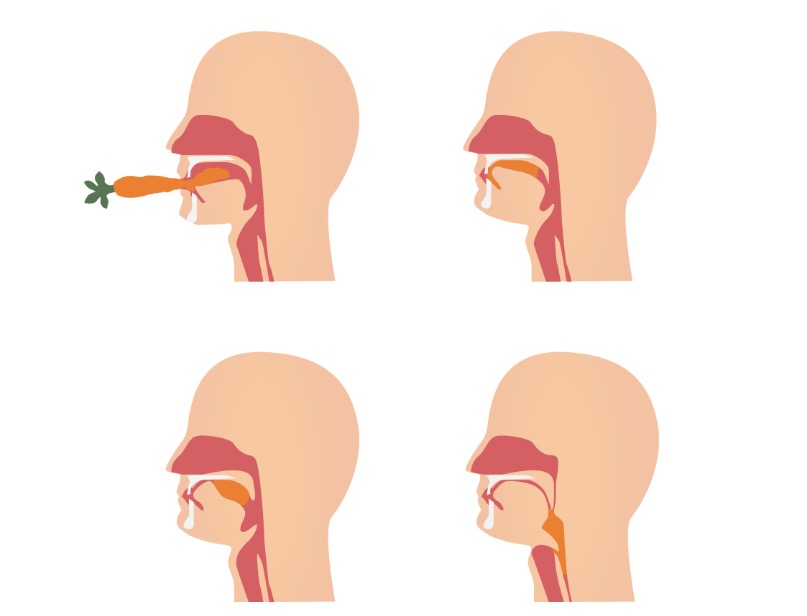
Normal physiology
SwallowingSwallowingThe act of taking solids and liquids into the gastrointestinal tract through the mouth and throat.Gastrointestinal Motility consists of 3 phases:
Food bolus is chewed and directed to the posterior tongueTongueThe tongue, on the other hand, is a complex muscular structure that permits tasting and facilitates the process of mastication and communication. The blood supply of the tongue originates from the external carotid artery, and the innervation is through cranial nerves.Lips and Tongue: Anatomy.
Pharyngeal phase:
Involuntary swallow response
Food bolus is advanced through the pharynxPharynxThe pharynx is a component of the digestive system that lies posterior to the nasal cavity, oral cavity, and larynx. The pharynx can be divided into the oropharynx, nasopharynx, and laryngopharynx. Pharyngeal muscles play an integral role in vital processes such as breathing, swallowing, and speaking. Pharynx: Anatomy.
Esophageal phase:
Involuntary esophageal peristalsisPeristalsisA movement, caused by sequential muscle contraction, that pushes the contents of the intestines or other tubular organs in one direction.Gastrointestinal Motility
Food bolus is advanced through the esophagusEsophagusThe esophagus is a muscular tube-shaped organ of around 25 centimeters in length that connects the pharynx to the stomach. The organ extends from approximately the 6th cervical vertebra to the 11th thoracic vertebra and can be divided grossly into 3 parts: the cervical part, the thoracic part, and the abdominal part. Esophagus: Anatomy.
Movement of a food bolus through the oral and pharyngeal phases of swallowing
Image by Lecturio.
Classification
There are 2 categories of dysphagia:
Oropharyngeal dysphagia:
Difficulty initiating a swallow due to a disorder in the oral or pharyngeal phase
PatientsPatientsIndividuals participating in the health care system for the purpose of receiving therapeutic, diagnostic, or preventive procedures.Clinician–Patient Relationship have difficulty transferring food from the mouth through the pharynxPharynxThe pharynx is a component of the digestive system that lies posterior to the nasal cavity, oral cavity, and larynx. The pharynx can be divided into the oropharynx, nasopharynx, and laryngopharynx. Pharyngeal muscles play an integral role in vital processes such as breathing, swallowing, and speaking. Pharynx: Anatomy.
Esophageal dysphagia:
Disorder in the esophageal phase of swallowingSwallowingThe act of taking solids and liquids into the gastrointestinal tract through the mouth and throat.Gastrointestinal Motility
Difficulty swallowingSwallowingThe act of taking solids and liquids into the gastrointestinal tract through the mouth and throat.Gastrointestinal Motility that presents several seconds after initiating a swallow
May be accompanied by the feeling of food getting “stuck”
PrevalencePrevalenceThe total number of cases of a given disease in a specified population at a designated time. It is differentiated from incidence, which refers to the number of new cases in the population at a given time.Measures of Disease Frequency in those ≥ 65 years of age:
14%–33% in the community
Approximately 40% in hospitalized patientsPatientsIndividuals participating in the health care system for the purpose of receiving therapeutic, diagnostic, or preventive procedures.Clinician–Patient Relationship
Occurs in ⅓ of patientsPatientsIndividuals participating in the health care system for the purpose of receiving therapeutic, diagnostic, or preventive procedures.Clinician–Patient Relationship with Parkinson diseaseParkinson diseaseParkinson’s disease (PD) is a chronic, progressive neurodegenerative disorder. Although the cause is unknown, several genetic and environmental risk factors are currently being studied. Individuals present clinically with resting tremor, bradykinesia, rigidity, and postural instability.Parkinson’s Disease
Etiology
Disorders of the oral phase:
Poor dentition
Decreased salivary glandSalivary glandGlands that secrete saliva in the mouth. There are three pairs of salivary glands (parotid gland; sublingual gland; submandibular gland).Diseases of the Salivary Glands production:
Sjogren’s syndrome
From head and neckNeckThe part of a human or animal body connecting the head to the rest of the body.Peritonsillar AbscessradiationRadiationEmission or propagation of acoustic waves (sound), electromagnetic energy waves (such as light; radio waves; gamma rays; or x-rays), or a stream of subatomic particles (such as electrons; neutrons; protons; or alpha particles).Osteosarcoma
Medications (anticholinergicsAnticholinergicsAnticholinergic drugs block the effect of the neurotransmitter acetylcholine at the muscarinic receptors in the central and peripheral nervous systems. Anticholinergic agents inhibit the parasympathetic nervous system, resulting in effects on the smooth muscle in the respiratory tract, vascular system, urinary tract, GI tract, and pupils of the eyes. Anticholinergic Drugs, antihistaminesAntihistaminesAntihistamines are drugs that target histamine receptors, particularly H1 and H2 receptors. H1 antagonists are competitive and reversible inhibitors of H1 receptors. First-generation antihistamines cross the blood-brain barrier and can cause sedation. Antihistamines)
Disruptions in the oropharyngeal mucosa:
MucositisMucositisStomatitis is a general term referring to inflammation of the mucous membranes of the mouth, which may include sores. Stomatitis can be caused by infections, autoimmune disorders, allergic reactions, or exposure to irritants. The typical presentation may be either solitary or a group of painful oral lesions.Stomatitis
Late-stage Parkinson diseaseParkinson diseaseParkinson’s disease (PD) is a chronic, progressive neurodegenerative disorder. Although the cause is unknown, several genetic and environmental risk factors are currently being studied. Individuals present clinically with resting tremor, bradykinesia, rigidity, and postural instability.Parkinson’s Disease
Myasthenia gravisMyasthenia GravisMyasthenia gravis (MG) is an autoimmune neuromuscular disorder characterized by weakness and fatigability of skeletal muscles caused by dysfunction/destruction of acetylcholine receptors at the neuromuscular junction. MG presents with fatigue, ptosis, diplopia, dysphagia, respiratory difficulties, and progressive weakness in the limbs, leading to difficulty in movement. Myasthenia Gravis
Disorders of the pharyngeal phase:
Structural abnormalities within the oropharynxOropharynxThe middle portion of the pharynx that lies posterior to the mouth, inferior to the soft palate, and superior to the base of the tongue and epiglottis. It has a digestive function as food passes from the mouth into the oropharynx before entering esophagus.Pharynx: Anatomy:
Amyotrophic lateral sclerosisSclerosisA pathological process consisting of hardening or fibrosis of an anatomical structure, often a vessel or a nerve.Wilms Tumor
Myasthenia gravisMyasthenia GravisMyasthenia gravis (MG) is an autoimmune neuromuscular disorder characterized by weakness and fatigability of skeletal muscles caused by dysfunction/destruction of acetylcholine receptors at the neuromuscular junction. MG presents with fatigue, ptosis, diplopia, dysphagia, respiratory difficulties, and progressive weakness in the limbs, leading to difficulty in movement. Myasthenia Gravis
Polio and post-polio syndrome
Parkinson diseaseParkinson diseaseParkinson’s disease (PD) is a chronic, progressive neurodegenerative disorder. Although the cause is unknown, several genetic and environmental risk factors are currently being studied. Individuals present clinically with resting tremor, bradykinesia, rigidity, and postural instability.Parkinson’s Disease
Multiple sclerosisSclerosisA pathological process consisting of hardening or fibrosis of an anatomical structure, often a vessel or a nerve.Wilms Tumor
DermatomyositisDermatomyositisA subacute or chronic inflammatory disease of muscle and skin, marked by proximal muscle weakness and a characteristic skin rash. The illness occurs with approximately equal frequency in children and adults. The skin lesions usually take the form of a purplish rash (or less often an exfoliative dermatitis) involving the nose, cheeks, forehead, upper trunk, and arms. The disease is associated with a complement mediated intramuscular microangiopathy, leading to loss of capillaries, muscle ischemia, muscle-fiber necrosis, and perifascicular atrophy. The childhood form of this disease tends to evolve into a systemic vasculitis. Dermatomyositis may occur in association with malignant neoplasms.Paraneoplastic Syndromes
Clinical presentation
Symptoms:
Feeling of an obstruction in the neckNeckThe part of a human or animal body connecting the head to the rest of the body.Peritonsillar Abscess
Recurrent aspiration pneumoniaAspiration pneumoniaA type of lung inflammation resulting from the aspiration of food, liquid, or gastric contents into the upper respiratory tract.Pneumonia
RadiationRadiationEmission or propagation of acoustic waves (sound), electromagnetic energy waves (such as light; radio waves; gamma rays; or x-rays), or a stream of subatomic particles (such as electrons; neutrons; protons; or alpha particles).Osteosarcoma therapy
Neuromuscular dysfunction:
Weak cough
DysarthriaDysarthriaDisorders of speech articulation caused by imperfect coordination of pharynx, larynx, tongue, or face muscles. This may result from cranial nerve diseases; neuromuscular diseases; cerebellar diseases; basal ganglia diseases; brain stem diseases; or diseases of the corticobulbar tracts. The cortical language centers are intact in this condition.Wilson Disease, dysphoniaDysphoniaDifficulty and/or pain in phonation or speaking.Epiglottitis, or nasal speech
HalitosisHalitosisAn offensive, foul breath odor resulting from a variety of causes such as poor oral hygiene, dental or oral infections, or the ingestion of certain foods.Oral Cancer (Zenker diverticulumDiverticulumA pouch or sac opening from the colon.Diverticular Disease)
Fullness in the neckNeckThe part of a human or animal body connecting the head to the rest of the body.Peritonsillar Abscess
Physical exam:
Head and neckNeckThe part of a human or animal body connecting the head to the rest of the body.Peritonsillar Abscess abnormalities to look for:
LymphadenopathyLymphadenopathyLymphadenopathy is lymph node enlargement (> 1 cm) and is benign and self-limited in most patients. Etiologies include malignancy, infection, and autoimmune disorders, as well as iatrogenic causes such as the use of certain medications. Generalized lymphadenopathy often indicates underlying systemic disease. Lymphadenopathy
Poor dentition
Masses
Facial muscle weakness
Neurologic abnormalities to test for:
SensorySensoryNeurons which conduct nerve impulses to the central nervous system.Nervous System: Histologycranial nervesCranial nervesThere are 12 pairs of cranial nerves (CNs), which run from the brain to various parts of the head, neck, and trunk. The CNs can be sensory or motor or both. The CNs are named and numbered in Roman numerals according to their location, from the front to the back of the brain.The 12 Cranial Nerves: Overview and Functions (V, IX, X)
MotorMotorNeurons which send impulses peripherally to activate muscles or secretory cells.Nervous System: Histologycranial nervesCranial nervesThere are 12 pairs of cranial nerves (CNs), which run from the brain to various parts of the head, neck, and trunk. The CNs can be sensory or motor or both. The CNs are named and numbered in Roman numerals according to their location, from the front to the back of the brain.The 12 Cranial Nerves: Overview and Functions (V, VII, IX, X, XII)
Decreased head/neckNeckThe part of a human or animal body connecting the head to the rest of the body.Peritonsillar Abscessmuscle strengthMuscle strengthThe amount of force generated by muscle contraction. Muscle strength can be measured during isometric, isotonic, or isokinetic contraction, either manually or using a device such as a muscle strength dynamometer.Neurological Examination
Based on the patient’s history and symptoms, an appropriate workup should be initiated to diagnose an underlying etiology (e.g., Sjogren’s, dermatomyositisDermatomyositisA subacute or chronic inflammatory disease of muscle and skin, marked by proximal muscle weakness and a characteristic skin rash. The illness occurs with approximately equal frequency in children and adults. The skin lesions usually take the form of a purplish rash (or less often an exfoliative dermatitis) involving the nose, cheeks, forehead, upper trunk, and arms. The disease is associated with a complement mediated intramuscular microangiopathy, leading to loss of capillaries, muscle ischemia, muscle-fiber necrosis, and perifascicular atrophy. The childhood form of this disease tends to evolve into a systemic vasculitis. Dermatomyositis may occur in association with malignant neoplasms.Paraneoplastic Syndromes, stroke, myasthenia gravisMyasthenia GravisMyasthenia gravis (MG) is an autoimmune neuromuscular disorder characterized by weakness and fatigability of skeletal muscles caused by dysfunction/destruction of acetylcholine receptors at the neuromuscular junction. MG presents with fatigue, ptosis, diplopia, dysphagia, respiratory difficulties, and progressive weakness in the limbs, leading to difficulty in movement. Myasthenia Gravis, Parkinson diseaseParkinson diseaseParkinson’s disease (PD) is a chronic, progressive neurodegenerative disorder. Although the cause is unknown, several genetic and environmental risk factors are currently being studied. Individuals present clinically with resting tremor, bradykinesia, rigidity, and postural instability.Parkinson’s Disease).
Next, the following are used to determine the severity and mechanism of swallowingSwallowingThe act of taking solids and liquids into the gastrointestinal tract through the mouth and throat.Gastrointestinal Motility dysfunction:
Can test the effects of different barium consistencies
Analyzes the movement of anatomic structures
Detects oropharyngeal dysfunction and determines severity
Assesses for aspiration
ManometryManometryMeasurement of the pressure or tension of liquids or gases with a manometer.Achalasia: evaluates the pressure with pharyngeal contraction and in the upper esophageal sphincterUpper esophageal sphincterThe structure at the pharyngoesophageal junction consisting chiefly of the cricopharyngeus muscle. It normally occludes the lumen of the esophagus, except during swallowing.Esophagus: Anatomy (UES function)
If no systemic process is suspected, or found, as an etiology:
Nasopharyngeal laryngoscopy:
Can evaluate for a structural lesion (tumors, Zenker diverticulumDiverticulumA pouch or sac opening from the colon.Diverticular Disease)
Visualizes the oropharynxOropharynxThe middle portion of the pharynx that lies posterior to the mouth, inferior to the soft palate, and superior to the base of the tongue and epiglottis. It has a digestive function as food passes from the mouth into the oropharynx before entering esophagus.Pharynx: Anatomy, hypopharynx, and larynxLarynxThe larynx, also commonly called the voice box, is a cylindrical space located in the neck at the level of the C3-C6 vertebrae. The major structures forming the framework of the larynx are the thyroid cartilage, cricoid cartilage, and epiglottis. The larynx serves to produce sound (phonation), conducts air to the trachea, and prevents large molecules from reaching the lungs.Larynx: Anatomy
Fiberoptic endoscopic evaluation of swallowingSwallowingThe act of taking solids and liquids into the gastrointestinal tract through the mouth and throat.Gastrointestinal Motility (FEES):
Similar to nasopharyngeal laryngoscopy, but can also assess function
Directly visualizes food and liquids as they are swallowed
SensorySensoryNeurons which conduct nerve impulses to the central nervous system.Nervous System: Histology testing can also be performed.
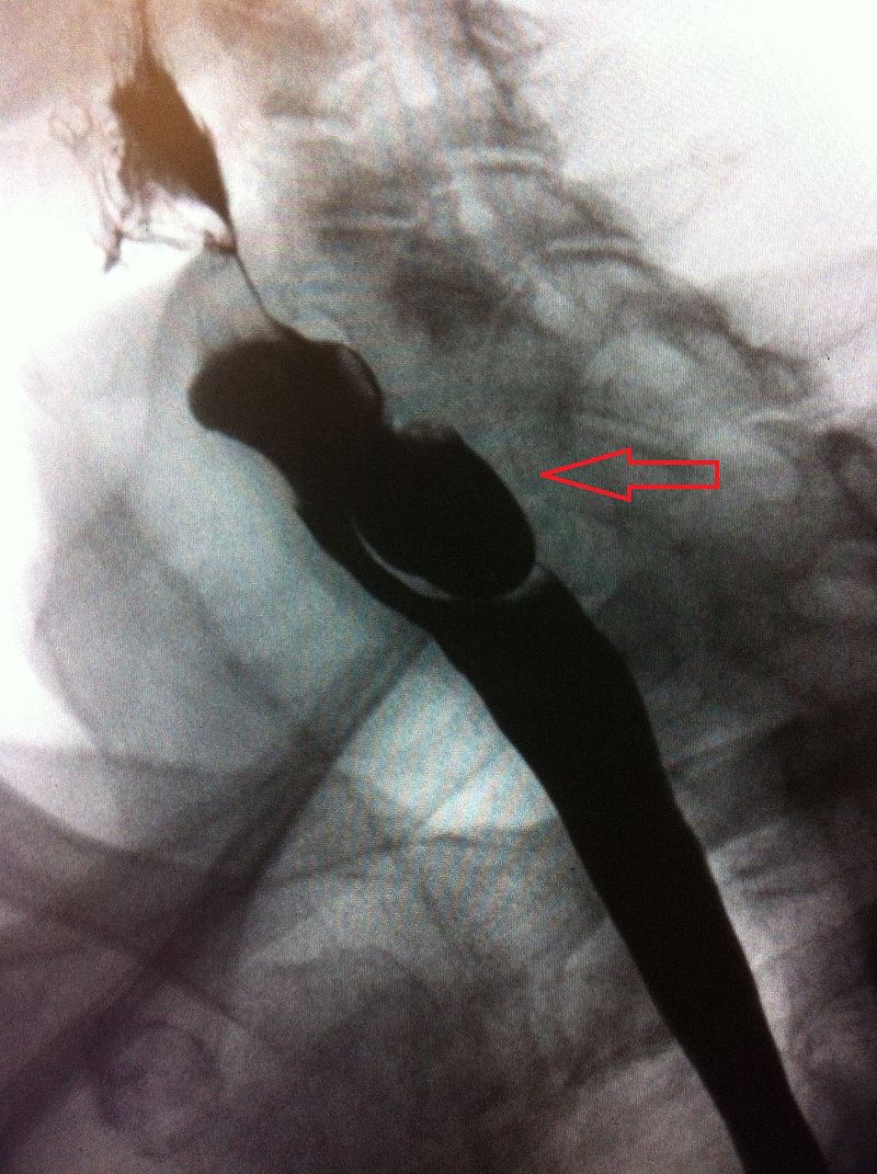
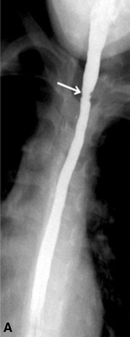
A barium swallow study identifying a false diverticula arising from the posterior wall of the upper esophagus: This is consistent with a Zenker diverticulum.
Image: “Lateral X-ray of a Zenker’s diverticula” by James Heilman, MD. License: CC BY-SA 4.0
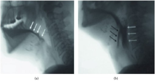
Video fluoroscopic view in a patient who has undergone a near-total glossectomy for advanced head and neck cancer. The patient has poor oral bolus control and early loss into the oropharynx (A, white arrows). He has lost the ability to pull the hyoid and the larynx up and forward to open the upper esophageal sphincter, resulting in pharyngeal dysphagia and food remaining in the pharynx (B, white arrows) with penetration above the vocal folds (black arrows). This patient was determined to be at high risk of aspiration.
Image: “Lateral fluoroscopic view” by Caterina Giannitto. License: CC BY 4.0.
Management
Management is guided by the diagnostic workup, and is focused on improving food transfer and preventing aspiration:
Treatment of any underlying disorder
SwallowingSwallowingThe act of taking solids and liquids into the gastrointestinal tract through the mouth and throat.Gastrointestinal Motility rehabilitation:
MotorMotorNeurons which send impulses peripherally to activate muscles or secretory cells.Nervous System: Histology exercises
Head placement while eating
Dietary modifications:
Thickened liquids
Smaller bites of food
Alternating solids and liquids when eating
Nutrition supplementation
Enteral nutritionEnteral nutritionNutritional support given via the alimentary canal or any route connected to the gastrointestinal system (i.e., the enteral route). This includes oral feeding, sip feeding, and tube feeding using nasogastric, gastrostomy, and jejunostomy tubes.Short Bowel Syndrome may need to be considered for severe dysfunction and aspiration risk.
Esophageal Dysphagia: Functional and Motility Disorders
Esophageal functional and motilityMotilityThe motor activity of the gastrointestinal tract.Gastrointestinal Motility disorders typically occur due to pathology of the muscles of the esophagusEsophagusThe esophagus is a muscular tube-shaped organ of around 25 centimeters in length that connects the pharynx to the stomach. The organ extends from approximately the 6th cervical vertebra to the 11th thoracic vertebra and can be divided grossly into 3 parts: the cervical part, the thoracic part, and the abdominal part. Esophagus: Anatomy, causing a disruption in peristalsisPeristalsisA movement, caused by sequential muscle contraction, that pushes the contents of the intestines or other tubular organs in one direction.Gastrointestinal Motility. All functional and motilityMotilityThe motor activity of the gastrointestinal tract.Gastrointestinal Motility disorders exhibit dysphagia with liquids and solids at the onset.
Impaired inhibitory innervation of the muscles of the esophagusEsophagusThe esophagus is a muscular tube-shaped organ of around 25 centimeters in length that connects the pharynx to the stomach. The organ extends from approximately the 6th cervical vertebra to the 11th thoracic vertebra and can be divided grossly into 3 parts: the cervical part, the thoracic part, and the abdominal part. Esophagus: Anatomy and ↓ endogenous nitric oxideNitric OxideA free radical gas produced endogenously by a variety of mammalian cells, synthesized from arginine by nitric oxide synthase. Nitric oxide is one of the endothelium-dependent relaxing factors released by the vascular endothelium and mediates vasodilation. It also inhibits platelet aggregation, induces disaggregation of aggregated platelets, and inhibits platelet adhesion to the vascular endothelium. Nitric oxide activates cytosolic guanylate cyclase and thus elevates intracellular levels of cyclic gmp.Pulmonary Hypertension DrugssynthesisSynthesisPolymerase Chain Reaction (PCR) → frequent, high-pressure, non-peristaltic contractions occurring spontaneously with normal peristalsisPeristalsisA movement, caused by sequential muscle contraction, that pushes the contents of the intestines or other tubular organs in one direction.Gastrointestinal Motility
Clinical presentation:
May be asymptomatic
Intermittent, non-progressive dysphagia
Non-cardiac chest painPainAn unpleasant sensation induced by noxious stimuli which are detected by nerve endings of nociceptive neurons.Pain: Types and Pathways
Can be aggravated with hot or cold liquids
Some patientsPatientsIndividuals participating in the health care system for the purpose of receiving therapeutic, diagnostic, or preventive procedures.Clinician–Patient Relationship may have associated gastroesophageal reflux diseaseGastroesophageal Reflux DiseaseGastroesophageal reflux disease (GERD) occurs when the stomach acid frequently flows back into the esophagus. This backwash (acid reflux) can irritate the lining of the esophagus, causing symptoms such as retrosternal burning pain (heartburn). Gastroesophageal Reflux Disease (GERD) (GERDGERDGastroesophageal reflux disease (GERD) occurs when the stomach acid frequently flows back into the esophagus. This backwash (acid reflux) can irritate the lining of the esophagus, causing symptoms such as retrosternal burning pain (heartburn). Gastroesophageal Reflux Disease (GERD)).
Diagnosis:
ManometryManometryMeasurement of the pressure or tension of liquids or gases with a manometer.Achalasia: diagnosis established by presence of prematurePrematureChildbirth before 37 weeks of pregnancy (259 days from the first day of the mother’s last menstrual period, or 245 days after fertilization).Necrotizing Enterocolitis contractions (≥ 20% prematurePrematureChildbirth before 37 weeks of pregnancy (259 days from the first day of the mother’s last menstrual period, or 245 days after fertilization).Necrotizing Enterocolitis contractions based on Chicago Classification criteria)
Barium esophagram: severe, non-peristaltic contractions with a “corkscrew” pattern
CalciumCalciumA basic element found in nearly all tissues. It is a member of the alkaline earth family of metals with the atomic symbol ca, atomic number 20, and atomic weight 40. Calcium is the most abundant mineral in the body and combines with phosphorus to form calcium phosphate in the bones and teeth. It is essential for the normal functioning of nerves and muscles and plays a role in blood coagulation (as factor IV) and in many enzymatic processes.Electrolytes channel blockers
Proton pumpPumpACES and RUSH: Resuscitation Ultrasound Protocols inhibitors (PPIs) for GERDGERDGastroesophageal reflux disease (GERD) occurs when the stomach acid frequently flows back into the esophagus. This backwash (acid reflux) can irritate the lining of the esophagus, causing symptoms such as retrosternal burning pain (heartburn). Gastroesophageal Reflux Disease (GERD) symptoms
Botulinum toxinBotulinum toxinToxic proteins produced from the species Clostridium botulinum. The toxins are synthesized as a single peptide chain which is processed into a mature protein consisting of a heavy chain and light chain joined via a disulfide bond. The botulinum toxin light chain is a zinc-dependent protease which is released from the heavy chain upon endocytosis into presynaptic nerve endings. Once inside the cell the botulinum toxin light chain cleaves specific snare proteins which are essential for secretion of acetylcholine by synaptic vesicles. This inhibition of acetylcholine release results in muscular paralysis.Botulism injection into the lower esophageal sphincterLower Esophageal SphincterEsophagus: Anatomy (LES) for refractory cases
Diffuse esophageal spasm: barium esophagram showing the typical “corkscrew” pattern
Image: “Smooth short stricture in the distal esophagus” by Chui Man Carmen Hui et al. License: CC BY 4.0.
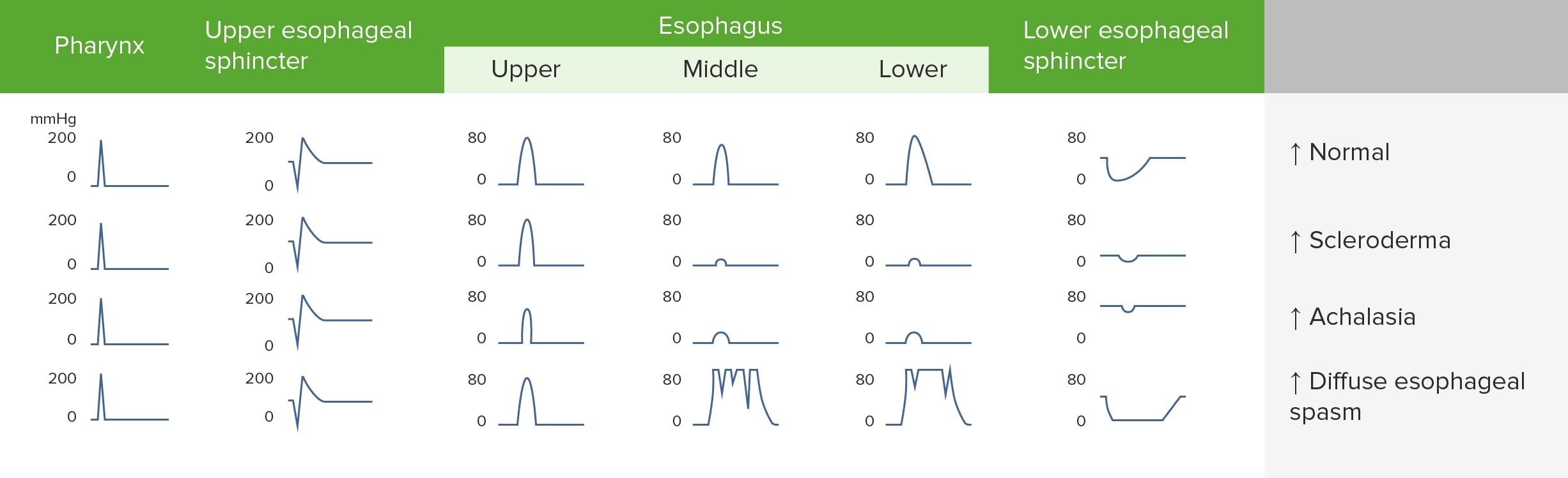
Manometry findings in esophageal motility disorders
Image by Lecturio.
SclerodermaSclerodermaScleroderma (systemic sclerosis) is an autoimmune condition characterized by diffuse collagen deposition and fibrosis. The clinical presentation varies from limited skin involvement to diffuse involvement of internal organs. Scleroderma
SclerodermaSclerodermaScleroderma (systemic sclerosis) is an autoimmune condition characterized by diffuse collagen deposition and fibrosis. The clinical presentation varies from limited skin involvement to diffuse involvement of internal organs. Scleroderma is an autoimmune disorderAutoimmune DisorderSeptic Arthritis that can cause atrophyAtrophyDecrease in the size of a cell, tissue, organ, or multiple organs, associated with a variety of pathological conditions such as abnormal cellular changes, ischemia, malnutrition, or hormonal changes.Cellular Adaptation and sclerosisSclerosisA pathological process consisting of hardening or fibrosis of an anatomical structure, often a vessel or a nerve.Wilms Tumor of the distal esophagusEsophagusThe esophagus is a muscular tube-shaped organ of around 25 centimeters in length that connects the pharynx to the stomach. The organ extends from approximately the 6th cervical vertebra to the 11th thoracic vertebra and can be divided grossly into 3 parts: the cervical part, the thoracic part, and the abdominal part. Esophagus: Anatomy, resulting in diminished (or absent) peristalsisPeristalsisA movement, caused by sequential muscle contraction, that pushes the contents of the intestines or other tubular organs in one direction.Gastrointestinal Motility and LES pressure.
Pathophysiology:
AtrophyAtrophyDecrease in the size of a cell, tissue, organ, or multiple organs, associated with a variety of pathological conditions such as abnormal cellular changes, ischemia, malnutrition, or hormonal changes.Cellular Adaptation and fibrosisFibrosisAny pathological condition where fibrous connective tissue invades any organ, usually as a consequence of inflammation or other injury.Bronchiolitis Obliterans of the smooth muscle in the distal ⅔ of the esophagusEsophagusThe esophagus is a muscular tube-shaped organ of around 25 centimeters in length that connects the pharynx to the stomach. The organ extends from approximately the 6th cervical vertebra to the 11th thoracic vertebra and can be divided grossly into 3 parts: the cervical part, the thoracic part, and the abdominal part. Esophagus: Anatomy → ↓ peristalsisPeristalsisA movement, caused by sequential muscle contraction, that pushes the contents of the intestines or other tubular organs in one direction.Gastrointestinal Motility and loss of LES tone
GERDGERDGastroesophageal reflux disease (GERD) occurs when the stomach acid frequently flows back into the esophagus. This backwash (acid reflux) can irritate the lining of the esophagus, causing symptoms such as retrosternal burning pain (heartburn). Gastroesophageal Reflux Disease (GERD), Barrett’s esophagusEsophagusThe esophagus is a muscular tube-shaped organ of around 25 centimeters in length that connects the pharynx to the stomach. The organ extends from approximately the 6th cervical vertebra to the 11th thoracic vertebra and can be divided grossly into 3 parts: the cervical part, the thoracic part, and the abdominal part. Esophagus: Anatomy, reflux esophagitisEsophagitisEsophagitis is the inflammation or irritation of the esophagus. The major types of esophagitis are medication-induced, infectious, eosinophilic, corrosive, and acid reflux. Patients typically present with odynophagia, dysphagia, and retrosternal chest pain. Esophagitis, and subsequent strictures may develop.
Clinical presentation:
Acid reflux
Possible associated systemic symptoms:
SkinSkinThe skin, also referred to as the integumentary system, is the largest organ of the body. The skin is primarily composed of the epidermis (outer layer) and dermis (deep layer). The epidermis is primarily composed of keratinocytes that undergo rapid turnover, while the dermis contains dense layers of connective tissue.Skin: Structure and Functions thickening, sclerodactylySclerodactylyScleroderma, calcinosisCalcinosisPathologic deposition of calcium salts in tissues.Scleroderma, telangiectases
Raynaud’s phenomenon
Interstitial lung disease or fibrosisFibrosisAny pathological condition where fibrous connective tissue invades any organ, usually as a consequence of inflammation or other injury.Bronchiolitis Obliterans
Pulmonary hypertensionPulmonary HypertensionPulmonary hypertension (PH) or pulmonary arterial hypertension (PAH) is characterized by elevated pulmonary arterial pressure, which can lead to chronic progressive right heart failure. Pulmonary hypertension is grouped into 5 categories based on etiology, which include primary PAH, and PH due to cardiac disease, lung or hypoxic disease, chronic thromboembolic disease, and multifactorial or unclear etiologies. Pulmonary Hypertension, pericarditisPericarditisPericarditis is an inflammation of the pericardium, often with fluid accumulation. It can be caused by infection (often viral), myocardial infarction, drugs, malignancies, metabolic disorders, autoimmune disorders, or trauma. Acute, subacute, and chronic forms exist. Pericarditis
Renal disease
Diagnosis:
Diagnosis of sclerodermaSclerodermaScleroderma (systemic sclerosis) is an autoimmune condition characterized by diffuse collagen deposition and fibrosis. The clinical presentation varies from limited skin involvement to diffuse involvement of internal organs. Scleroderma is based on the clinical features and detection of anti-Scl-70 (anti-topoisomerase), anticentromere, or anti-RNA polymerase IIIAnti-RNA polymerase IIISclerodermaantibodiesAntibodiesImmunoglobulins (Igs), also known as antibodies, are glycoprotein molecules produced by plasma cells that act in immune responses by recognizing and binding particular antigens. The various Ig classes are IgG (the most abundant), IgM, IgE, IgD, and IgA, which differ in their biologic features, structure, target specificity, and distribution.Immunoglobulins: Types and Functions.
ManometryManometryMeasurement of the pressure or tension of liquids or gases with a manometer.Achalasia: ↓ LES tone and absence of peristalsisPeristalsisA movement, caused by sequential muscle contraction, that pushes the contents of the intestines or other tubular organs in one direction.Gastrointestinal Motility in the body of the esophagusEsophagusThe esophagus is a muscular tube-shaped organ of around 25 centimeters in length that connects the pharynx to the stomach. The organ extends from approximately the 6th cervical vertebra to the 11th thoracic vertebra and can be divided grossly into 3 parts: the cervical part, the thoracic part, and the abdominal part. Esophagus: Anatomy
EndoscopyEndoscopyProcedures of applying endoscopes for disease diagnosis and treatment. Endoscopy involves passing an optical instrument through a small incision in the skin i.e., percutaneous; or through a natural orifice and along natural body pathways such as the digestive tract; and/or through an incision in the wall of a tubular structure or organ, i.e. Transluminal, to examine or perform surgery on the interior parts of the body.Gastroesophageal Reflux Disease (GERD): reflux esophagitisEsophagitisEsophagitis is the inflammation or irritation of the esophagus. The major types of esophagitis are medication-induced, infectious, eosinophilic, corrosive, and acid reflux. Patients typically present with odynophagia, dysphagia, and retrosternal chest pain. Esophagitis, Barrett’s esophagusEsophagusThe esophagus is a muscular tube-shaped organ of around 25 centimeters in length that connects the pharynx to the stomach. The organ extends from approximately the 6th cervical vertebra to the 11th thoracic vertebra and can be divided grossly into 3 parts: the cervical part, the thoracic part, and the abdominal part. Esophagus: Anatomy, or esophageal strictureStricturePrimary Sclerosing Cholangitis
Management:
PPIs
Endoscopic dilation for strictures
Gastroplasty or fundoplication for refractory GERDGERDGastroesophageal reflux disease (GERD) occurs when the stomach acid frequently flows back into the esophagus. This backwash (acid reflux) can irritate the lining of the esophagus, causing symptoms such as retrosternal burning pain (heartburn). Gastroesophageal Reflux Disease (GERD)
A: upper endoscopy demonstrating reflux esophagitis; B: upper endoscopy demonstrating an esophageal stricture in a patient with scleroderma
Image: “EGD in systemic sclerosis” by the Postgraduate Department of Dermatology, STDs & Leprosy, Government Medical College, Srinagar, Jammu and Kashmir, India. License: CC BY 4.0.
AchalasiaAchalasiaAchalasia is a primary esophageal motility disorder that develops from the degeneration of the myenteric plexus. This condition results in impaired lower esophageal sphincter relaxation and absence of normal esophageal peristalsis. Patients typically present with dysphagia to solids and liquids along with regurgitation. Achalasia
A neurogenic esophageal motilityEsophageal MotilityGastrointestinal Motility disorder resulting in impaired LES relaxation and diminished peristalsisPeristalsisA movement, caused by sequential muscle contraction, that pushes the contents of the intestines or other tubular organs in one direction.Gastrointestinal Motility:
Secondary: due to malignancyMalignancyHemothorax or Chagas diseaseChagas diseaseInfection with the protozoan parasite trypanosoma cruzi, a form of trypanosomiasis endemic in central and south america. It is named after the brazilian physician carlos chagas, who discovered the parasite. Infection by the parasite (positive serologic result only) is distinguished from the clinical manifestations that develop years later, such as destruction of parasympathetic ganglia; chagas cardiomyopathy; and dysfunction of the esophagus or colon.Trypanosoma cruzi/Chagas disease
Pathophysiology:
Degeneration of ganglion cellsGanglion CellsThe Visual Pathway and Related Disorders in Auerbach’s plexus → failure of smooth muscle relaxation at the LES → progressive loss of peristaltic function in the distal esophagusEsophagusThe esophagus is a muscular tube-shaped organ of around 25 centimeters in length that connects the pharynx to the stomach. The organ extends from approximately the 6th cervical vertebra to the 11th thoracic vertebra and can be divided grossly into 3 parts: the cervical part, the thoracic part, and the abdominal part. Esophagus: Anatomy
↑ LES pressure → obstruction and secondary esophageal dilation → retention of foods and liquids in the esophagusEsophagusThe esophagus is a muscular tube-shaped organ of around 25 centimeters in length that connects the pharynx to the stomach. The organ extends from approximately the 6th cervical vertebra to the 11th thoracic vertebra and can be divided grossly into 3 parts: the cervical part, the thoracic part, and the abdominal part. Esophagus: Anatomy
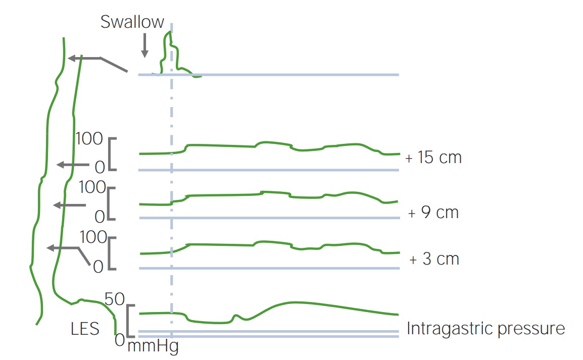
Esophageal manometryManometryMeasurement of the pressure or tension of liquids or gases with a manometer.Achalasia (preferred test): incomplete relaxation of the LES, ↑ resting LES pressure, and aperistalsis of the distal ⅔ of the esophagusEsophagusThe esophagus is a muscular tube-shaped organ of around 25 centimeters in length that connects the pharynx to the stomach. The organ extends from approximately the 6th cervical vertebra to the 11th thoracic vertebra and can be divided grossly into 3 parts: the cervical part, the thoracic part, and the abdominal part. Esophagus: Anatomy
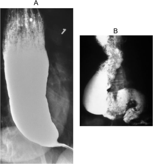
Barium esophagram: suggestive findings of a “bird’s beak” esophagusEsophagusThe esophagus is a muscular tube-shaped organ of around 25 centimeters in length that connects the pharynx to the stomach. The organ extends from approximately the 6th cervical vertebra to the 11th thoracic vertebra and can be divided grossly into 3 parts: the cervical part, the thoracic part, and the abdominal part. Esophagus: Anatomy
EndoscopyEndoscopyProcedures of applying endoscopes for disease diagnosis and treatment. Endoscopy involves passing an optical instrument through a small incision in the skin i.e., percutaneous; or through a natural orifice and along natural body pathways such as the digestive tract; and/or through an incision in the wall of a tubular structure or organ, i.e. Transluminal, to examine or perform surgery on the interior parts of the body.Gastroesophageal Reflux Disease (GERD) with biopsyBiopsyRemoval and pathologic examination of specimens from the living body.Ewing Sarcoma: Rule out secondary causes, such as malignancyMalignancyHemothorax and Chagas diseaseChagas diseaseInfection with the protozoan parasite trypanosoma cruzi, a form of trypanosomiasis endemic in central and south america. It is named after the brazilian physician carlos chagas, who discovered the parasite. Infection by the parasite (positive serologic result only) is distinguished from the clinical manifestations that develop years later, such as destruction of parasympathetic ganglia; chagas cardiomyopathy; and dysfunction of the esophagus or colon.Trypanosoma cruzi/Chagas disease.
Management:
Treatment is aimed at ↓ LES pressure.
Mechanical stretching of muscle fibers:
Pneumatic balloon dilation
Surgical myotomy
Chemical reduction of LES pressure:
Botulinum toxinBotulinum toxinToxic proteins produced from the species Clostridium botulinum. The toxins are synthesized as a single peptide chain which is processed into a mature protein consisting of a heavy chain and light chain joined via a disulfide bond. The botulinum toxin light chain is a zinc-dependent protease which is released from the heavy chain upon endocytosis into presynaptic nerve endings. Once inside the cell the botulinum toxin light chain cleaves specific snare proteins which are essential for secretion of acetylcholine by synaptic vesicles. This inhibition of acetylcholine release results in muscular paralysis.Botulism injection into the LES
Oral nitratesNitratesNitrates are a class of medications that cause systemic vasodilation (veins > arteries) by smooth muscle relaxation. Nitrates are primarily indicated for the treatment of angina, where preferential venodilation causes pooling of blood, decreased preload, and ultimately decreased myocardial O2 demand.Nitrates
CalciumCalciumA basic element found in nearly all tissues. It is a member of the alkaline earth family of metals with the atomic symbol ca, atomic number 20, and atomic weight 40. Calcium is the most abundant mineral in the body and combines with phosphorus to form calcium phosphate in the bones and teeth. It is essential for the normal functioning of nerves and muscles and plays a role in blood coagulation (as factor IV) and in many enzymatic processes.Electrolytes channel blockers
Achalasia: “bird’s beak” appearance on a barium esophagram
Image: “Barium swallow” by the Department of Internal Medicine, Nashville, TN, USA. License: CC BY 4.0.
Achalasia: manometry showing high LES pressure (top line)
Esophageal Dysphagia: Mechanical and Obstructive Disorders
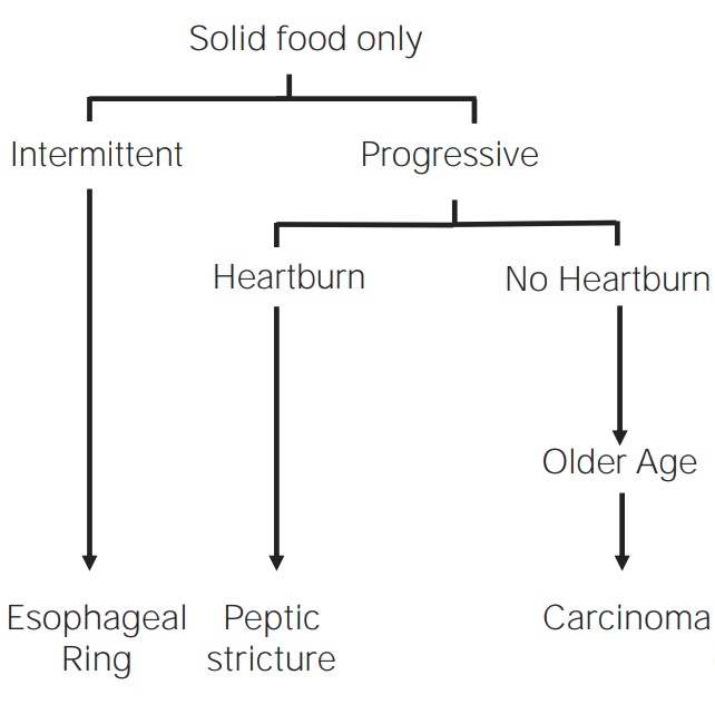
Esophageal mechanical and obstructive disorders typically occur due to obstruction of the esophageal lumen. All mechanical and obstructive disorders exhibit dysphagia with solids with progression to liquids.
Overview of mechanical and obstructive disorders
Image by Lecturio.
Esophageal rings and webs
Rings and webs are thin mucosal strictures that partially occlude the esophageal lumen:
Definitions:
Esophageal ring:
Concentric tissue protruding into the esophageal lumen
Most commonly in the distal esophagusEsophagusThe esophagus is a muscular tube-shaped organ of around 25 centimeters in length that connects the pharynx to the stomach. The organ extends from approximately the 6th cervical vertebra to the 11th thoracic vertebra and can be divided grossly into 3 parts: the cervical part, the thoracic part, and the abdominal part. Esophagus: Anatomy
Usually mucosal, but can be due to hypertrophyHypertrophyGeneral increase in bulk of a part or organ due to cell enlargement and accumulation of fluids and secretions, not due to tumor formation, nor to an increase in the number of cells (hyperplasia).Cellular Adaptation of smooth muscle
Esophageal web:
Eccentric membranes that protrude into the esophageal lumen
Most commonly occur anteriorly in the proximal esophagusEsophagusThe esophagus is a muscular tube-shaped organ of around 25 centimeters in length that connects the pharynx to the stomach. The organ extends from approximately the 6th cervical vertebra to the 11th thoracic vertebra and can be divided grossly into 3 parts: the cervical part, the thoracic part, and the abdominal part. Esophagus: Anatomy
Pathophysiology:
Etiology is unknown.
Rings may be due to chronic damage from GERDGERDGastroesophageal reflux disease (GERD) occurs when the stomach acid frequently flows back into the esophagus. This backwash (acid reflux) can irritate the lining of the esophagus, causing symptoms such as retrosternal burning pain (heartburn). Gastroesophageal Reflux Disease (GERD).
Clinical presentation:
Usually asymptomatic
Dysphagia is intermittent.
Diagnosis:
Barium esophagram: Rings and webs appear as thin, circumferential or eccentric narrowing in the esophagusEsophagusThe esophagus is a muscular tube-shaped organ of around 25 centimeters in length that connects the pharynx to the stomach. The organ extends from approximately the 6th cervical vertebra to the 11th thoracic vertebra and can be divided grossly into 3 parts: the cervical part, the thoracic part, and the abdominal part. Esophagus: Anatomy.
Specific conditions:
Schatzki ring:
Associated with GERDGERDGastroesophageal reflux disease (GERD) occurs when the stomach acid frequently flows back into the esophagus. This backwash (acid reflux) can irritate the lining of the esophagus, causing symptoms such as retrosternal burning pain (heartburn). Gastroesophageal Reflux Disease (GERD), eosinophilic esophagitisEosinophilic esophagitisChronic esophagitis characterized by esophageal mucosal eosinophilia. It is diagnosed when an increase in eosinophils are present over the entire esophagus. The reflux symptoms fail to respond to proton pump inhibitors treatment, unlike in gastroesophageal reflux disease. The symptoms are associated with ige-mediated hypersensitivity to food or inhalant allergens.Esophagitis, and hiatal herniaHiatal herniaStomach herniation located at or near the diaphragmatic opening for the esophagus, the esophageal hiatus.Congenital Diaphragmatic Hernias
Located in the distal esophagusEsophagusThe esophagus is a muscular tube-shaped organ of around 25 centimeters in length that connects the pharynx to the stomach. The organ extends from approximately the 6th cervical vertebra to the 11th thoracic vertebra and can be divided grossly into 3 parts: the cervical part, the thoracic part, and the abdominal part. Esophagus: Anatomy (proximal to the LES)
More common in patientsPatientsIndividuals participating in the health care system for the purpose of receiving therapeutic, diagnostic, or preventive procedures.Clinician–Patient Relationship > 40 years of age
Treatment: esophageal dilation and PPIs
Plummer-Vinson syndromePlummer-Vinson syndromeA syndrome of dysphagia with iron-deficiency anemia that is due to congenital anomalies in the esophagus (such as cervical esophageal webs). It is known as patterson-kelly syndrome in the united kingdom.Iron Deficiency Anemia:
Clinical triad:ironIronA metallic element with atomic symbol fe, atomic number 26, and atomic weight 55. 85. It is an essential constituent of hemoglobins; cytochromes; and iron-binding proteins. It plays a role in cellular redox reactions and in the transport of oxygen.Trace Elements deficiency anemiaAnemiaAnemia is a condition in which individuals have low Hb levels, which can arise from various causes. Anemia is accompanied by a reduced number of RBCs and may manifest with fatigue, shortness of breath, pallor, and weakness. Subtypes are classified by the size of RBCs, chronicity, and etiology. Anemia: Overview and Types, dysphagia, and cervical esophageal webs
More common in middle-aged women
Associated with an increased risk of esophageal squamous cell carcinomaEsophageal Squamous Cell CarcinomaA carcinoma that originates usually from cells on the surface of the middle and lower third of the esophagus. Tumor cells exhibit typical squamous morphology and form large polypoid lesions. Mutations in rnf6, lzts1, TGFbr2, dec1, and wwox1 genes are associated with this cancer.Esophageal Cancer (SCC)
Chronic inflammationChronic InflammationInflammation from long-standing GERDGERDGastroesophageal reflux disease (GERD) occurs when the stomach acid frequently flows back into the esophagus. This backwash (acid reflux) can irritate the lining of the esophagus, causing symptoms such as retrosternal burning pain (heartburn). Gastroesophageal Reflux Disease (GERD)
Direct damage caused by the ingestion of acidic or alkali agents
Pathophysiology:
Circumferential scarringScarringInflammation → reduced lumen of the esophagusEsophagusThe esophagus is a muscular tube-shaped organ of around 25 centimeters in length that connects the pharynx to the stomach. The organ extends from approximately the 6th cervical vertebra to the 11th thoracic vertebra and can be divided grossly into 3 parts: the cervical part, the thoracic part, and the abdominal part. Esophagus: Anatomy → progressive dysphagia of solids
Often located in the distal esophagusEsophagusThe esophagus is a muscular tube-shaped organ of around 25 centimeters in length that connects the pharynx to the stomach. The organ extends from approximately the 6th cervical vertebra to the 11th thoracic vertebra and can be divided grossly into 3 parts: the cervical part, the thoracic part, and the abdominal part. Esophagus: Anatomy
Clinical presentation:
Dysphagia is gradually progressive.
History of GERDGERDGastroesophageal reflux disease (GERD) occurs when the stomach acid frequently flows back into the esophagus. This backwash (acid reflux) can irritate the lining of the esophagus, causing symptoms such as retrosternal burning pain (heartburn). Gastroesophageal Reflux Disease (GERD)
Remote history of ingestion of acid or alkali agents
Barium esophagram: can establish the location, length, and number of strictures
Confirmed with endoscopyEndoscopyProcedures of applying endoscopes for disease diagnosis and treatment. Endoscopy involves passing an optical instrument through a small incision in the skin i.e., percutaneous; or through a natural orifice and along natural body pathways such as the digestive tract; and/or through an incision in the wall of a tubular structure or organ, i.e. Transluminal, to examine or perform surgery on the interior parts of the body.Gastroesophageal Reflux Disease (GERD) and biopsyBiopsyRemoval and pathologic examination of specimens from the living body.Ewing Sarcoma (to rule out cancer)
Management:
High-dose PPIs
Periodic esophageal dilation
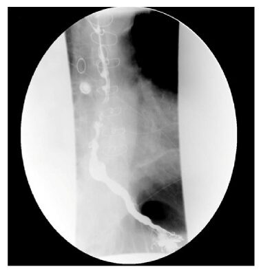
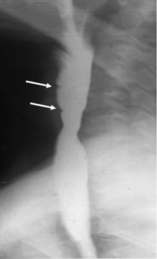
Barium esophagram demonstrating an esophageal stricture (arrow), which shows a stricture in the proximal esophagus, but strictures will usually be in the distal esophagus
Image: “Barium esophagogram” by the Division of Pediatric Gastroenterology, Children’s Hospital, King Fahad Medical City, P. O. Box 59046, Riyadh, 11525, Kingdom of Saudi Arabia. License: CC BY 4.0.
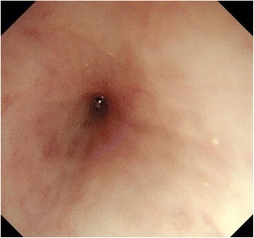
Endoscopy image showing a significant esophageal stricture
Image: “Esophagogastroduodenoscopy” by the Department of Gastroenterological Surgery, Tokai University School of Medicine, 143 Shimokasuya, Isehara, Kanagawa, 259-1193, Japan. License: CC BY 4.0.
Esophageal cancerEsophageal cancerEsophageal cancer is 1 of the most common causes of cancer-related deaths worldwide. Nearly all esophageal cancers are either adenocarcinoma (commonly affecting the distal esophagus) or squamous cell carcinoma (affecting the proximal two-thirds of the esophagus). Esophageal Cancer
Etiology:
SCC: cancer of the upper ⅔ of the esophagusEsophagusThe esophagus is a muscular tube-shaped organ of around 25 centimeters in length that connects the pharynx to the stomach. The organ extends from approximately the 6th cervical vertebra to the 11th thoracic vertebra and can be divided grossly into 3 parts: the cervical part, the thoracic part, and the abdominal part. Esophagus: Anatomy, commonly seen in:
Smokers
Alcohol intake
AchalasiaAchalasiaAchalasia is a primary esophageal motility disorder that develops from the degeneration of the myenteric plexus. This condition results in impaired lower esophageal sphincter relaxation and absence of normal esophageal peristalsis. Patients typically present with dysphagia to solids and liquids along with regurgitation. Achalasia
Adenocarcinoma: cancer of the distal esophagusEsophagusThe esophagus is a muscular tube-shaped organ of around 25 centimeters in length that connects the pharynx to the stomach. The organ extends from approximately the 6th cervical vertebra to the 11th thoracic vertebra and can be divided grossly into 3 parts: the cervical part, the thoracic part, and the abdominal part. Esophagus: Anatomy, commonly seen in older patientsPatientsIndividuals participating in the health care system for the purpose of receiving therapeutic, diagnostic, or preventive procedures.Clinician–Patient Relationship with Barrett’s esophagusEsophagusThe esophagus is a muscular tube-shaped organ of around 25 centimeters in length that connects the pharynx to the stomach. The organ extends from approximately the 6th cervical vertebra to the 11th thoracic vertebra and can be divided grossly into 3 parts: the cervical part, the thoracic part, and the abdominal part. Esophagus: Anatomy
Pathophysiology: cancer overgrowth → reduced lumen of the esophagusEsophagusThe esophagus is a muscular tube-shaped organ of around 25 centimeters in length that connects the pharynx to the stomach. The organ extends from approximately the 6th cervical vertebra to the 11th thoracic vertebra and can be divided grossly into 3 parts: the cervical part, the thoracic part, and the abdominal part. Esophagus: Anatomy → progressive dysphagia
Clinical presentation:
Progressive dysphagia
OdynophagiaOdynophagiaEpiglottitis (painPainAn unpleasant sensation induced by noxious stimuli which are detected by nerve endings of nociceptive neurons.Pain: Types and Pathways with swallowingSwallowingThe act of taking solids and liquids into the gastrointestinal tract through the mouth and throat.Gastrointestinal Motility) in 20% of patientsPatientsIndividuals participating in the health care system for the purpose of receiving therapeutic, diagnostic, or preventive procedures.Clinician–Patient Relationship
IronIronA metallic element with atomic symbol fe, atomic number 26, and atomic weight 55. 85. It is an essential constituent of hemoglobins; cytochromes; and iron-binding proteins. It plays a role in cellular redox reactions and in the transport of oxygen.Trace Elements deficiency anemiaAnemiaAnemia is a condition in which individuals have low Hb levels, which can arise from various causes. Anemia is accompanied by a reduced number of RBCs and may manifest with fatigue, shortness of breath, pallor, and weakness. Subtypes are classified by the size of RBCs, chronicity, and etiology. Anemia: Overview and Types (from chronic upper gastrointestinal blood loss)
EndoscopyEndoscopyProcedures of applying endoscopes for disease diagnosis and treatment. Endoscopy involves passing an optical instrument through a small incision in the skin i.e., percutaneous; or through a natural orifice and along natural body pathways such as the digestive tract; and/or through an incision in the wall of a tubular structure or organ, i.e. Transluminal, to examine or perform surgery on the interior parts of the body.Gastroesophageal Reflux Disease (GERD) with biopsyBiopsyRemoval and pathologic examination of specimens from the living body.Ewing Sarcoma: for definitive diagnosis
Positron emission tomography (PETPETAn imaging technique that combines a positron-emission tomography (PET) scanner and a ct X ray scanner. This establishes a precise anatomic localization in the same session.Nuclear Imaging)
Computed tomography (CT)
Resection is the only curative therapy.
Chemoradiation
Palliative treatments for advanced disease
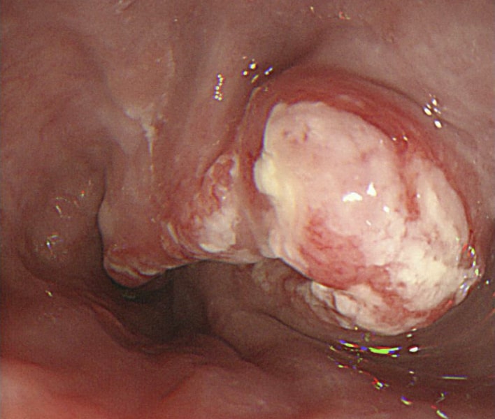
Esophagogastroduodenoscopy (EGD) showing cancer outgrowth (SCC), causing a narrowing of the lumen
Image: “A Late-Stage Squamous Cell Carcinoma” by Brooks PJ, Enoch M-A, Goldman D, Li T-K, Yokoyama A. License: C BY 2.5.
Rohof, W. O. A., & Bredenoord, A. J. (2017). Chicago Classification of Esophageal Motility Disorders: Lessons learned. Current Gastroenterology Reports, 19(8), Article 37. https://doi.org/10.1007/s11894-017-0576-7
Create your free account or log in to continue reading!