Epidemiology
- United States:
- Annual incidence in adults: 1.6 per 100,000; in children: 0.6-0.8 per 100,000
- Before the introduction of the Haemophilus influenzae type b (Hib) conjugate vaccine in 1987, the annual incidence of epiglottitis among children < 5 years old was 3 times that of adults (5 per 100.000 versus 1.6 per 100,000). That ratio is now reversed. The median age of children with epiglottitis is now 6–12 years of age.
- Worldwide:
- More common in adult men, with a male-to-female ratio of 3:1
- No seasonal differences in incidence
- More common in nations with no immunization for H. influenzae type b
- Risk factors:
- Children:
- Incomplete or lack of immunization for Hib
- Immune deficiency
- Adults:
- Immune deficiency
- Substance abuse
- Hypertension
- Diabetes mellitus
- Body mass index > 25
- Concurrent pneumonia
- Children:
Etiology
- Epiglottitis can be bacterial, viral, fungal, or noninfectious. In previously healthy children, most causes are bacterial.
- Most common bacterial causes:
- H. influenzae, accounting for approximately 25% of cases (especially in nonimmunized children, but still seen (rarely) in immunized children because the vaccine is not 100% effective)
- Streptococcus pneumonia (most common in adults)
- H. parainfluenza
- Group A Streptococci
- The disease is less commonly caused by the following pathogens, and most commonly in immunocompromised patients:
- Staphylococcus aureus
- Mycobacteria
- Bacteroides
- Escherichia coli
- Candida
- Pseudomonas aeruginosa
- Serratia
- Enterobacter
- Non-infectious causes: trauma from thermal injury, foreign body ingestion, caustic ingestion, crack cocaine smoking, chemotherapy to head and neck region
- Other causes: lymphoproliferative disorders (e.g., graft-versus-host disease)
Signs and Symptoms
- Hallmarks in children are the “4 Ds”: dysphagia, dysphonia, drooling, and distress, with rapid progression.
- In adults, the clinical progression is slower.
- Difficulty in swallowing (dysphagia)
- Painful swallowing (odynophagia)
- Sudden onset of high fever
- Inspiratory stridor: abnormal, high-pitched sound while breathing
- Excessive drooling
- Dysphonia (hoarseness): and/or muffled speech, often described as a “hot potato” voice
- Difficulty breathing → respiratory distress → cyanosis
- Restlessness, anxiety, irritability
- Pharyngitis
- Severe sore throat: the most common complaint in adults
- Refusal to lie flat
- Insisting on sitting up and leaning forward (tripod position) with neck hyperextended and chin thrust forward (sniffing posture): to maximize the diameter of the narrowed airway.
Diagnosis
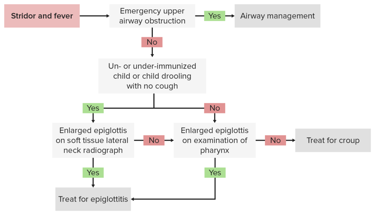
- Upon clinical suspicion, proceed to airway management before further testing.
- Suspected epiglottitis is a medical emergency!
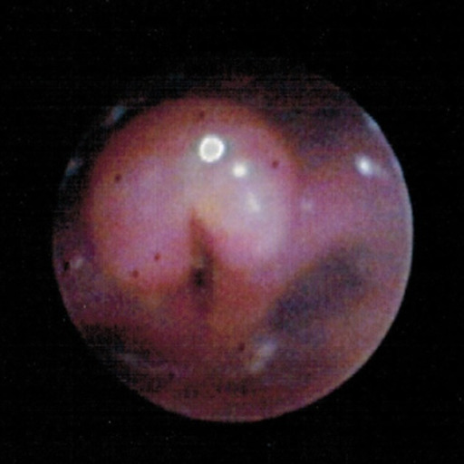
- Diagnosis is made by visualization of the epiglottis via indirect or direct fiberoptic laryngoscopy (should happen under general anesthesia in the operating room).
- Epiglottis appears to be cherry red in color and swollen.
- Inflammation and edema of the supraglottic structures
- Tenderness to palpation to the hyoid bone and region
- Inspection by a tongue depressor is discouraged as it may provoke airway spasm or distress in children.
- Laboratory: CBC with differential, blood culture, epiglottal culture if intubated
- Not required for diagnosis
- Most patients will have an elevated WBC count, but this is a nonspecific finding.
- Do not perform needle sticks in young children with suspected epiglottitis unless already intubated.
- Lateral neck X-ray:
- Disease can be confirmed by the “thumb sign” (enlarged, swollen epiglottis protruding from the anterior wall of the hypopharynx).
- Radiographs are not necessary to make the diagnosis.
- Computed tomography (CT) scan imaging:
- Halloween sign describes the CT appearance of a normal-thickness epiglottis in the axial plane so it excludes acute epiglottitis but CT can be useful to diagnose other conditions such as a peritonsillar abscess or a retropharyngeal abscess which have similar clinical features.
- Radiographs are not necessary to make the diagnosis.
Bronchoscopy image shows a swollen and inflated epiglottis and arytenoids along with aryepiglottic folds consistent with acute epiglottitis (supraglottitis).
Image: “Bronchoscopy” by Department of Pediatrics, University of Toledo Toledo, Ohio, United States of America. License: CC BY 3.0Acute epiglottitis presenting with the “thumb sign” in a lateral neck x-ray
Image: “Acute epiglottitis; Lateral view in X-ray imaging.” by Med Chaos – Own work. License: CC0
Treatment and Prevention
- Airway management
- Tracheal intubation should be performed in case of airway compromise.
- Extubation usually takes place 2–3 days after starting antibiotics.
- If intubation fails, tracheostomy should be performed.
- Intubation of a child should be performed in the operating room as unsuccessful intubation makes tracheostomy necessary.
- Monitor in an intensive care unit.
- Antibiotic therapy
- Third-generation cephalosporin (ceftriaxone or cefotaxime) AND an antistaphylococcal agent (vancomycin) for 7–10 days
- Severe hypersensitivity reaction to penicillin or cephalosporin antibiotics: vancomycin + quinolone or carbapenem
- Consult infectious disease specialist for recommendations and follow-up.
- Adjust accordingly to blood culture and epiglottis culture if epiglottis culture was possible to obtain.
- Prevention
- Hib conjugate vaccine and pneumococcal vaccine
- In case of exposure to infected individuals, rifampicin can be given as prophylaxis to all close contacts.
Complications
Prognosis is good if diagnosed and treated immediately but the disease can lead to death if there is acute and untreated airway obstruction.
Complications of epiglottitis
- Airway obstruction → asphyxia → death
- Epiglottic abscess
- Secondary infection
- Pneumonia
- Cervical adenitis
- Cellulitis
- Septic arthritis
- Empyema
- Meningitis
- Bacteremia/septic shock
- Vocal granuloma
- Cartilaginous metaplasia of epiglottis
- Necrotizing epiglottitis with immunodeficiencies
Differential Diagnosis
- Tuberculosis: a disease caused by Mycobacterium tuberculosis, an acid-fast bacteria that grows slowly and can survive in macrophages
- Pertussis: a bacterial infection caused by Bordetella pertussis; infected patients present with paroxysmal whooping cough, which persists for 2 weeks or more
- Pharyngitis: inflammation of the back of the throat or pharynx, usually caused by an upper respiratory tract infection. Pharyngitis typically results in a sore throat and fever. Other symptoms may include a runny nose, cough, headache, and hoarseness.
- Pneumonia: acute or chronic inflammation of lung tissue caused by infection with bacteria, viruses, or fungi. Pneumonia can also be due to toxic triggers through inhalation of toxic substances, immunological processes, or in the course of radiotherapy.
- Peritonsillar abscess: a bacterial infection with an onset of untreated strep throat or tonsillitis, which involves a pus-filled pocket that forms near a tonsil
- Retropharyngeal abscess: a collection of pus in the back of the throat caused by a bacterial infection, which clinically manifests as pain when swallowing, fever, stiff neck, and noisy breathing
- Croup: also called laryngotracheobronchitis; caused by a viral infection or, rarely, by a bacterial infection that results in swelling inside the trachea and that interferes with normal breathing. Patients develop a characteristic barking cough and stridor.
- Laryngomalacia: laryngomalacia and tracheomalacia are the most common upper airway conditions that produce stridor in newborns and have characteristic stridor to respiratory distress
- Measles: measles morbillivirus is a virus of the genus Morbillivirus within the family Paramyxoviridae. Humans are the natural hosts of this virus and no other animal reservoirs are known to exist.