Hemolytic Reactions
Acute hemolytic transfusion reaction
- Etiology:
- Type II hypersensitivity reaction→ targets the transfused RBCs
- ABO blood group incompatibility causes intravascular hemolysis.
- Host’s own antibodies (immunoglobulin G (IgG)) detect and bind to antigens on donor RBCs, causing extravascular hemolysis.
- Signs and symptoms:
- Start during the transfusion or within 1 hour after
- Tachycardia
- Tachypnea
- Hypotension
- Flank pain
- Hemoglobinuria due to intravascular hemolysis
- Jaundice due to the extravascular hemolysis (yellowing of the sclera and skin)
- Fever and chills
- Management:
- Immediate cessation of transfusion
- Repeat typing and crossmatching to identify cause and record to prevent future reactions.
- Supportive care:
- Infusion of IV fluids to aid diuresis
- Hypotension managed with IV fluids and vasopressors
- In case of disseminated intravascular coagulation (DIC): Perform coagulation studies and administer of fresh frozen plasma (FFP) and platelets.
- Complications:
- Hemoglobinuria can lead to renal tubular necrosis and renal failure.
- DIC: severe hemolysis can lead to an accumulation of cytokines in the blood → hypercoagulation → microthrombi and hemorrhage → impaired perfusion of organs and tissue necrosis → multi-system organ failure
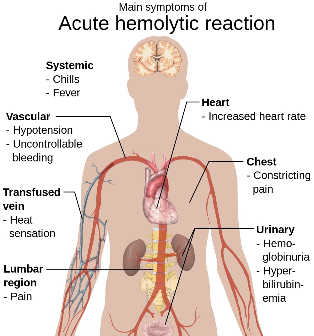
Signs and symptoms of acute hemolytic transfusion reactions
Image: “Main symptoms of acute hemolytic reaction” by Mikael Häggström. License: Public Domain, edited by Lecturio.Delayed hemolytic transfusion reaction
- Etiology:
- Occurs in patients who have been sensitized to specific RBC antigens (previous transfusions, pregnancy, or transplantations)
- Re-exposure stimulates rapid antibody production and leads to extravascular hemolysis.
- Signs and symptoms:
- Starts days or weeks after the transfusion
- Fever and chills
- Jaundice
- Anemia
- Hemoglobinuria
- Management:
- No treatment required
- Antibody testing should be performed to prevent future reactions.
Nonhemolytic Reaction
Febrile nonhemolytic transfusion reaction (FNHTR)
- Etiology:
- Type II hypersensitivity reaction in which host antibodies target donor leukocytes
- Accumulation of cytokines (IL-1, IL-6, IL-8, and tumor necrosis factor (TNF)) in the donor blood during storage produces an immune reaction in the recipient.
- Signs and symptoms:
- Start within 1–6 hours after transfusion
- Fever and chills
- Headaches
- Flushing
- General malaise
- Management:
- Cessation of transfusion
- CBC to rule out acute hemolytic reaction (anemia, hyperbilirubinemia, thrombocytopenia)
- Acetaminophen to aid with fever
- Prevention by leukoreduction of blood products
Allergic Reactions
- Etiology:
- Type I hypersensitivity reaction against plasma proteins in donor blood
- In the case of IgA deficiency: the anti-IgA IgG antibodies of the recipient detect the IgA on the surface of RBCs in the donor blood, triggering a mass release of cytokines → anaphylaxis
- Signs and symptoms:
- Start during the transfusion or within 2 hours after
- Minor allergic reactions: urticaria or pruritus
- Life-threatening reaction (anaphylaxis):
- Hypotension
- Bronchospasm, wheezing, or stridor
- Nausea and/or vomiting
- Shock
- Cardiac and/or respiratory arrest
- Management:
- Minor allergic reactions: antihistamines (e.g., diphenhydramine)
- Anaphylaxis:
- Epinephrine
- Hemodynamic stabilization
- Airway management
Other Types of Transfusion Reactions
Transfusion-related acute lung injury (TRALI)
- Etiology:
- Antibodies in the donor blood products target the neutrophils and pulmonary endothelial cells of the recipient.
- Causes non-cardiogenic pulmonary edema
- Can lead to acute hypoxemia
- Activated neutrophils in the lungs may also secrete proteolytic enzymes, leading to more tissue damage.
- Signs and symptoms:
- Start during the transfusion or within 6 hours after
- Respiratory distress or dyspnea
- Hypotonia
- Fever
- Hypotension (hypovolemia due to the accumulation of fluid in the lungs)
- X-ray shows bilateral infiltrates suggesting pulmonary edema.
- Management:
- Cessation of transfusion
- Supportive care
- Assisted ventilation or oxygen administration
- Monitoring and regulation of hemodynamic parameters
- IV steroids to aid with inflammation
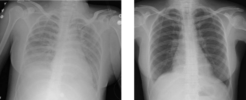
Chest X-ray of TRALI showing bilateral diffuse infiltrates (left); chest X-ray of the same subject after treatment (right)
Image: “Two chest X-rays” by Altaf Gauhar Haji et al. License: CC BY 2.0, edited by Lecturio.Graft-versus-host disease (GvHD)
- Etiology:
- Delayed transfusion reaction
- Donor’s T lymphocytes target the recipient’s tissues and organs and produce → systemic inflammatory reaction.
- Most commonly affected organs: skin, intestines, and liver
- Immunocompromised or immunocompetent patients are most likely to develop this condition.
- Can present acutely (< 100 days after transfusion/transplant) or chronically (> 100 days after transfusion/transplant)
- Signs and symptoms:
- Acute reaction
- Starts < 100 days after transfusion/transplant
- Pruritic, painful maculopapular rash
- Nausea and/or vomiting
- Diarrhea and cramping abdominal pain
- Jaundice
- Chronic reaction
- Starts > 100 days after transfusion/transplant
- All symptoms seen in the acute reaction may occur.
- Scleroderma-like skin changes (lichenoid skin changes)
- Xerostomia (dry mouth) and xerophthalmia (dry eyes)
- Bloody diarrhea (chronic enteritis similar to inflammatory bowel disease (IBD))
- Weight loss
- Muscle pain and weakness
- Bronchiolitis obliterans (chronic/persistent cough, wheezing, and dyspnea)
- Acute reaction
- Diagnosis:
- CBC shows anemia, thrombocytopenia, and/or leukopenia (acute reaction).
- Spirometry may show obstructive lung disease (chronic reaction).
- Biopsy of the transplanted tissue to confirm diagnosis
- Management:
- 1st-line: corticosteroids
- Topical in the case of < 50% skin involvement
- Systemic in the case of > 50% skin involvement, gastrointestinal (GI) involvement, liver involvement
- 2nd-line: increased corticosteroid dose + immunosuppressant (e.g., cyclosporine)
- 1st-line: corticosteroids
Transfusion-associated circulatory overlaad (TACO)
- Etiology:
- Large transfusion volume
- Fast infusion rate
- Underlying renal or cardiovascular disease
- Low body weight
- Infant or elderly patients
- Signs and symptoms:
- Start during the transfusion or within 6 hours after
- Signs of hypervolemia
- S3 gallop
- Shortness of breath
- Jugular venous distention
- Sudden hypertension
- Tachycardia
- Widened pulse pressure
- Cough and/or dyspnea
- X-ray shows bilateral infiltrates suggesting pulmonary edema.
- Management:
- Diuretics (only method to counteract elevated blood volume)
- Oxygen supplementation or assisted ventilation if necessary