Achieve Mastery of Medical Concepts

Study for medical school and boards with Lecturio

Sepsis in Children

Sepsis is defined as a life-threatening systemic response syndrome caused by viruses, bacteria, fungi, and parasites, or toxins released from these organisms. Neonates and children are particularly prone to sepsis owing to their immature immune systems. Affected individuals may present with localizing symptoms specific to a source of infection but may often complain of nonspecific systemic symptoms, such as confusion, lethargy, and fever. Diagnosis is based on history, exam, laboratory testing, and imaging studies. A high index of suspicion, rapid diagnosis, and clinical intervention with appropriate therapy are mandatory to achieve favorable outcomes.

Last updated: Feb 20, 2023

Editorial responsibility: Stanley Oiseth, Lindsay Jones, Evelin Maza

Overview

Definition

  • Systemic inflammatory response syndrome (SIRS): a disseminated inflammatory response that is identified by a combination of symptomatic (e.g., fever Fever Fever is defined as a measured body temperature of at least 38°C (100.4°F). Fever is caused by circulating endogenous and/or exogenous pyrogens that increase levels of prostaglandin E2 in the hypothalamus. Fever is commonly associated with chills, rigors, sweating, and flushing of the skin. Fever, tachycardia, tachypnea Tachypnea Increased respiratory rate. Pulmonary Examination) and lab (e.g., white blood cell count) criteria.
  • Sepsis Sepsis Systemic inflammatory response syndrome with a proven or suspected infectious etiology. When sepsis is associated with organ dysfunction distant from the site of infection, it is called severe sepsis. When sepsis is accompanied by hypotension despite adequate fluid infusion, it is called septic shock. Sepsis and Septic Shock: SIRS with the addition of a known pathogen source of infection (e.g., SIRS criteria and positive urine culture Urine culture Urinary tract infections (UTIs))

Systematic inflammatory response syndrome criteria

Sepsis Sepsis Systemic inflammatory response syndrome with a proven or suspected infectious etiology. When sepsis is associated with organ dysfunction distant from the site of infection, it is called severe sepsis. When sepsis is accompanied by hypotension despite adequate fluid infusion, it is called septic shock. Sepsis and Septic Shock:

  • 2 of the SIRS criteria (note: cutoff values are dependent on age):
    • Tachycardia or bradycardia Bradycardia Bradyarrhythmia is a rhythm in which the heart rate is less than 60/min. Bradyarrhythmia can be physiologic, without symptoms or hemodynamic change. Pathologic bradyarrhythmia results in reduced cardiac output and hemodynamic instability causing syncope, dizziness, or dyspnea. Bradyarrhythmias (in children < 1 year of age)
    • Tachypnea Tachypnea Increased respiratory rate. Pulmonary Examination
    • Fever Fever Fever is defined as a measured body temperature of at least 38°C (100.4°F). Fever is caused by circulating endogenous and/or exogenous pyrogens that increase levels of prostaglandin E2 in the hypothalamus. Fever is commonly associated with chills, rigors, sweating, and flushing of the skin. Fever or hypothermia Hypothermia Hypothermia can be defined as a drop in the core body temperature below 35°C (95°F) and is classified into mild, moderate, severe, and profound forms based on the degree of temperature decrease. Hypothermia: temperature > 38°C or < 36°C (> 100.4°F or < 95.0°F)
    • Leukocytosis Leukocytosis A transient increase in the number of leukocytes in a body fluid. West Nile Virus, leukopenia, or bandemia ≥ 10%
  • Plus, a suspected or confirmed infectious source

Severe sepsis Sepsis Systemic inflammatory response syndrome with a proven or suspected infectious etiology. When sepsis is associated with organ dysfunction distant from the site of infection, it is called severe sepsis. When sepsis is accompanied by hypotension despite adequate fluid infusion, it is called septic shock. Sepsis and Septic Shock:

  • Must meet the criteria for sepsis Sepsis Systemic inflammatory response syndrome with a proven or suspected infectious etiology. When sepsis is associated with organ dysfunction distant from the site of infection, it is called severe sepsis. When sepsis is accompanied by hypotension despite adequate fluid infusion, it is called septic shock. Sepsis and Septic Shock
  • Plus, 1 of the following:
    • Hypotension Hypotension Hypotension is defined as low blood pressure, specifically < 90/60 mm Hg, and is most commonly a physiologic response. Hypotension may be mild, serious, or life threatening, depending on the cause. Hypotension (systolic blood pressure < 90 mm MM Multiple myeloma (MM) is a malignant condition of plasma cells (activated B lymphocytes) primarily seen in the elderly. Monoclonal proliferation of plasma cells results in cytokine-driven osteoclastic activity and excessive secretion of IgG antibodies. Multiple Myeloma Hg)
    • Organ dysfunction:
      • Cardiovascular dysfunction
      • ARDS
      • Renal failure Renal failure Conditions in which the kidneys perform below the normal level in the ability to remove wastes, concentrate urine, and maintain electrolyte balance; blood pressure; and calcium metabolism. Renal insufficiency can be classified by the degree of kidney damage (as measured by the level of proteinuria) and reduction in glomerular filtration rate. Crush Syndrome
      • Altered mental status
    • Lactic acid > 4 mmol

Septic shock Septic shock Sepsis associated with hypotension or hypoperfusion despite adequate fluid resuscitation. Perfusion abnormalities may include, but are not limited to lactic acidosis; oliguria; or acute alteration in mental status. Sepsis and Septic Shock is severe sepsis Sepsis Systemic inflammatory response syndrome with a proven or suspected infectious etiology. When sepsis is associated with organ dysfunction distant from the site of infection, it is called severe sepsis. When sepsis is accompanied by hypotension despite adequate fluid infusion, it is called septic shock. Sepsis and Septic Shock that is unresponsive to fluid resuscitation Resuscitation The restoration to life or consciousness of one apparently dead. . Neonatal Respiratory Distress Syndrome.

Mortality-rates-in-shock

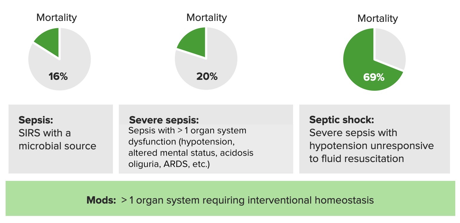
Mortality rates in sepsis, severe sepsis, and septic shock:
SIRS: systemic inflammatory response syndrome
ARDS: acute respiratory distress syndrome

Image by Lecturio.

Other sepsis-scoring criteria

  • The sequential Sequential Computed Tomography (CT) organ failure assessment ( SOFA SOFA Sepsis and Septic Shock) score and the abbreviated version ( qSOFA qSOFA Presence of 2 of the following 3 criteria indicates a worse outcome in a patient suspected of having sepsis and triggers an immediate diagnostic workup and treatment as appropriate. Sbp < 100 mm hg, respiratory rate > 22/min, altered mental status. Types of Shock) are a newer set of criteria.
  • SOFA SOFA Sepsis and Septic Shock and qSOFA qSOFA Presence of 2 of the following 3 criteria indicates a worse outcome in a patient suspected of having sepsis and triggers an immediate diagnostic workup and treatment as appropriate. Sbp < 100 mm hg, respiratory rate > 22/min, altered mental status. Types of Shock help predict mortality Mortality All deaths reported in a given population. Measures of Health Status associated with sepsis Sepsis Systemic inflammatory response syndrome with a proven or suspected infectious etiology. When sepsis is associated with organ dysfunction distant from the site of infection, it is called severe sepsis. When sepsis is accompanied by hypotension despite adequate fluid infusion, it is called septic shock. Sepsis and Septic Shock.
  • Can be used as an alternative to SIRS
  • Sepsis Sepsis Systemic inflammatory response syndrome with a proven or suspected infectious etiology. When sepsis is associated with organ dysfunction distant from the site of infection, it is called severe sepsis. When sepsis is accompanied by hypotension despite adequate fluid infusion, it is called septic shock. Sepsis and Septic Shock is suspected when 2 of the qSOFA qSOFA Presence of 2 of the following 3 criteria indicates a worse outcome in a patient suspected of having sepsis and triggers an immediate diagnostic workup and treatment as appropriate. Sbp < 100 mm hg, respiratory rate > 22/min, altered mental status. Types of Shock criteria are met MET Preoperative Care.
  • The 3 criteria for the qSOFA qSOFA Presence of 2 of the following 3 criteria indicates a worse outcome in a patient suspected of having sepsis and triggers an immediate diagnostic workup and treatment as appropriate. Sbp < 100 mm hg, respiratory rate > 22/min, altered mental status. Types of Shock score include:
    • RR RR Relative risk (RR) is the risk of a disease or condition occurring in a group or population with a particular exposure relative to a control (unexposed) group. Measures of Risk > 22 breaths/min
    • Systolic blood pressure < 100 mm MM Multiple myeloma (MM) is a malignant condition of plasma cells (activated B lymphocytes) primarily seen in the elderly. Monoclonal proliferation of plasma cells results in cytokine-driven osteoclastic activity and excessive secretion of IgG antibodies. Multiple Myeloma Hg
    • Altered mental status

Epidemiology and Etiology

Epidemiology

  • The incidence Incidence The number of new cases of a given disease during a given period in a specified population. It also is used for the rate at which new events occur in a defined population. It is differentiated from prevalence, which refers to all cases in the population at a given time. Measures of Disease Frequency of sepsis Sepsis Systemic inflammatory response syndrome with a proven or suspected infectious etiology. When sepsis is associated with organ dysfunction distant from the site of infection, it is called severe sepsis. When sepsis is accompanied by hypotension despite adequate fluid infusion, it is called septic shock. Sepsis and Septic Shock is inversely related to age in children.
  • Neonatal:
    • Up to 10% of newborns have an infection within the 1st month of life.
    • More common in children with birth weight < 1,500 grams
    • The most common mode of pathogen transmission is intrapartum or through an infected birth canal Birth canal Pelvis: Anatomy.
    • Vertical transmission Vertical transmission The transmission of infectious disease or pathogens from one generation to another. It includes transmission in utero or intrapartum by exposure to blood and secretions, and postpartum exposure via breastfeeding. Congenital TORCH Infections has ↓ due to early detection and the treatment of group B strep during pregnancy Pregnancy The status during which female mammals carry their developing young (embryos or fetuses) in utero before birth, beginning from fertilization to birth. Pregnancy: Diagnosis, Physiology, and Care
  • Infants and children:
    • In the USA, 75,000 children are hospitalized annually for severe sepsis Sepsis Systemic inflammatory response syndrome with a proven or suspected infectious etiology. When sepsis is associated with organ dysfunction distant from the site of infection, it is called severe sepsis. When sepsis is accompanied by hypotension despite adequate fluid infusion, it is called septic shock. Sepsis and Septic Shock.
    • 65% of cases are associated with respiratory or bloodstream infections Infections Invasion of the host organism by microorganisms or their toxins or by parasites that can cause pathological conditions or diseases. Chronic Granulomatous Disease.
    • Immunizations against Streptococcus Streptococcus Streptococcus is one of the two medically important genera of gram-positive cocci, the other being Staphylococcus. Streptococci are identified as different species on blood agar on the basis of their hemolytic pattern and sensitivity to optochin and bacitracin. There are many pathogenic species of streptococci, including S. pyogenes, S. agalactiae, S. pneumoniae, and the viridans streptococci. Streptococcus pneumoniae and Haemophilus influenzae Haemophilus Influenzae A species of Haemophilus found on the mucous membranes of humans and a variety of animals. The species is further divided into biotypes I through viii. Haemophilus have ↓ the incidence Incidence The number of new cases of a given disease during a given period in a specified population. It also is used for the rate at which new events occur in a defined population. It is differentiated from prevalence, which refers to all cases in the population at a given time. Measures of Disease Frequency

Risk factors

  • Neonatal:
    • Immunological immaturity
    • Exposure to maternal genital tract pathogens
    • Trauma to the scalp during childbirth
    • Invasive procedures (e.g., prolonged endotracheal intubation Intubation Peritonsillar Abscess, parenteral feeding)
    • Poor mechanical defenses (e.g., easily erodible skin Skin The skin, also referred to as the integumentary system, is the largest organ of the body. The skin is primarily composed of the epidermis (outer layer) and dermis (deep layer). The epidermis is primarily composed of keratinocytes that undergo rapid turnover, while the dermis contains dense layers of connective tissue. Skin: Structure and Functions)
    • Resistant microorganisms
  • Infants and children:
    • Age < 1 month
    • Serious injury (e.g., penetrating trauma)
    • Chronic and debilitating conditions
    • Immunosuppression (e.g., HIV HIV Anti-HIV Drugs infection)
    • Large surgical incisions
    • Indwelling or invasive devices (e.g., chest tube)
    • Frequent urinary tract Urinary tract The urinary tract is located in the abdomen and pelvis and consists of the kidneys, ureters, urinary bladder, and urethra. The structures permit the excretion of urine from the body. Urine flows from the kidneys through the ureters to the urinary bladder and out through the urethra. Urinary Tract: Anatomy infections Infections Invasion of the host organism by microorganisms or their toxins or by parasites that can cause pathological conditions or diseases. Chronic Granulomatous Disease

Etiology

  • Neonates:
    • Most common bacteria Bacteria Bacteria are prokaryotic single-celled microorganisms that are metabolically active and divide by binary fission. Some of these organisms play a significant role in the pathogenesis of diseases. Bacteriology:
      • Group B Streptococcus Group B Streptococcus Meningitis in Children (in neonates)
      • S. agalactiae
      • MRSA MRSA A strain of Staphylococcus aureus that is non-susceptible to the action of methicillin. The mechanism of resistance usually involves modification of normal or the presence of acquired penicillin binding proteins. Staphylococcus
      • Escherichia coli Escherichia coli The gram-negative bacterium Escherichia coli is a key component of the human gut microbiota. Most strains of E. coli are avirulent, but occasionally they escape the GI tract, infecting the urinary tract and other sites. Less common strains of E. coli are able to cause disease within the GI tract, most commonly presenting as abdominal pain and diarrhea. Escherichia coli
      • Enterococcus Enterococcus Enterococcus is a genus of oval-shaped gram-positive cocci that are arranged in pairs or short chains. Distinguishing factors include optochin resistance and the presence of pyrrolidonyl arylamidase (PYR) and Lancefield D antigen. Enterococcus is part of the normal flora of the human GI tract. Enterococcus faecalis
    • Uncommon bacteria Bacteria Bacteria are prokaryotic single-celled microorganisms that are metabolically active and divide by binary fission. Some of these organisms play a significant role in the pathogenesis of diseases. Bacteriology:
      • S. pneumoniae
      • Pseudomonas aeruginosa Pseudomonas aeruginosa A species of gram-negative, aerobic, rod-shaped bacteria commonly isolated from clinical specimens (wound, burn, and urinary tract infections). It is also found widely distributed in soil and water. P. Aeruginosa is a major agent of nosocomial infection. Pseudomonas
      • Listeria Listeria Listeria spp. are motile, flagellated, gram-positive, facultative intracellular bacilli. The major pathogenic species is Listeria monocytogenes. Listeria are part of the normal gastrointestinal flora of domestic mammals and poultry and are transmitted to humans through the ingestion of contaminated food, especially unpasteurized dairy products. Listeria Monocytogenes/Listeriosis monocytogenes
      • Klebsiella Klebsiella Klebsiella are encapsulated gram-negative, lactose-fermenting bacilli. They form pink colonies on MacConkey agar due to lactose fermentation. The main virulence factor is a polysaccharide capsule. Klebsiella pneumoniae is the most important pathogenic species. Klebsiella
      • H. influenza Influenza Influenza viruses are members of the Orthomyxoviridae family and the causative organisms of influenza, a highly contagious febrile respiratory disease. There are 3 primary influenza viruses (A, B, and C) and various subtypes, which are classified based on their virulent surface antigens, hemagglutinin (HA) and neuraminidase (NA). Influenza typically presents with a fever, myalgia, headache, and symptoms of an upper respiratory infection. Influenza Viruses/Influenzae
      • Mycoplasma hominis Mycoplasma hominis A common inhabitant of the vagina and cervix and a potential human pathogen, causing infections of the male and female reproductive tracts. It has also been associated with respiratory disease and pharyngitis. Mycoplasma
    • Viruses Viruses Minute infectious agents whose genomes are composed of DNA or RNA, but not both. They are characterized by a lack of independent metabolism and the inability to replicate outside living host cells. Virology:
      • Adenovirus Adenovirus Adenovirus (member of the family Adenoviridae) is a nonenveloped, double-stranded DNA virus. Adenovirus is transmitted in a variety of ways, and it can have various presentations based on the site of entry. Presentation can include febrile pharyngitis, conjunctivitis, acute respiratory disease, atypical pneumonia, and gastroenteritis. Adenovirus
      • CMV
      • Enteroviruses
      • Parechoviruses
      • Hepatitis B Hepatitis B Hepatitis B virus (HBV) is a partially double-stranded DNA virus, which belongs to the Orthohepadnavirus genus and the Hepadnaviridae family. Most individuals with acute HBV infection are asymptomatic or have mild, self-limiting symptoms. Chronic infection can be asymptomatic or create hepatic inflammation, leading to liver cirrhosis and hepatocellular carcinoma (HCC). Hepatitis B Virus and C viruses Viruses Minute infectious agents whose genomes are composed of DNA or RNA, but not both. They are characterized by a lack of independent metabolism and the inability to replicate outside living host cells. Virology
      • HSV HSV Herpes simplex virus (HSV) is a double-stranded DNA virus belonging to the family Herpesviridae. Herpes simplex virus commonly causes recurrent infections involving the skin and mucosal surfaces, including the mouth, lips, eyes, and genitals. Herpes Simplex Virus 1 and 2
      • Human immunodeficiency Immunodeficiency Chédiak-Higashi Syndrome virus Virus Viruses are infectious, obligate intracellular parasites composed of a nucleic acid core surrounded by a protein capsid. Viruses can be either naked (non-enveloped) or enveloped. The classification of viruses is complex and based on many factors, including type and structure of the nucleoid and capsid, the presence of an envelope, the replication cycle, and the host range. Virology ( HIV HIV Anti-HIV Drugs)
      • Parvovirus
      • Rubella Rubella An acute infectious disease caused by the rubella virus. The virus enters the respiratory tract via airborne droplet and spreads to the lymphatic system. Rubella Virus virus Virus Viruses are infectious, obligate intracellular parasites composed of a nucleic acid core surrounded by a protein capsid. Viruses can be either naked (non-enveloped) or enveloped. The classification of viruses is complex and based on many factors, including type and structure of the nucleoid and capsid, the presence of an envelope, the replication cycle, and the host range. Virology
      • Varicella-zoster virus Varicella-Zoster Virus Varicella-zoster virus (VZV) is a linear, double-stranded DNA virus in the Herpesviridae family. Varicella-zoster infections are highly contagious and transmitted through aerosolized respiratory droplets or contact with infected skin lesions. Varicella-Zoster Virus/Chickenpox (VZV)
      • EBV EBV Epstein-barr virus (EBV) is a linear, double-stranded DNA virus belonging to the herpesviridae family. This highly prevalent virus is mostly transmitted through contact with oropharyngeal secretions from an infected individual. The virus can infect epithelial cells and B lymphocytes, where it can undergo lytic replication or latency. Epstein-Barr Virus (in immunocompromised immunocompromised A human or animal whose immunologic mechanism is deficient because of an immunodeficiency disorder or other disease or as the result of the administration of immunosuppressive drugs or radiation. Gastroenteritis individuals)
    • Fungi Fungi A kingdom of eukaryotic, heterotrophic organisms that live parasitically as saprobes, including mushrooms; yeasts; smuts, molds, etc. They reproduce either sexually or asexually, and have life cycles that range from simple to complex. Filamentous fungi, commonly known as molds, refer to those that grow as multicellular colonies. Mycology:
      • Candida Candida Candida is a genus of dimorphic, opportunistic fungi. Candida albicans is part of the normal human flora and is the most common cause of candidiasis. The clinical presentation varies and can include localized mucocutaneous infections (e.g., oropharyngeal, esophageal, intertriginous, and vulvovaginal candidiasis) and invasive disease (e.g., candidemia, intraabdominal abscess, pericarditis, and meningitis). Candida/Candidiasis spp.
      • Malassezia Malassezia Malassezia is a lipophilic yeast commonly found on the skin surfaces of many animals, including humans. In the presence of certain environments or triggers, this fungus can cause pathologic diseases ranging from superficial skin conditions (tinea versicolor and dermatitis) to invasive disease (e.g., Malassezia folliculitis, catheter-associated fungemia, meningitis, and urinary tract infections). Malassezia Fungi spp.
    • Protozoa Protozoa Nitroimidazoles:
      • Plasmodium Plasmodium A genus of protozoa that comprise the malaria parasites of mammals. Four species infect humans (although occasional infections with primate malarias may occur). These are plasmodium falciparum; plasmodium malariae; plasmodium ovale, and plasmodium vivax. Species causing infection in vertebrates other than man include: plasmodium berghei; plasmodium chabaudi; p. Vinckei, and plasmodium yoelii in rodents; p. Brasilianum, plasmodium cynomolgi; and plasmodium knowlesi in monkeys; and plasmodium gallinaceum in chickens. Antimalarial Drugs species
      • Toxoplasma Toxoplasma Toxoplasmosis is an infectious disease caused by Toxoplasma gondii, an obligate intracellular protozoan parasite. Felines are the definitive host, but transmission to humans can occur through contact with cat feces or the consumption of contaminated foods. The clinical presentation and complications depend on the host’s immune status. Toxoplasma/Toxoplasmosis gondii
      • Trypanosoma cruzi Trypanosoma cruzi Chagas disease is an infection caused by the American trypanosome Trypanosoma cruzi. This parasitic protozoan is transmitted in the feces of reduviid bugs in South and Central America. Acute infection may present with inflammation at the inoculation site (chagoma), fever, and lymphadenopathy. Untreated, chronic infection can progress to severe complications. Trypanosoma cruzi/Chagas disease
  • Infants and children:
    • E. coli
    • S. aureus S. aureus Potentially pathogenic bacteria found in nasal membranes, skin, hair follicles, and perineum of warm-blooded animals. They may cause a wide range of infections and intoxications. Staphylococcus
    • S. pneumoniae
    • H. Influenzae H. influenzae A species of Haemophilus found on the mucous membranes of humans and a variety of animals. The species is further divided into biotypes I through VIII. Haemophilus type b
    • Neisseria meningitidis Neisseria meningitidis A species of gram-negative, aerobic bacteria. It is a commensal and pathogen only of humans, and can be carried asymptomatically in the nasopharynx. When found in cerebrospinal fluid it is the causative agent of cerebrospinal meningitis. It is also found in venereal discharges and blood. There are at least 13 serogroups based on antigenic differences in the capsular polysaccharides; the ones causing most meningitis infections being a, b, c, y, and w-135. Each serogroup can be further classified by serotype, serosubtype, and immunotype. Neisseria
    • Salmonella Salmonella Salmonellae are gram-negative bacilli of the family Enterobacteriaceae. Salmonellae are flagellated, non-lactose-fermenting, and hydrogen sulfide-producing microbes. Salmonella enterica, the most common disease-causing species in humans, is further classified based on serotype as typhoidal (S. typhi and paratyphi) and nontyphoidal (S. enteritidis and typhimurium). Salmonella spp.
    • Viruses Viruses Minute infectious agents whose genomes are composed of DNA or RNA, but not both. They are characterized by a lack of independent metabolism and the inability to replicate outside living host cells. Virology

Pathophysiology

The host’s ability to resist direct damage by the pathogen and immune system Immune system The body’s defense mechanism against foreign organisms or substances and deviant native cells. It includes the humoral immune response and the cell-mediated response and consists of a complex of interrelated cellular, molecular, and genetic components. Primary Lymphatic Organs response determines if an infection can be controlled or will result in sepsis Sepsis Systemic inflammatory response syndrome with a proven or suspected infectious etiology. When sepsis is associated with organ dysfunction distant from the site of infection, it is called severe sepsis. When sepsis is accompanied by hypotension despite adequate fluid infusion, it is called septic shock. Sepsis and Septic Shock.

  • Antigen Antigen Substances that are recognized by the immune system and induce an immune reaction. Vaccination recognition → initiates the innate immune response Innate Immune Response Immunity to pathogens is divided into innate and adaptive immune responses. The innate immune response is the 1st line of defense against a variety of pathogens, including bacteria, fungi, viruses, and parasites. In essentially the same form, the innate type of immunity is present in all multicellular organisms. Innate Immunity: Barriers, Complement, and Cytokines secretion Secretion Coagulation Studies of cytokines Cytokines Non-antibody proteins secreted by inflammatory leukocytes and some non-leukocytic cells, that act as intercellular mediators. They differ from classical hormones in that they are produced by a number of tissue or cell types rather than by specialized glands. They generally act locally in a paracrine or autocrine rather than endocrine manner. Adaptive Immune Response → activation of the complement system Complement system Serum glycoproteins participating in the host defense mechanism of complement activation that creates the complement membrane attack complex. Included are glycoproteins in the various pathways of complement activation (classical complement pathway; alternative complement pathway; and lectin complement pathway). Innate Immunity: Barriers, Complement, and Cytokines, platelet-activation-factor, arachidonic acid Arachidonic Acid An unsaturated, essential fatty acid. It is found in animal and human fat as well as in the liver, brain, and glandular organs, and is a constituent of animal phosphatides. It is formed by the synthesis from dietary linoleic acid and is a precursor in the biosynthesis of prostaglandins, thromboxanes, and leukotrienes. Nonsteroidal Antiinflammatory Drugs (NSAIDs), and inflammatory pathways, and NO release
  • Activated coagulation factors Coagulation factors Endogenous substances, usually proteins, that are involved in the blood coagulation process. Hemostasis and fibrinolysis → DIC DIC Disseminated intravascular coagulation (DIC) is a condition characterized by systemic bodywide activation of the coagulation cascade. This cascade results in both widespread microvascular thrombi contributing to multiple organ dysfunction and consumption of clotting factors and platelets, leading to hemorrhage. Disseminated Intravascular Coagulation
  • System-wide inflammation Inflammation Inflammation is a complex set of responses to infection and injury involving leukocytes as the principal cellular mediators in the body’s defense against pathogenic organisms. Inflammation is also seen as a response to tissue injury in the process of wound healing. The 5 cardinal signs of inflammation are pain, heat, redness, swelling, and loss of function. Inflammation → generalized vasodilation Vasodilation The physiological widening of blood vessels by relaxing the underlying vascular smooth muscle. Pulmonary Hypertension Drugs and poor organ perfusion anaerobic metabolism → lactic acidosis Lactic Acidosis Oxazolidinones
  • ↓ Perfusion → multiorgan dysfunction syndrome
  • Generalized vasodilation Vasodilation The physiological widening of blood vessels by relaxing the underlying vascular smooth muscle. Pulmonary Hypertension Drugs → relative hypovolemia and, eventually, distributive shock Distributive Shock Types of Shock

Clinical Presentation

Nonspecific

  • Poor feeding
  • Change in behavior
  • Lethargy Lethargy A general state of sluggishness, listless, or uninterested, with being tired, and having difficulty concentrating and doing simple tasks. It may be related to depression or drug addiction. Hyponatremia
  • Altered mental status
  • ↑/↓ HR
  • Hypotension Hypotension Hypotension is defined as low blood pressure, specifically < 90/60 mm Hg, and is most commonly a physiologic response. Hypotension may be mild, serious, or life threatening, depending on the cause. Hypotension
  • Temperature instability

System specific

  • Circulatory
    • Poor perfusion:
      • Pallor and cyanosis Cyanosis A bluish or purplish discoloration of the skin and mucous membranes due to an increase in the amount of deoxygenated hemoglobin in the blood or a structural defect in the hemoglobin molecule. Pulmonary Examination
      • ↑ Capillary refill
      • Edema Edema Edema is a condition in which excess serous fluid accumulates in the body cavity or interstitial space of connective tissues. Edema is a symptom observed in several medical conditions. It can be categorized into 2 types, namely, peripheral (in the extremities) and internal (in an organ or body cavity). Edema
      • Dehydration Dehydration The condition that results from excessive loss of water from a living organism. Volume Depletion and Dehydration
    • Septic shock Septic shock Sepsis associated with hypotension or hypoperfusion despite adequate fluid resuscitation. Perfusion abnormalities may include, but are not limited to lactic acidosis; oliguria; or acute alteration in mental status. Sepsis and Septic Shock:
      • “Warm” shock Shock Shock is a life-threatening condition associated with impaired circulation that results in tissue hypoxia. The different types of shock are based on the underlying cause: distributive (↑ cardiac output (CO), ↓ systemic vascular resistance (SVR)), cardiogenic (↓ CO, ↑ SVR), hypovolemic (↓ CO, ↑ SVR), obstructive (↓ CO), and mixed. Types of Shock:
        • Instant capillary refill
        • Bounding pulses (wide pulse pressure)
        • Warm extremities
      • “Cold” shock Shock Shock is a life-threatening condition associated with impaired circulation that results in tissue hypoxia. The different types of shock are based on the underlying cause: distributive (↑ cardiac output (CO), ↓ systemic vascular resistance (SVR)), cardiogenic (↓ CO, ↑ SVR), hypovolemic (↓ CO, ↑ SVR), obstructive (↓ CO), and mixed. Types of Shock:
        • ↓ Capillary refill (> 2 seconds)
        • ↓ Pulse
        • Cold extremities
        • Most common in pediatrics
  • Digestive:
    • Vomiting Vomiting The forcible expulsion of the contents of the stomach through the mouth. Hypokalemia/ diarrhea Diarrhea Diarrhea is defined as ≥ 3 watery or loose stools in a 24-hour period. There are a multitude of etiologies, which can be classified based on the underlying mechanism of disease. The duration of symptoms (acute or chronic) and characteristics of the stools (e.g., watery, bloody, steatorrheic, mucoid) can help guide further diagnostic evaluation. Diarrhea
    • Abdominal distention Abdominal distention Megacolon
    • Hepatomegaly
  • Respiratory:
    • ↑/↓ RR RR Relative risk (RR) is the risk of a disease or condition occurring in a group or population with a particular exposure relative to a control (unexposed) group. Measures of Risk
    • Retractions
    • Grunting Grunting Physical Examination of the Newborn
    • Nasal flaring
    • Dyspnea Dyspnea Dyspnea is the subjective sensation of breathing discomfort. Dyspnea is a normal manifestation of heavy physical or psychological exertion, but also may be caused by underlying conditions (both pulmonary and extrapulmonary). Dyspnea
    • Cyanosis Cyanosis A bluish or purplish discoloration of the skin and mucous membranes due to an increase in the amount of deoxygenated hemoglobin in the blood or a structural defect in the hemoglobin molecule. Pulmonary Examination
    • Hypoxia Hypoxia Sub-optimal oxygen levels in the ambient air of living organisms. Ischemic Cell Damage (< 90% saturation)
  • Urologic:
    • Oliguria Oliguria Decreased urine output that is below the normal range. Oliguria can be defined as urine output of less than or equal to 0. 5 or 1 ml/kg/hr depending on the age. Renal Potassium Regulation
    • Anuria Anuria Absence of urine formation. It is usually associated with complete bilateral ureteral (ureter) obstruction, complete lower urinary tract obstruction, or unilateral ureteral obstruction when a solitary kidney is present. Acute Kidney Injury
  • Neurologic:
    • Seizures Seizures A seizure is abnormal electrical activity of the neurons in the cerebral cortex that can manifest in numerous ways depending on the region of the brain affected. Seizures consist of a sudden imbalance that occurs between the excitatory and inhibitory signals in cortical neurons, creating a net excitation. The 2 major classes of seizures are focal and generalized. Seizures or tremors
    • Apathy Apathy Lack of emotion or emotional expression; a disorder of motivation that persists over time. Wernicke Encephalopathy and Korsakoff Syndrome
    • Irritability
    • Lethargy Lethargy A general state of sluggishness, listless, or uninterested, with being tired, and having difficulty concentrating and doing simple tasks. It may be related to depression or drug addiction. Hyponatremia
    • Hypotonia Hypotonia Duchenne Muscular Dystrophy/ hypertonia Hypertonia Abnormal increase in skeletal or smooth muscle tone. Skeletal muscle hypertonicity may be associated with pyramidal tract lesions or basal ganglia diseases. Neurological Examination
    • Hyperreflexia
    • Full fontanelle Fontanelle Any of six membrane-covered openings between the cranial sutures in the incompletely ossified skull of the fetus or newborn infant. The fontanelles normally close sometime after birth. Skull: Anatomy
    • Abnormal Moro reflex Moro Reflex Primitive Reflexes
    • Irregular breathing
    • High-pitched crying
  • Hematologic:
    • Jaundice Jaundice Jaundice is the abnormal yellowing of the skin and/or sclera caused by the accumulation of bilirubin. Hyperbilirubinemia is caused by either an increase in bilirubin production or a decrease in the hepatic uptake, conjugation, or excretion of bilirubin. Jaundice
    • Hepatosplenomegaly Hepatosplenomegaly Cytomegalovirus
    • Bleeding
  • Dermatologic:

Clinical progression in neonatal sepsis Sepsis Systemic inflammatory response syndrome with a proven or suspected infectious etiology. When sepsis is associated with organ dysfunction distant from the site of infection, it is called severe sepsis. When sepsis is accompanied by hypotension despite adequate fluid infusion, it is called septic shock. Sepsis and Septic Shock

Table: Clinical progression in neonatal sepsis Sepsis Systemic inflammatory response syndrome with a proven or suspected infectious etiology. When sepsis is associated with organ dysfunction distant from the site of infection, it is called severe sepsis. When sepsis is accompanied by hypotension despite adequate fluid infusion, it is called septic shock. Sepsis and Septic Shock
Initial phase Septic phase Late phase
  • The child appears ill.
  • Poor feeding
  • Lethargy Lethargy A general state of sluggishness, listless, or uninterested, with being tired, and having difficulty concentrating and doing simple tasks. It may be related to depression or drug addiction. Hyponatremia
  • Unexplained tachycardia
  • GI symptoms
  • Respiratory symptoms
  • Neurologic signs
  • Urologic symptoms
  • Circulatory collapse
  • Hematologic signs

Diagnosis

Laboratory analysis

  • CBC:
    • ↑/↓ WBC count
    • ↓ Platelet count
    • Blasts Blasts Injuries resulting when a person is struck by particles impelled with violent force from an explosion. Blast causes pulmonary contusion and hemorrhage, laceration of other thoracic and abdominal viscera, ruptured ear drums, and minor effects in the central nervous system. Blunt Chest Trauma
  • Electrolytes Electrolytes Electrolytes are mineral salts that dissolve in water and dissociate into charged particles called ions, which can be either be positively (cations) or negatively (anions) charged. Electrolytes are distributed in the extracellular and intracellular compartments in different concentrations. Electrolytes are essential for various basic life-sustaining functions. Electrolytes:
    • ↓ Na
    • ↓/↑ K
    • ↓ Phosphorus
    • Glucose Glucose A primary source of energy for living organisms. It is naturally occurring and is found in fruits and other parts of plants in its free state. It is used therapeutically in fluid and nutrient replacement. Lactose Intolerance due to ↑ metabolic demand and ↓ caloric intake
    • Serum creatinine and BUN: prerenal azotemia Azotemia A biochemical abnormality referring to an elevation of blood urea nitrogen and creatinine. Azotemia can be produced by kidney diseases or other extrarenal disorders. When azotemia becomes associated with a constellation of clinical signs, it is termed uremia. Acute Kidney Injury due to AKI AKI Acute kidney injury refers to sudden and often reversible loss of renal function, which develops over days or weeks. Azotemia refers to elevated levels of nitrogen-containing substances in the blood that accompany AKI, which include BUN and creatinine. Acute Kidney Injury
  • Blood gas:
  • Total bilirubin Bilirubin A bile pigment that is a degradation product of heme. Heme Metabolism:
    • ↑ (> 4 mg/dL) ALT ALT An enzyme that catalyzes the conversion of l-alanine and 2-oxoglutarate to pyruvate and l-glutamate. Liver Function Tests
    • Transaminases Transaminases A subclass of enzymes of the transferase class that catalyze the transfer of an amino group from a donor (generally an amino acid) to an acceptor (generally a 2-keto acid). Most of these enzymes are pyridoxyl phosphate proteins. Autoimmune Hepatitis may indicate liver Liver The liver is the largest gland in the human body. The liver is found in the superior right quadrant of the abdomen and weighs approximately 1.5 kilograms. Its main functions are detoxification, metabolism, nutrient storage (e.g., iron and vitamins), synthesis of coagulation factors, formation of bile, filtration, and storage of blood. Liver: Anatomy dysfunction, HSV HSV Herpes simplex virus (HSV) is a double-stranded DNA virus belonging to the family Herpesviridae. Herpes simplex virus commonly causes recurrent infections involving the skin and mucosal surfaces, including the mouth, lips, eyes, and genitals. Herpes Simplex Virus 1 and 2, or enterovirus Enterovirus A genus of the family picornaviridae whose members preferentially inhabit the intestinal tract of a variety of hosts. The genus contains many species. Newly described members of human enteroviruses are assigned continuous numbers with the species designated ‘human enterovirus’. Coxsackievirus infection
  • Blood cultures x 2 to identify the causative agent
  • PT, PTT, and INR due to coagulation disorder
  • Fibrinogen Fibrinogen Plasma glycoprotein clotted by thrombin, composed of a dimer of three non-identical pairs of polypeptide chains (alpha, beta, gamma) held together by disulfide bonds. Fibrinogen clotting is a sol-gel change involving complex molecular arrangements: whereas fibrinogen is cleaved by thrombin to form polypeptides a and b, the proteolytic action of other enzymes yields different fibrinogen degradation products. Hemostasis
  • D-dimer D-dimer Deep Vein Thrombosis: DIC DIC Disseminated intravascular coagulation (DIC) is a condition characterized by systemic bodywide activation of the coagulation cascade. This cascade results in both widespread microvascular thrombi contributing to multiple organ dysfunction and consumption of clotting factors and platelets, leading to hemorrhage. Disseminated Intravascular Coagulation
  • Urinalysis Urinalysis Examination of urine by chemical, physical, or microscopic means. Routine urinalysis usually includes performing chemical screening tests, determining specific gravity, observing any unusual color or odor, screening for bacteriuria, and examining the sediment microscopically. Urinary Tract Infections (UTIs) in Children and culture

Diagnostic imaging

  • Chest X-ray X-ray Penetrating electromagnetic radiation emitted when the inner orbital electrons of an atom are excited and release radiant energy. X-ray wavelengths range from 1 pm to 10 nm. Hard x-rays are the higher energy, shorter wavelength x-rays. Soft x-rays or grenz rays are less energetic and longer in wavelength. The short wavelength end of the x-ray spectrum overlaps the gamma rays wavelength range. The distinction between gamma rays and x-rays is based on their radiation source. Pulmonary Function Tests: if suspected pneumonia Pneumonia Pneumonia or pulmonary inflammation is an acute or chronic inflammation of lung tissue. Causes include infection with bacteria, viruses, or fungi. In more rare cases, pneumonia can also be caused through toxic triggers through inhalation of toxic substances, immunological processes, or in the course of radiotherapy. Pneumonia or fluid overload
  • Ultrasound or CT scan of the abdomen: +/- if etiology not apparent
  • Head CT: warranted in individuals with coagulopathy and altered mental status

Management and Prevention

Management

  • Begins with stabilizing the affected individual
  • Goal-directed therapy:
    • IV access within 5 minutes
    • Fluid resuscitation Resuscitation The restoration to life or consciousness of one apparently dead. . Neonatal Respiratory Distress Syndrome within 30 minutes
    • Broad-spectrum Broad-Spectrum Fluoroquinolones antibiotic therapy within 60 minutes
    • Inotropic infusion within 60 minutes (in fluid-refractory shock Shock Shock is a life-threatening condition associated with impaired circulation that results in tissue hypoxia. The different types of shock are based on the underlying cause: distributive (↑ cardiac output (CO), ↓ systemic vascular resistance (SVR)), cardiogenic (↓ CO, ↑ SVR), hypovolemic (↓ CO, ↑ SVR), obstructive (↓ CO), and mixed. Types of Shock)
    • Periodic reevaluation of:
      • Central and peripheral pulses
      • Skin Skin The skin, also referred to as the integumentary system, is the largest organ of the body. The skin is primarily composed of the epidermis (outer layer) and dermis (deep layer). The epidermis is primarily composed of keratinocytes that undergo rapid turnover, while the dermis contains dense layers of connective tissue. Skin: Structure and Functions perfusion (capillary refill and skin Skin The skin, also referred to as the integumentary system, is the largest organ of the body. The skin is primarily composed of the epidermis (outer layer) and dermis (deep layer). The epidermis is primarily composed of keratinocytes that undergo rapid turnover, while the dermis contains dense layers of connective tissue. Skin: Structure and Functions temperature)
      • Fluid status
      • Urine output (> 1 mL/kg/hour)
      • Systolic blood pressure
      • Mental status
      • Serum lactate levels
      • Central venous oxygen saturation Oxygen Saturation Basic Procedures (ScvO₂)
  • Resuscitation Resuscitation The restoration to life or consciousness of one apparently dead. . Neonatal Respiratory Distress Syndrome goals:
    • Mean arterial pressure Mean Arterial Pressure Mean arterial pressure (MAP) is the average systemic arterial pressure and is directly related to cardiac output (CO) and systemic vascular resistance (SVR). The SVR and MAP are affected by the vascular anatomy as well as a number of local and neurohumoral factors. Vascular Resistance, Flow, and Mean Arterial Pressure > 65 mm MM Multiple myeloma (MM) is a malignant condition of plasma cells (activated B lymphocytes) primarily seen in the elderly. Monoclonal proliferation of plasma cells results in cytokine-driven osteoclastic activity and excessive secretion of IgG antibodies. Multiple Myeloma Hg
    • Urine output > 0.5 mL/kg/hour
    • Central venous pressure Central venous pressure The blood pressure in the central large veins of the body. It is distinguished from peripheral venous pressure which occurs in an extremity. Central Venous Catheter ( CVP CVP The blood pressure in the central large veins of the body. It is distinguished from peripheral venous pressure which occurs in an extremity. Central Venous Catheter) 8–10 mm MM Multiple myeloma (MM) is a malignant condition of plasma cells (activated B lymphocytes) primarily seen in the elderly. Monoclonal proliferation of plasma cells results in cytokine-driven osteoclastic activity and excessive secretion of IgG antibodies. Multiple Myeloma Hg
    • ScvO₂ > 70% or SvO₂ > 65%
  • Therapies:
    • Isotonic Isotonic Solutions having the same osmotic pressure as blood serum, or another solution with which they are compared. Renal Sodium and Water Regulation IV solutions: starting bolus of 20 cc/kg for older children with no cardiac or renal pathology
    • Vasopressors (e.g., norepinephrine Norepinephrine Precursor of epinephrine that is secreted by the adrenal medulla and is a widespread central and autonomic neurotransmitter. Norepinephrine is the principal transmitter of most postganglionic sympathetic fibers, and of the diffuse projection system in the brain that arises from the locus ceruleus. Receptors and Neurotransmitters of the CNS, epinephrine Epinephrine The active sympathomimetic hormone from the adrenal medulla. It stimulates both the alpha- and beta- adrenergic systems, causes systemic vasoconstriction and gastrointestinal relaxation, stimulates the heart, and dilates bronchi and cerebral vessels. Sympathomimetic Drugs)
    • Inotropic therapy (e.g., epinephrine Epinephrine The active sympathomimetic hormone from the adrenal medulla. It stimulates both the alpha- and beta- adrenergic systems, causes systemic vasoconstriction and gastrointestinal relaxation, stimulates the heart, and dilates bronchi and cerebral vessels. Sympathomimetic Drugs, dopamine Dopamine One of the catecholamine neurotransmitters in the brain. It is derived from tyrosine and is the precursor to norepinephrine and epinephrine. Dopamine is a major transmitter in the extrapyramidal system of the brain, and important in regulating movement. Receptors and Neurotransmitters of the CNS)
    • Calculation of the rate of infusion: dose/volume of dilution / child’s weight/time of infusion

Antibiotic therapy

  • Ideally, start as soon as possible.
    • Within the 1st hour of presentation (golden hour)
    • After obtaining culture samples
  • Empirical broad-spread antibiotics at 1st; then, tailor to cultures and antibiograms
  • Factors to consider:
    • Age
    • History
    • Comorbidities Comorbidities The presence of co-existing or additional diseases with reference to an initial diagnosis or with reference to the index condition that is the subject of study. Comorbidity may affect the ability of affected individuals to function and also their survival; it may be used as a prognostic indicator for length of hospital stay, cost factors, and outcome or survival. St. Louis Encephalitis Virus
    • Clinical situation
    • Type of infection
    • Gram staining Gram staining Bacteriology results
  • Antibiotic therapy options:
    • < 28-weeks old: ampicillin Ampicillin Semi-synthetic derivative of penicillin that functions as an orally active broad-spectrum antibiotic. Penicillins (50 mg/kg) or vancomycin Vancomycin Antibacterial obtained from streptomyces orientalis. It is a glycopeptide related to ristocetin that inhibits bacterial cell wall assembly and is toxic to kidneys and the inner ear. Glycopeptides (15 mg/kg) + cefotaxime Cefotaxime Semisynthetic broad-spectrum cephalosporin. Cephalosporins (100 mg/kg) + gentamicin Gentamicin Aminoglycosides (2.5 mg/kg) and acyclovir Acyclovir A guanosine analog that acts as an antimetabolite. Viruses are especially susceptible. Used especially against herpes. Herpes Zoster (Shingles) (20 mg/kg) if suspecting HSV HSV Herpes simplex virus (HSV) is a double-stranded DNA virus belonging to the family Herpesviridae. Herpes simplex virus commonly causes recurrent infections involving the skin and mucosal surfaces, including the mouth, lips, eyes, and genitals. Herpes Simplex Virus 1 and 2
    • > 28-weeks old:
      • Vancomycin Vancomycin Antibacterial obtained from streptomyces orientalis. It is a glycopeptide related to ristocetin that inhibits bacterial cell wall assembly and is toxic to kidneys and the inner ear. Glycopeptides (15 mg/kg) + ceftriaxone Ceftriaxone A broad-spectrum cephalosporin antibiotic and cefotaxime derivative with a very long half-life and high penetrability to meninges, eyes and inner ears. Cephalosporins (75 mg/kg) or cefotaxime Cefotaxime Semisynthetic broad-spectrum cephalosporin. Cephalosporins (100 mg/kg)
      • In immunosuppresion: vancomycin Vancomycin Antibacterial obtained from streptomyces orientalis. It is a glycopeptide related to ristocetin that inhibits bacterial cell wall assembly and is toxic to kidneys and the inner ear. Glycopeptides (15 mg/kg) + cefepime Cefepime A fourth-generation cephalosporin antibacterial agent that is used in the treatment of infections, including those of the abdomen, urinary tract, respiratory tract, and skin. It is effective against pseudomonas aeruginosa and may also be used in the empiric treatment of febrile neutropenia. Cephalosporins (75 mg/kg) or meropenem Meropenem A thienamycin derivative antibacterial agent that is more stable to renal dehydropeptidase I than imipenem, but does not need to be given with an enzyme inhibitor such as cilastatin. It is used in the treatment of bacterial infections, including infections in immunocompromised patients. Carbapenems and Aztreonam (20 mg/kg
    • In children who cannot receive penicillin Penicillin Rheumatic Fever: vancomycin Vancomycin Antibacterial obtained from streptomyces orientalis. It is a glycopeptide related to ristocetin that inhibits bacterial cell wall assembly and is toxic to kidneys and the inner ear. Glycopeptides (15 mg/kg) + meropenem Meropenem A thienamycin derivative antibacterial agent that is more stable to renal dehydropeptidase I than imipenem, but does not need to be given with an enzyme inhibitor such as cilastatin. It is used in the treatment of bacterial infections, including infections in immunocompromised patients. Carbapenems and Aztreonam (20 mg/kg) or aztreonam Aztreonam The carbapenems and aztreonam are both members of the bactericidal beta-lactam family of antibiotics (similar to penicillins). They work by preventing bacteria from producing their cell wall, ultimately leading to bacterial cell death. Carbapenems and Aztreonam (90–120 mg/kg/day) + clindamycin Clindamycin An antibacterial agent that is a semisynthetic analog of lincomycin. Lincosamides (40 mg/kg)
    • In children with fungal infections Infections Invasion of the host organism by microorganisms or their toxins or by parasites that can cause pathological conditions or diseases. Chronic Granulomatous Disease: amphotericin B Amphotericin B Macrolide antifungal antibiotic produced by streptomyces nodosus obtained from soil of the orinoco river region of venezuela. Polyenes + caspofungin Caspofungin A cyclic lipopeptide echinocandin and beta-(1, 3)-d-glucan synthase inhibitor that is used to treat internal or systemic mycoses. Echinocandins

Prevention

  • Follow vaccination Vaccination Vaccination is the administration of a substance to induce the immune system to develop protection against a disease. Unlike passive immunization, which involves the administration of pre-performed antibodies, active immunization constitutes the administration of a vaccine to stimulate the body to produce its own antibodies. Vaccination schedule to prevent infections Infections Invasion of the host organism by microorganisms or their toxins or by parasites that can cause pathological conditions or diseases. Chronic Granulomatous Disease.
  • Limit Limit A value (e.g., pressure or time) that should not be exceeded and which is specified by the operator to protect the lung Invasive Mechanical Ventilation the use of indwelling catheters Indwelling catheters Catheters designed to be left within an organ or passage for an extended period of time. Pseudomonas.
  • Screening Screening Preoperative Care for potential pathogens during pregnancy Pregnancy The status during which female mammals carry their developing young (embryos or fetuses) in utero before birth, beginning from fertilization to birth. Pregnancy: Diagnosis, Physiology, and Care, and management prior to birth.
  • Rectovaginal swab for group B Streptococcus Streptococcus Streptococcus is one of the two medically important genera of gram-positive cocci, the other being Staphylococcus. Streptococci are identified as different species on blood agar on the basis of their hemolytic pattern and sensitivity to optochin and bacitracin. There are many pathogenic species of streptococci, including S. pyogenes, S. agalactiae, S. pneumoniae, and the viridans streptococci. Streptococcus colonization Colonization Bacteriology at 35–37-weeks gestation, serum testing for:
    • Syphilis Syphilis Syphilis is a bacterial infection caused by the spirochete Treponema pallidum pallidum (T. p. pallidum), which is usually spread through sexual contact. Syphilis has 4 clinical stages: primary, secondary, latent, and tertiary. Syphilis
    • Hepatitis B surface antigen Hepatitis B surface antigen Those hepatitis B antigens found on the surface of the dane particle and on the 20 nm spherical and tubular particles. Several subspecificities of the surface antigen are known. These were formerly called the Australia antigen. Hepatitis B Virus testing
    • C. trachomatis
    • N. gonorrhea Gonorrhea Gonorrhea is a sexually transmitted infection (STI) caused by the gram-negative bacteria Neisseria gonorrhoeae (N. gonorrhoeae). Gonorrhea may be asymptomatic but commonly manifests as cervicitis or urethritis with less common presentations such as proctitis, conjunctivitis, or pharyngitis. Gonorrhea
    • Screening Screening Preoperative Care for hepatitis C Hepatitis C Hepatitis C is an infection of the liver caused by the hepatitis C virus (HCV). The infection can be transmitted through infectious blood or body fluids and may be transmitted during childbirth or through IV drug use or sexual intercourse. Hepatitis C virus can cause both acute and chronic hepatitis, ranging from a mild to a serious, lifelong illness including liver cirrhosis and hepatocellular carcinoma (HCC). Hepatitis C Virus antibodies Antibodies Immunoglobulins (Igs), also known as antibodies, are glycoprotein molecules produced by plasma cells that act in immune responses by recognizing and binding particular antigens. The various Ig classes are IgG (the most abundant), IgM, IgE, IgD, and IgA, which differ in their biologic features, structure, target specificity, and distribution. Immunoglobulins: Types and Functions in high-risk women

Related videos

Differential Diagnosis

  • Pediatric meningitis Meningitis Meningitis is inflammation of the meninges, the protective membranes of the brain, and spinal cord. The causes of meningitis are varied, with the most common being bacterial or viral infection. The classic presentation of meningitis is a triad of fever, altered mental status, and nuchal rigidity. Meningitis: inflammation Inflammation Inflammation is a complex set of responses to infection and injury involving leukocytes as the principal cellular mediators in the body’s defense against pathogenic organisms. Inflammation is also seen as a response to tissue injury in the process of wound healing. The 5 cardinal signs of inflammation are pain, heat, redness, swelling, and loss of function. Inflammation of the meninges Meninges The brain and the spinal cord are enveloped by 3 overlapping layers of connective tissue called the meninges. The layers are, from the most external layer to the most internal layer, the dura mater, arachnoid mater, and pia mater. Between these layers are 3 potential spaces called the epidural, subdural, and subarachnoid spaces. Meninges: Anatomy, which can be life threatening. Multiple microorganisms are responsible for meningitis Meningitis Meningitis is inflammation of the meninges, the protective membranes of the brain, and spinal cord. The causes of meningitis are varied, with the most common being bacterial or viral infection. The classic presentation of meningitis is a triad of fever, altered mental status, and nuchal rigidity. Meningitis. Symptoms vary according to age and range from bulging fontanelles Fontanelles Physical Examination of the Newborn in infants to nuchal rigidity Nuchal Rigidity Meningitis in older children. Fever Fever Fever is defined as a measured body temperature of at least 38°C (100.4°F). Fever is caused by circulating endogenous and/or exogenous pyrogens that increase levels of prostaglandin E2 in the hypothalamus. Fever is commonly associated with chills, rigors, sweating, and flushing of the skin. Fever, headache Headache The symptom of pain in the cranial region. It may be an isolated benign occurrence or manifestation of a wide variety of headache disorders. Brain Abscess, lethargy Lethargy A general state of sluggishness, listless, or uninterested, with being tired, and having difficulty concentrating and doing simple tasks. It may be related to depression or drug addiction. Hyponatremia, and vomiting Vomiting The forcible expulsion of the contents of the stomach through the mouth. Hypokalemia may also be present. A lumbar puncture Lumbar Puncture Febrile Infant is used for diagnosis. Management is using antibiotics.
  • Pediatric adrenal insufficiency Adrenal Insufficiency Conditions in which the production of adrenal corticosteroids falls below the requirement of the body. Adrenal insufficiency can be caused by defects in the adrenal glands, the pituitary gland, or the hypothalamus. Adrenal Insufficiency and Addison Disease: characterized as primary (affecting the adrenal gland) and central (hypothalamic-pituitary dysfunction), and may be congenital Congenital Chorioretinitis or acquired. Affected individuals present with hypovolemia, hypoglycemia Hypoglycemia Hypoglycemia is an emergency condition defined as a serum glucose level ≤ 70 mg/dL (≤ 3.9 mmol/L) in diabetic patients. In nondiabetic patients, there is no specific or defined limit for normal serum glucose levels, and hypoglycemia is defined mainly by its clinical features. Hypoglycemia, hyponatremia Hyponatremia Hyponatremia is defined as a decreased serum sodium (sNa+) concentration less than 135 mmol/L. Serum sodium is the greatest contributor to plasma osmolality, which is very tightly controlled via antidiuretic hormone (ADH) release from the hypothalamus and by the thirst mechanism. Hyponatremia, and hyperkalemia Hyperkalemia Hyperkalemia is defined as a serum potassium (K+) concentration >5.2 mEq/L. Homeostatic mechanisms maintain the serum K+ concentration between 3.5 and 5.2 mEq/L, despite marked variation in dietary intake. Hyperkalemia can be due to a variety of causes, which include transcellular shifts, tissue breakdown, inadequate renal excretion, and drugs. Hyperkalemia. Laboratory tests are used to confirm the diagnosis. Management is using saline and dextrose Dextrose Intravenous Fluids to regulate electrolytes Electrolytes Electrolytes are mineral salts that dissolve in water and dissociate into charged particles called ions, which can be either be positively (cations) or negatively (anions) charged. Electrolytes are distributed in the extracellular and intracellular compartments in different concentrations. Electrolytes are essential for various basic life-sustaining functions. Electrolytes.
  • Necrotizing enterocolitis Enterocolitis Inflammation of the mucosa of both the small intestine and the large intestine. Etiology includes ischemia, infections, allergic, and immune responses. Yersinia spp./Yersiniosis: a GI disease characterized by the intestinal necrosis Necrosis The death of cells in an organ or tissue due to disease, injury or failure of the blood supply. Ischemic Cell Damage of a part of the intestine. Common in preterm infants and newborns who are ill. Infants present with poor feeding, abdominal distension, and signs of sepsis Sepsis Systemic inflammatory response syndrome with a proven or suspected infectious etiology. When sepsis is associated with organ dysfunction distant from the site of infection, it is called severe sepsis. When sepsis is accompanied by hypotension despite adequate fluid infusion, it is called septic shock. Sepsis and Septic Shock. Necrotizing enterocolitis Enterocolitis Inflammation of the mucosa of both the small intestine and the large intestine. Etiology includes ischemia, infections, allergic, and immune responses. Yersinia spp./Yersiniosis is diagnosed using abdominal X-ray X-ray Penetrating electromagnetic radiation emitted when the inner orbital electrons of an atom are excited and release radiant energy. X-ray wavelengths range from 1 pm to 10 nm. Hard x-rays are the higher energy, shorter wavelength x-rays. Soft x-rays or grenz rays are less energetic and longer in wavelength. The short wavelength end of the x-ray spectrum overlaps the gamma rays wavelength range. The distinction between gamma rays and x-rays is based on their radiation source. Pulmonary Function Tests. Early diagnosis and antibiotics can improve outcomes. Surgical intervention may be needed.
  • Cardiogenic shock Cardiogenic shock Shock resulting from diminution of cardiac output in heart disease. Types of Shock: a condition that occurs when there is circulatory collapse secondary to cardiac insult. Affected individuals present with tachycardia, hepatomegaly, cardiac gallops, murmurs, precordial heaves, and jugular venous distension Jugular Venous Distension Cardiovascular Examination. Laboratory tests aid in the diagnosis. Chest X-ray X-ray Penetrating electromagnetic radiation emitted when the inner orbital electrons of an atom are excited and release radiant energy. X-ray wavelengths range from 1 pm to 10 nm. Hard x-rays are the higher energy, shorter wavelength x-rays. Soft x-rays or grenz rays are less energetic and longer in wavelength. The short wavelength end of the x-ray spectrum overlaps the gamma rays wavelength range. The distinction between gamma rays and x-rays is based on their radiation source. Pulmonary Function Tests and cardiac output Cardiac output The volume of blood passing through the heart per unit of time. It is usually expressed as liters (volume) per minute so as not to be confused with stroke volume (volume per beat). Cardiac Mechanics monitoring are useful. Management involves maintaining the airway Airway ABCDE Assessment, oxygenation, ventilation Ventilation The total volume of gas inspired or expired per unit of time, usually measured in liters per minute. Ventilation: Mechanics of Breathing, and circulation Circulation The movement of the blood as it is pumped through the cardiovascular system. ABCDE Assessment.
  • Pediatric infective pericarditis Pericarditis Pericarditis is an inflammation of the pericardium, often with fluid accumulation. It can be caused by infection (often viral), myocardial infarction, drugs, malignancies, metabolic disorders, autoimmune disorders, or trauma. Acute, subacute, and chronic forms exist. Pericarditis: an infection of the pericardium Pericardium A conical fibroserous sac surrounding the heart and the roots of the great vessels (aorta; venae cavae; pulmonary artery). Pericardium consists of two sacs: the outer fibrous pericardium and the inner serous pericardium. The latter consists of an outer parietal layer facing the fibrous pericardium, and an inner visceral layer (epicardium) resting next to the heart, and a pericardial cavity between these two layers. Heart: Anatomy by bacteria Bacteria Bacteria are prokaryotic single-celled microorganisms that are metabolically active and divide by binary fission. Some of these organisms play a significant role in the pathogenesis of diseases. Bacteriology, viruses Viruses Minute infectious agents whose genomes are composed of DNA or RNA, but not both. They are characterized by a lack of independent metabolism and the inability to replicate outside living host cells. Virology, or fungi Fungi A kingdom of eukaryotic, heterotrophic organisms that live parasitically as saprobes, including mushrooms; yeasts; smuts, molds, etc. They reproduce either sexually or asexually, and have life cycles that range from simple to complex. Filamentous fungi, commonly known as molds, refer to those that grow as multicellular colonies. Mycology. Affected individuals present with fever Fever Fever is defined as a measured body temperature of at least 38°C (100.4°F). Fever is caused by circulating endogenous and/or exogenous pyrogens that increase levels of prostaglandin E2 in the hypothalamus. Fever is commonly associated with chills, rigors, sweating, and flushing of the skin. Fever and chest pain Pain An unpleasant sensation induced by noxious stimuli which are detected by nerve endings of nociceptive neurons. Pain: Types and Pathways that worsen with palmar pressure to the sternum Sternum A long, narrow, and flat bone commonly known as breastbone occurring in the midsection of the anterior thoracic segment or chest region, which stabilizes the rib cage and serves as the point of origin for several muscles that move the arms, head, and neck. Chest Wall: Anatomy and may have prodromal syndrome. Pediatric infective pericarditis Pericarditis Pericarditis is an inflammation of the pericardium, often with fluid accumulation. It can be caused by infection (often viral), myocardial infarction, drugs, malignancies, metabolic disorders, autoimmune disorders, or trauma. Acute, subacute, and chronic forms exist. Pericarditis may cause pericardial effusion Pericardial effusion Fluid accumulation within the pericardium. Serous effusions are associated with pericardial diseases. Hemopericardium is associated with trauma. Lipid-containing effusion (chylopericardium) results from leakage of thoracic duct. Severe cases can lead to cardiac tamponade. Pericardial Effusion and Cardiac Tamponade or cardiac tamponade Tamponade Pericardial effusion, usually of rapid onset, exceeding ventricular filling pressures and causing collapse of the heart with a markedly reduced cardiac output. Pericarditis and death. Laboratory tests and cardiac imaging aid in the diagnosis. Management involves pain Pain An unpleasant sensation induced by noxious stimuli which are detected by nerve endings of nociceptive neurons. Pain: Types and Pathways control and antibiotic therapy or pericardiocentesis Pericardiocentesis Puncture and aspiration of fluid from the pericardium. Cardiac Surgery.
  • Pediatric metabolic acidosis Acidosis A pathologic condition of acid accumulation or depletion of base in the body. The two main types are respiratory acidosis and metabolic acidosis, due to metabolic acid build up. Respiratory Acidosis: an acid-base disorder that results from low serum HCO3 or high hydrogen ion concentrations. Various etiologies including hypovolemia, toxic ingestion, inborn errors of metabolism, cardiac failure Cardiac failure Congestive heart failure refers to the inability of the heart to supply the body with normal cardiac output to meet metabolic needs. Echocardiography can confirm the diagnosis and give information about the ejection fraction. Heart Failure, diabetic ketoacidosis Ketoacidosis A life-threatening complication of diabetes mellitus, primarily of type 1 diabetes mellitus with severe insulin deficiency and extreme hyperglycemia. It is characterized by ketosis; dehydration; and depressed consciousness leading to coma. Metabolic Acidosis, chronic renal failure Renal failure Conditions in which the kidneys perform below the normal level in the ability to remove wastes, concentrate urine, and maintain electrolyte balance; blood pressure; and calcium metabolism. Renal insufficiency can be classified by the degree of kidney damage (as measured by the level of proteinuria) and reduction in glomerular filtration rate. Crush Syndrome, and sepsis Sepsis Systemic inflammatory response syndrome with a proven or suspected infectious etiology. When sepsis is associated with organ dysfunction distant from the site of infection, it is called severe sepsis. When sepsis is accompanied by hypotension despite adequate fluid infusion, it is called septic shock. Sepsis and Septic Shock result in metabolic acidosis Acidosis A pathologic condition of acid accumulation or depletion of base in the body. The two main types are respiratory acidosis and metabolic acidosis, due to metabolic acid build up. Respiratory Acidosis. Identifying and treating the underlying etiology is important in management.

References

  1. Butt, W. (2001). Septic shock. Pediatric Clinics of North America, 48(3), 601.
  2. Haslam, D.B. (2020). Epidemiology of infections. In R.M. Kliegman MD, J.W. St Geme MD, N.J. Blum MD, Shah, Samir S., MD, MSCE, Tasker, Robert C., MBBS, MD, Wilson, Karen M., MD, MPH (Eds.), Nelson Textbook of Pediatrics (pp. 99–1005.e1)
  3. Mathias, B., Mira, J.C., Larson, S.D. (2016). Pediatric sepsis. Current Opinion in Pediatrics, 28(3), 380–387.
  4. Pasman, E.A. (2021). Shock in Pediatrics Treatment & Management: Approach Considerations, Initial Resuscitation, Glucose and Calcium Stabilization. Medscape. Retrieved August 8, 2021, from https://emedicine.medscape.com/article/1833578-treatment
  5. Rhodes, A., Evans, L.E., Alhazzani, W., Levy, M.M., Antonelli, M., Ferrer, R., Dellinger, R.P. (2017). Surviving sepsis campaign: international guidelines for the management of sepsis and septic shock. Intensive Care Medicine, 43(3), 304–377.
  6. Santhanam, S. (2021). Pediatric Sepsis: Practice Essentials, Background, Pathophysiology. Medscape. Retrieved August 8, 2021, from https://emedicine.medscape.com/article/972559-overview
  7. Sarmin, M., Afroze, F., Sharifuzzaman, Alam, T., Shaly, N.J., Ahmed, T., Chisti, M.J. (2019). Predictor of death in diarrheal children under 5 years of age having severe sepsis in an urban critical care ward in Bangladesh. Global Pediatric Health, 6, 2333794X19862716.
  8. Mathias, B., Mira, J.C., Larson, S.D. (2016). Pediatric sepsis. Current Opinion in Pediatrics, 28(3), 380–387.
  9. Wood, B.P. (2020). Necrotizing Enterocolitis Imaging: Practice Essentials, Radiography, Ultrasonography. Medscape. Retrieved August 8, 2021, from https://emedicine.medscape.com/article/411616-overview

Create your free account or log in to continue reading!

Sign up now and get free access to Lecturio with concept pages, medical videos, and questions for your medical education.

User Reviews

Details