Primitive reflexes are involuntary motor Motor Neurons which send impulses peripherally to activate muscles or secretory cells. Nervous System: Histology responses that can be elicited after birth. Although these reflexes are important for survival, they gradually disappear within the 1st year of life due to their inhibition by the developing frontal lobe Frontal lobe The part of the cerebral hemisphere anterior to the central sulcus, and anterior and superior to the lateral sulcus. Cerebral Cortex: Anatomy. Lesions in this area of the CNS may cause the restoration of primitive reflexes as a result of loss of inhibition.
Last updated: Dec 15, 2025
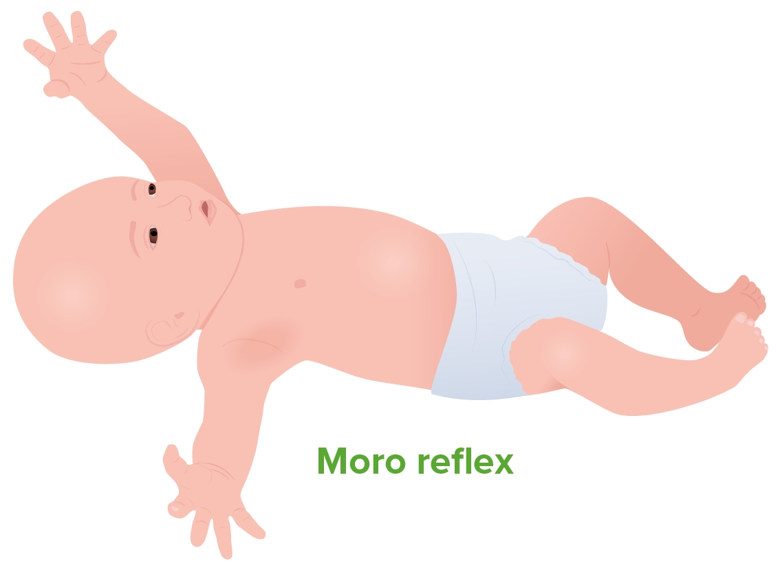
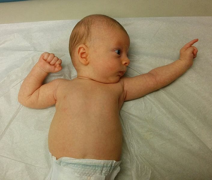
Moro reflex:
Sudden drop of an infant’s head causing the infant to extend their arms and then flex them as if grasping
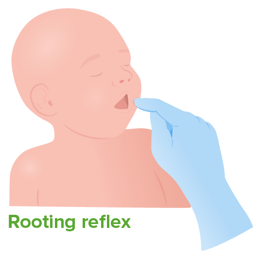
Rooting reflex:
When the infant’s cheek or mouth is stroked, they turn toward the side of the face that has been touched.
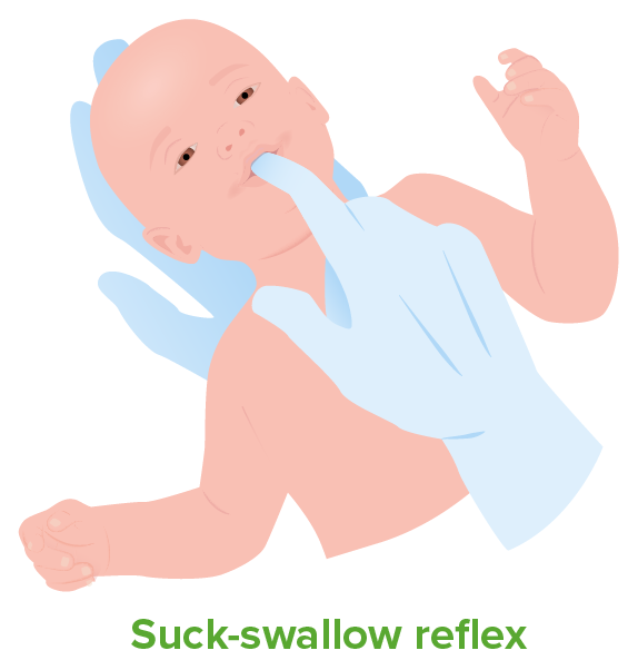
Suck-swallow reflex:
Contact with the infant’s soft palate stimulates suckling.
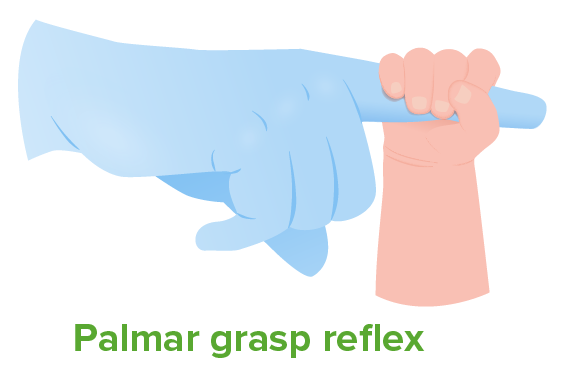
Palmar grasp reflex:
Sliding fingers across the infant’s palm elicits flexion of their fingers and grasping of the examiner’s finger.
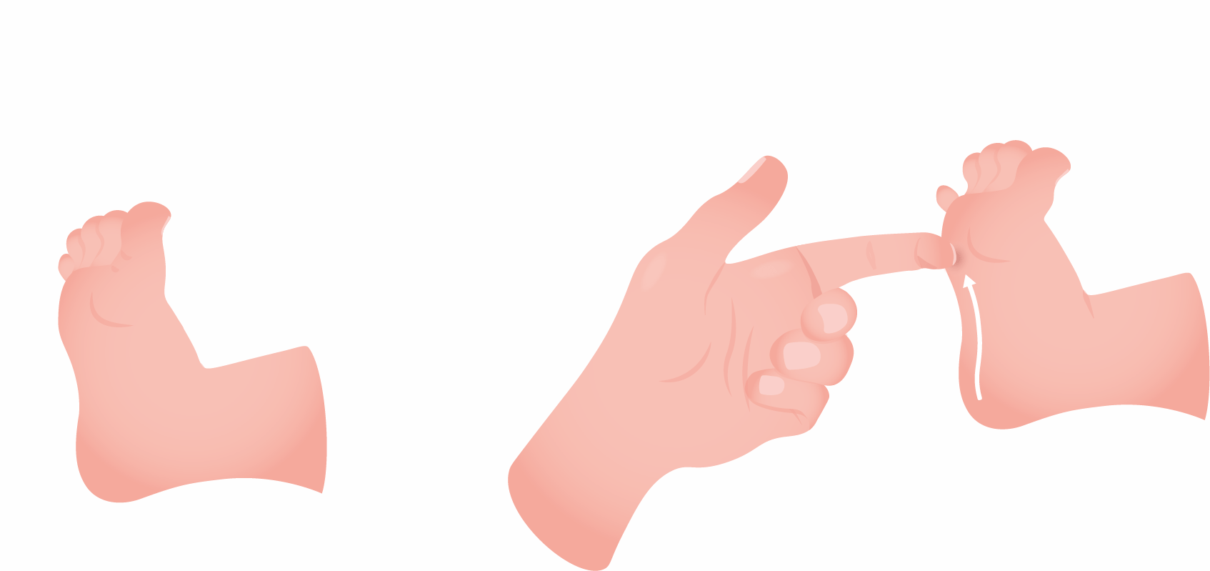
Babinski sign:
Plantar stimulation by firmly stroking the lateral aspect of the sole of the foot causes dorsiflexion of large toe and fanning of the other toes.
Asymmetrical tonic neck reflex in a 2-month-old baby:
Extension of the arm and leg on the face side with flexion of the arm and leg on the skull side
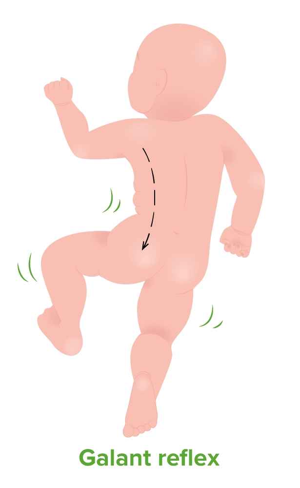
Galant reflex
Stroking along one side of the spine while the infant is held facing down causes lateral flexion of the lower limbs towards the side of stimulation.
| Reflex | Stimulus | Reaction | Disappears |
|---|---|---|---|
| Moro | Pull up → drop | Spreads arms → pulls them back | 3–6 months |
| Rooting | Stroke cheek/mouth. | Orients mouth toward the stimulus | 4 months |
| Palmar grasp | Slide finger across the palm. | Infant closes hand Hand The hand constitutes the distal part of the upper limb and provides the fine, precise movements needed in activities of daily living. It consists of 5 metacarpal bones and 14 phalanges, as well as numerous muscles innervated by the median and ulnar nerves. Hand: Anatomy around finger. | 6 months |
| Babinski | Firmly stroke the sole of the foot Foot The foot is the terminal portion of the lower limb, whose primary function is to bear weight and facilitate locomotion. The foot comprises 26 bones, including the tarsal bones, metatarsal bones, and phalanges. The bones of the foot form longitudinal and transverse arches and are supported by various muscles, ligaments, and tendons. Foot: Anatomy. | Dorsiflexion of large toe; fanning of the other toes | 12 months |
| Stepping | Placing infant’s soles flat on surface while holding infant | Walking motion | 2 months |
| Tonic neck Neck The part of a human or animal body connecting the head to the rest of the body. Peritonsillar Abscess | Turning neck Neck The part of a human or animal body connecting the head to the rest of the body. Peritonsillar Abscess to side | Fencing posture | 5–7 months |
| Galant | Stroke along 1 side of the spine Spine The human spine, or vertebral column, is the most important anatomical and functional axis of the human body. It consists of 7 cervical vertebrae, 12 thoracic vertebrae, and 5 lumbar vertebrae and is limited cranially by the skull and caudally by the sacrum. Vertebral Column: Anatomy while infant faces down. | Lateral movement of the limbs toward stimulated side | 6 months |
Upper motor Motor Neurons which send impulses peripherally to activate muscles or secretory cells. Nervous System: Histology neuron lesions can cause disinhibition and deficits in judgement, orientation Orientation Awareness of oneself in relation to time, place and person. Psychiatric Assessment, and concentration. This type of lesion is also characterized by the restoration of primitive reflexes. The various reflexes manifesting in an adult can be helpful in identifying the location of the lesion in the CNS.