The hand constitutes the distal part of the upper limb and provides the fine, precise movements needed in activities of daily living. It consists of 5 metacarpal bones and 14 phalanges, as well as numerous muscles innervated by the median and ulnar nerves. The muscles of the hand are classified as extrinsic (forearm-based) or intrinsic (hand-based) depending on the location of the muscle belly. These muscles are also grouped by area or type: thenar, hypothenar, lumbricals, and interossei.
Last updated: Dec 15, 2025
The bones of the hand consist of the following:
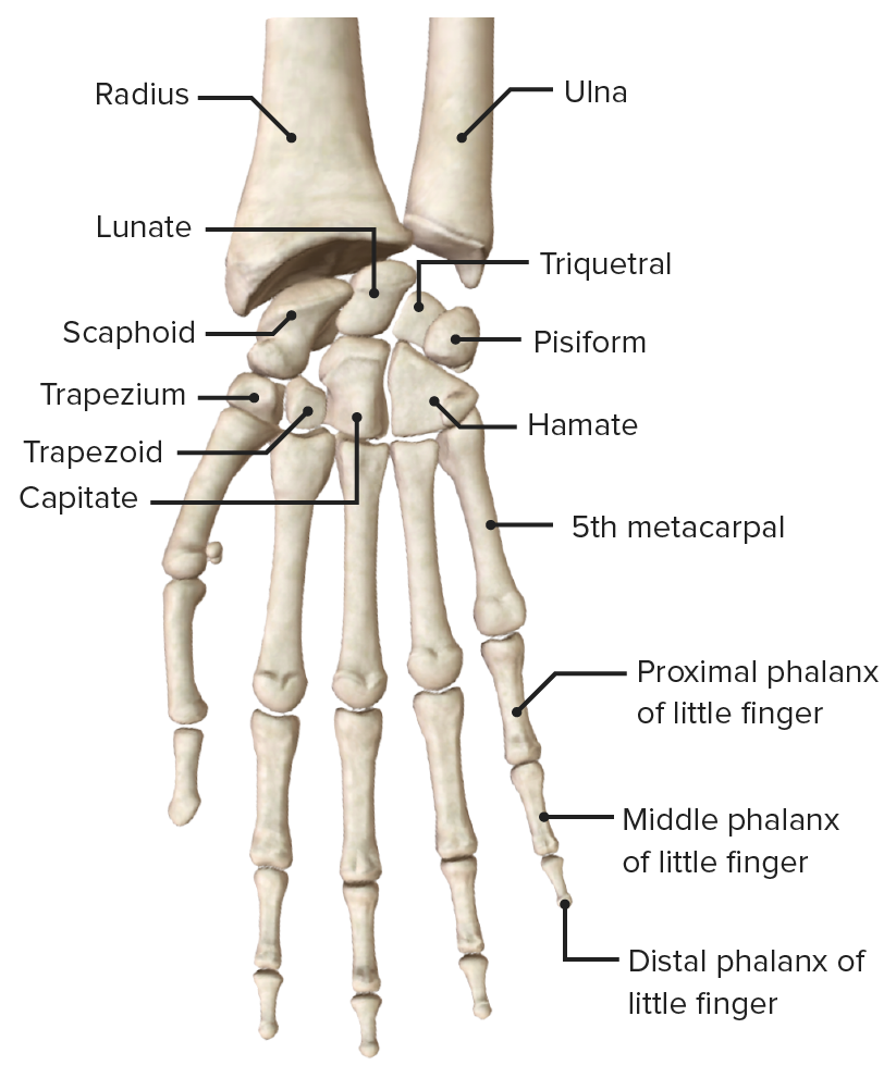
Anterior view of the hand and carpus, featuring the carpal bones, metacarpals, and phalanges
Image by BioDigital, edited by Lecturio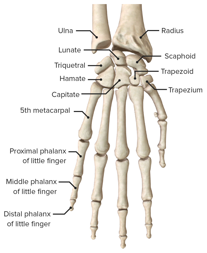
Posterior view of the hand and carpus, featuring the carpal bones, metacarpals, and phalanges
Image by BioDigital, edited by LecturioThe joints of the hand and fingers consist of:
| Type | Components | Function | Clinical relevance | |
|---|---|---|---|---|
| Interphalangeal | Hinge joint |
|
|
|
| Metacarpophalangeal | Thumb: hinge joint | Head of the 1st metacarpal and the proximal end of the proximal phalanx |
|
Affected early by rheumatoid arthritis Arthritis Acute or chronic inflammation of joints. Osteoarthritis |
| 2nd–5th digits: ellipsoid joints | Heads of the 2nd–5th metacarpals Metacarpals The five cylindrical bones of the metacarpus, articulating with the carpal bones proximally and the phalanges of fingers distally. Wrist Joint: Anatomy and the proximal end of the proximal phalanges |
|
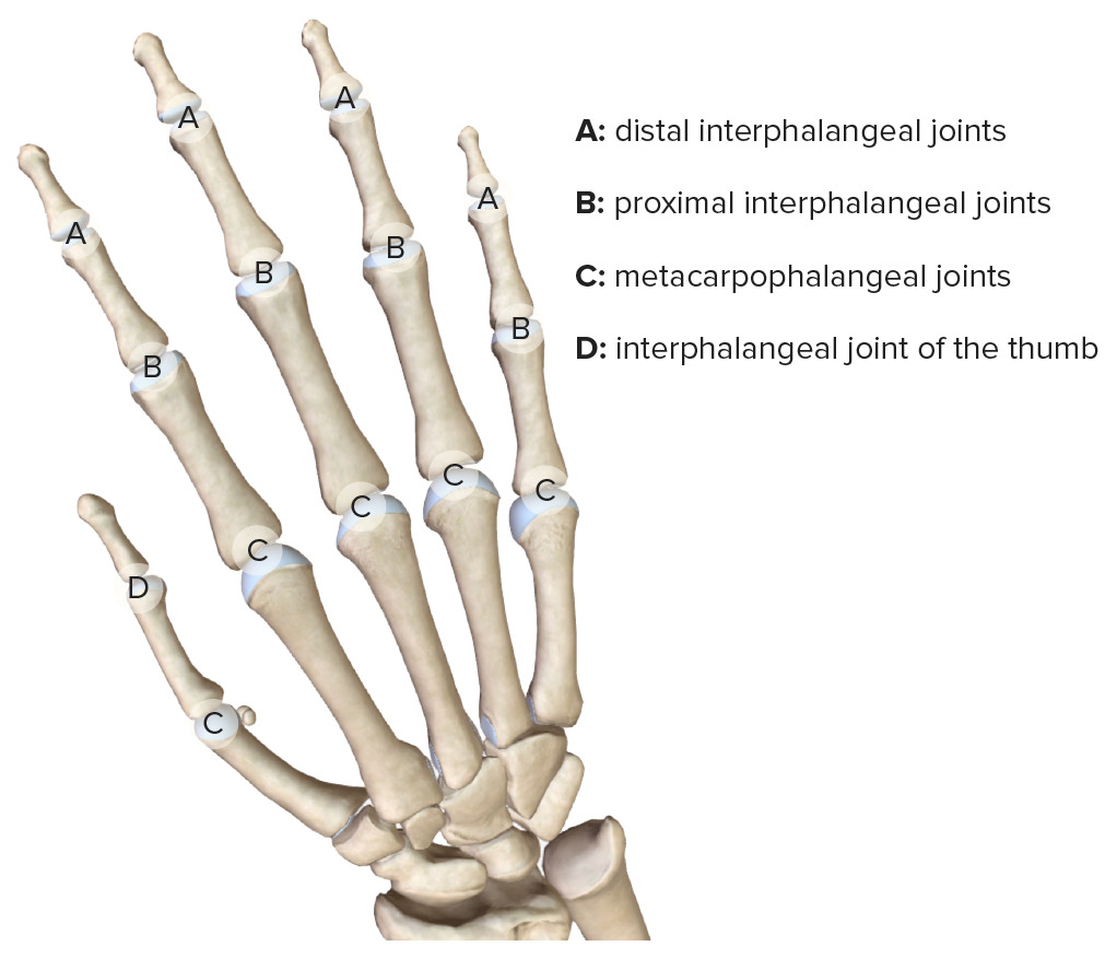
Joints of the digits, featuring the metacarpophalangeal, proximal interphalangeal, and distal interphalangeal joints
Image by BioDigital, edited by Lecturio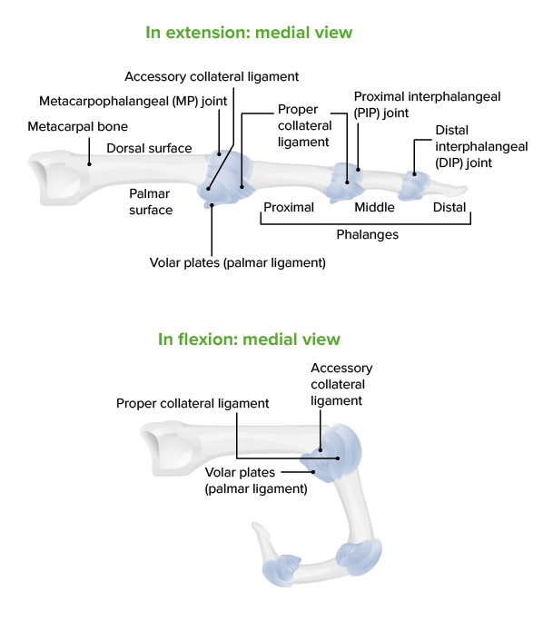
Diagram depicting the structure of the interphalangeal joints:
Note that ligaments of metacarpophalangeal and interphalangeal joints are similar.
The muscles of the hand are divided into 2 groups based on muscle belly location:
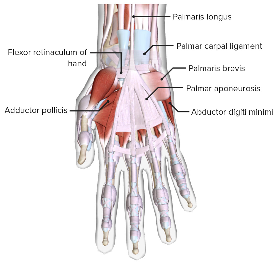
Anterior view of the right hand, featuring the palm, thenar, and hypothenar muscles making up the thenar and hypothenar eminences, and the palmar aponeurosis
Image by BioDigital, edited by Lecturio.| Muscle | Origin | Insertion | Innervation | Function |
|---|---|---|---|---|
| Opponens pollicis | Flexor retinaculum Flexor Retinaculum Ankle Joint: Anatomy and trapezium | Lateral side of the 1st metacarpal bone Bone Bone is a compact type of hardened connective tissue composed of bone cells, membranes, an extracellular mineralized matrix, and central bone marrow. The 2 primary types of bone are compact and spongy. Bones: Structure and Types | Recurrent branch of median nerve Median Nerve A major nerve of the upper extremity. In humans, the fibers of the median nerve originate in the lower cervical and upper thoracic spinal cord (usually C6 to T1), travel via the brachial plexus, and supply sensory and motor innervation to parts of the forearm and hand. Cubital Fossa: Anatomy (C8) | Opposes thumb |
| Abductor pollicis brevis | Flexor retinaculum Flexor Retinaculum Ankle Joint: Anatomy and tubercles of scaphoid and trapezium | Lateral side of proximal phalanx of 1st digit |
|
|
| Flexor pollicis brevis | Flexor retinaculum Flexor Retinaculum Ankle Joint: Anatomy and trapezium |
|
Flexes thumb | |
| Adductor pollicis |
|
Medial side of proximal phalanx of thumb | Deep branch of ulnar nerve Ulnar Nerve A major nerve of the upper extremity. In humans, the fibers of the ulnar nerve originate in the lower cervical and upper thoracic spinal cord (usually C7 to T1), travel via the medial cord of the brachial plexus, and supply sensory and motor innervation to parts of the hand and forearm. Axilla and Brachial Plexus: Anatomy (C8) | Adducts thumb |

Thenar muscles of hand
Image by Lecturio.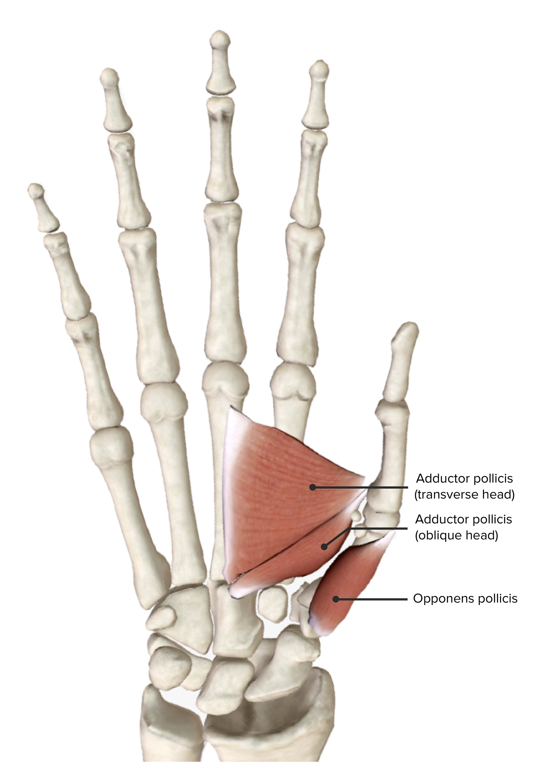
Thenar muscles, featuring the deepest muscles: adductor pollicis and opponens pollicis
Image by BioDigital, edited by Lecturio.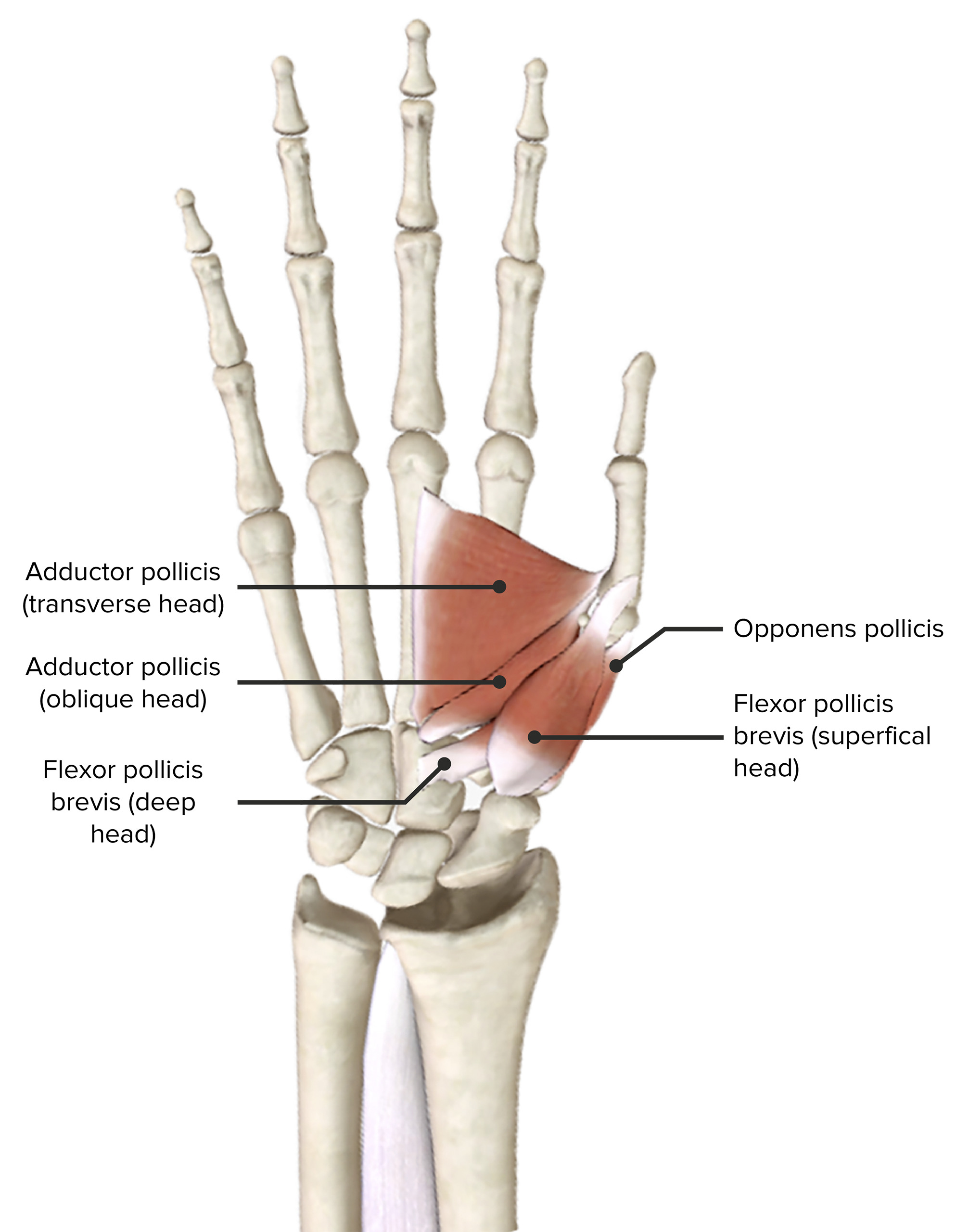
Thenar muscles, featuring the intermediate muscle: flexor pollicis brevis
Image by BioDigital, edited by Lecturio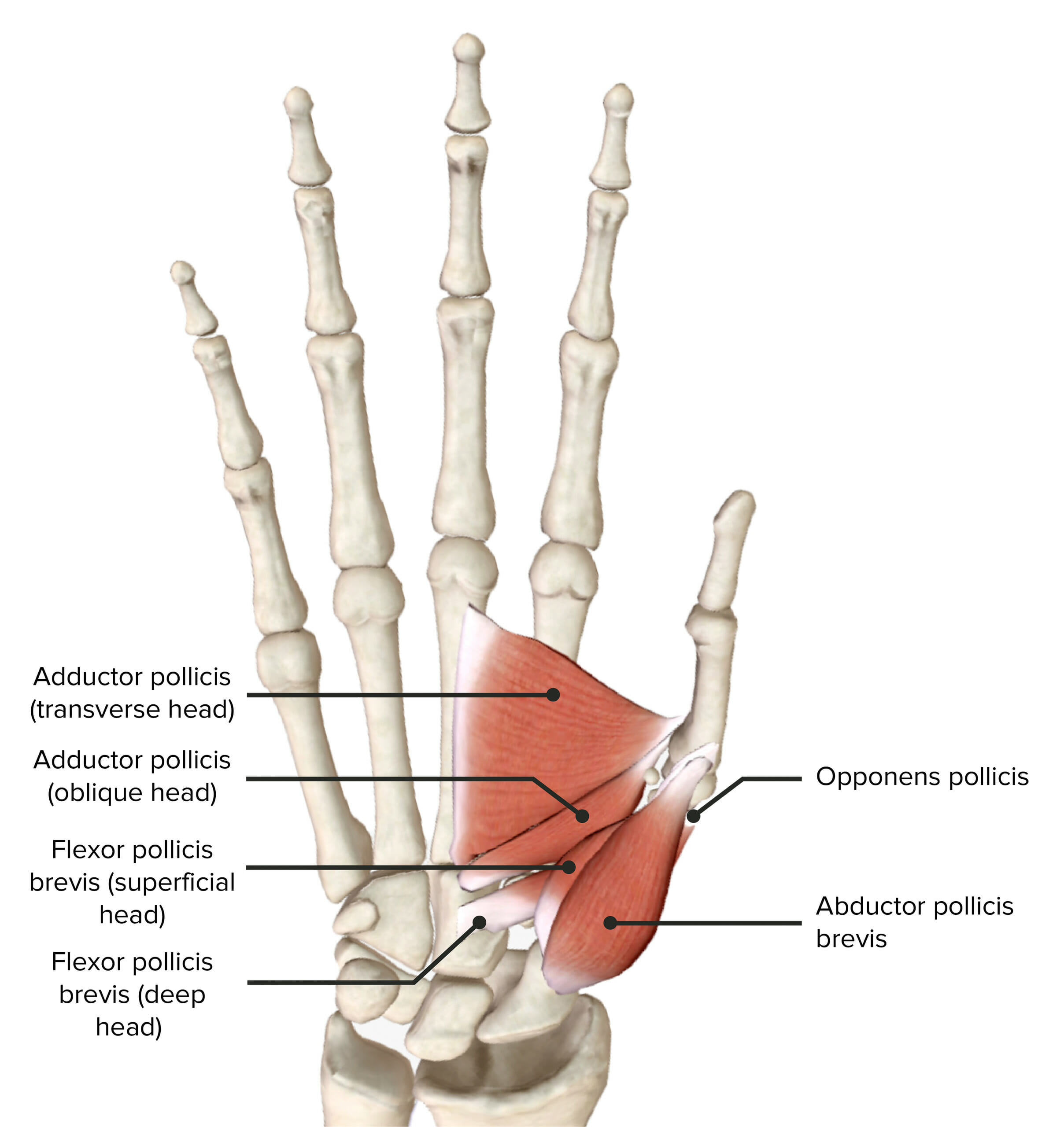
Thenar muscles, featuring the most superficial muscle: abductor pollicis brevis
Image by BioDigital, edited by Lecturio| Muscle | Origin | Insertion | Innervation | Function |
|---|---|---|---|---|
| Palmaris brevis | Flexor retinaculum Flexor Retinaculum Ankle Joint: Anatomy and palmar aponeurosis | Skin Skin The skin, also referred to as the integumentary system, is the largest organ of the body. The skin is primarily composed of the epidermis (outer layer) and dermis (deep layer). The epidermis is primarily composed of keratinocytes that undergo rapid turnover, while the dermis contains dense layers of connective tissue. Skin: Structure and Functions of the hypothenar eminence | Ulnar nerve Ulnar Nerve A major nerve of the upper extremity. In humans, the fibers of the ulnar nerve originate in the lower cervical and upper thoracic spinal cord (usually C7 to T1), travel via the medial cord of the brachial plexus, and supply sensory and motor innervation to parts of the hand and forearm. Axilla and Brachial Plexus: Anatomy | Strengthens palmar grip by wrinkling the skin Skin The skin, also referred to as the integumentary system, is the largest organ of the body. The skin is primarily composed of the epidermis (outer layer) and dermis (deep layer). The epidermis is primarily composed of keratinocytes that undergo rapid turnover, while the dermis contains dense layers of connective tissue. Skin: Structure and Functions of the ulnar palm |
| Abductor digiti minimi | Pisiform and flexor carpi ulnaris Flexor carpi ulnaris Forearm: Anatomy | Medial side of proximal phalanx of the 5th digit | Deep branch of ulnar nerve Ulnar Nerve A major nerve of the upper extremity. In humans, the fibers of the ulnar nerve originate in the lower cervical and upper thoracic spinal cord (usually C7 to T1), travel via the medial cord of the brachial plexus, and supply sensory and motor innervation to parts of the hand and forearm. Axilla and Brachial Plexus: Anatomy (T1) | Abducts the 5th digit |
| Flexor digiti minimi brevis | Hook of hamate and flexor retinaculum Flexor Retinaculum Ankle Joint: Anatomy | Flexes proximal phalanx of the 5th digit | ||
| Opponens digiti minimi | Medial border of 5th metacarpal | Opposes the 5th digit |
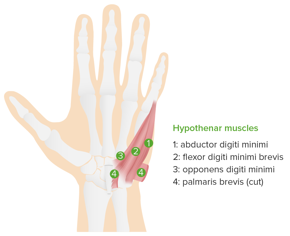
Hypothenar muscles
Image by Lecturio.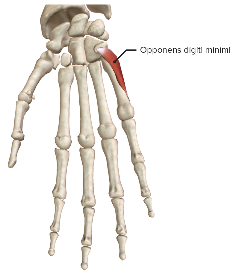
Hypothenar muscles, featuring the deepest muscle: opponens digiti minimi
Image by BioDigital, edited by Lecturio.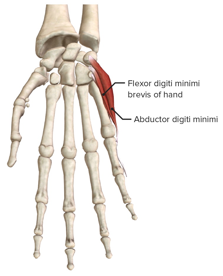
Hypothenar muscles, featuring the intermediate muscles: flexor digiti minimi brevis and abductor digiti minimi
Image by BioDigital, edited by Lecturio.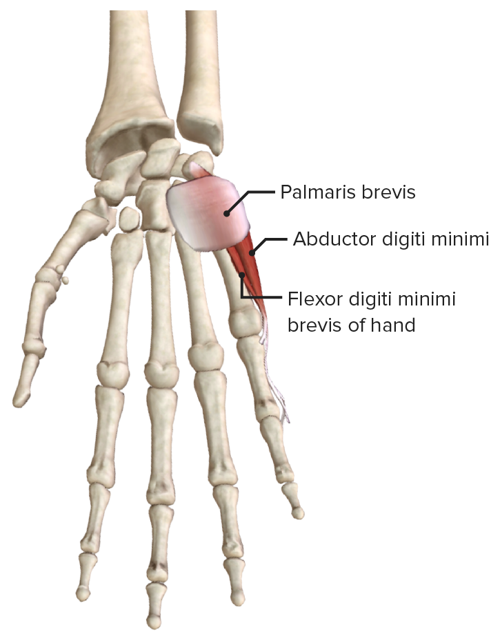
Hypothenar muscles, featuring the most superficial muscle: palmaris brevis
Image by BioDigital, edited by Lecturio.| Muscle | Origin | Insertion | Innervation | Function |
|---|---|---|---|---|
| Lumbricals (I–II) | Lateral 2 tendons of flexor digitorum profundus Flexor digitorum profundus Forearm: Anatomy | Lateral surfaces of extensor expansions of the 2nd–5th digits | Median nerve Median Nerve A major nerve of the upper extremity. In humans, the fibers of the median nerve originate in the lower cervical and upper thoracic spinal cord (usually C6 to T1), travel via the brachial plexus, and supply sensory and motor innervation to parts of the forearm and hand. Cubital Fossa: Anatomy (T1) | Flex metacarpophalangeal and extend interphalangeal joints of 2nd–5th digits |
| Lumbricals (III–IV) | Medial 2 tendons of flexor digitorum profundus Flexor digitorum profundus Forearm: Anatomy | Deep branch of ulnar nerve Ulnar Nerve A major nerve of the upper extremity. In humans, the fibers of the ulnar nerve originate in the lower cervical and upper thoracic spinal cord (usually C7 to T1), travel via the medial cord of the brachial plexus, and supply sensory and motor innervation to parts of the hand and forearm. Axilla and Brachial Plexus: Anatomy (T1) |
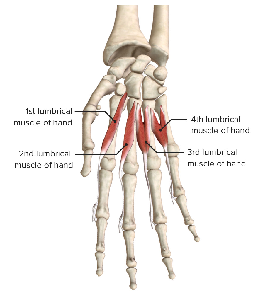
Lumbrical muscles
Image by BioDigital, edited by Lecturio| Muscle | Origin | Insertion | Innervation | Function |
|---|---|---|---|---|
| Dorsal interossei (4 muscles) | Radial and ulnar side of each pair of consecutive metacarpals Metacarpals The five cylindrical bones of the metacarpus, articulating with the carpal bones proximally and the phalanges of fingers distally. Wrist Joint: Anatomy | Base of proximal phalanges and extensor expansions (dorsal: 2nd–4th digits; palmar: 2nd, 4th, and 5th digits) | Deep branch of ulnar nerve Ulnar Nerve A major nerve of the upper extremity. In humans, the fibers of the ulnar nerve originate in the lower cervical and upper thoracic spinal cord (usually C7 to T1), travel via the medial cord of the brachial plexus, and supply sensory and motor innervation to parts of the hand and forearm. Axilla and Brachial Plexus: Anatomy (T1) | Abduct 2nd–4th digits |
| Palmar interossei (3 muscles) | Sides of the metacarpals Metacarpals The five cylindrical bones of the metacarpus, articulating with the carpal bones proximally and the phalanges of fingers distally. Wrist Joint: Anatomy facing midline | Adduct 2nd, 4th, and 5th digits |

Palmar interossei (3 muscles)
Image by BioDigital, edited by Lecturio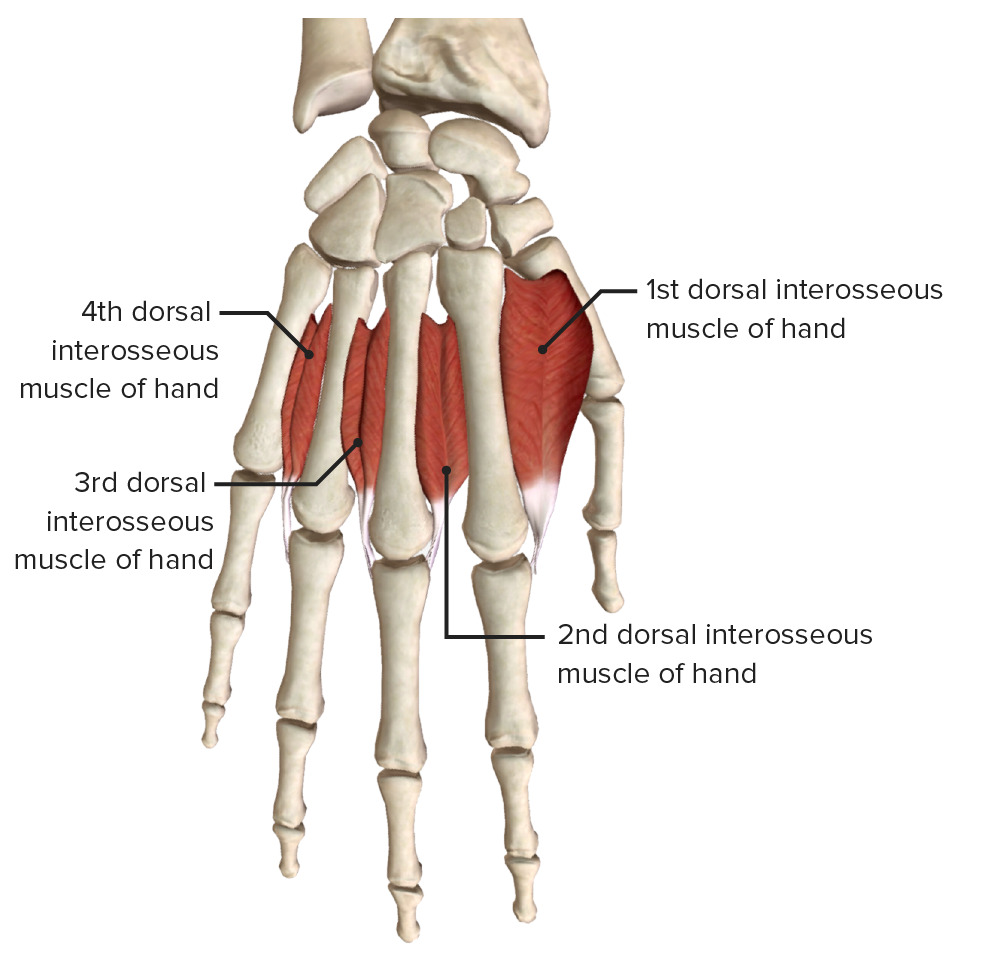
Dorsal interossei (4 muscles)
Image by BioDigital, edited by LecturioThe extrinsic muscles of the hand have their origin and muscle bellies in the forearm Forearm The forearm is the region of the upper limb between the elbow and the wrist. The term “forearm” is used in anatomy to distinguish this area from the arm, a term that is commonly used to describe the entire upper limb. The forearm consists of 2 long bones (the radius and the ulna), the interosseous membrane, and multiple arteries, nerves, and muscles. Forearm: Anatomy and are divided into flexor (anterior compartment) and extensor (posterior compartment) muscles.
| Muscle | Origin | Insertion | Innervation | Function |
|---|---|---|---|---|
| Extensor carpi radialis longus Extensor carpi radialis longus Forearm: Anatomy | Lateral supracondylar ridge of the humerus Humerus Bone in humans and primates extending from the shoulder joint to the elbow joint. Arm: Anatomy | Dorsal aspect of base of 2nd metacarpal | Radial nerve Radial Nerve A major nerve of the upper extremity. In humans the fibers of the radial nerve originate in the lower cervical and upper thoracic spinal cord (usually C5 to T1), travel via the posterior cord of the brachial plexus, and supply motor innervation to extensor muscles of the arm and cutaneous sensory fibers to extensor regions of the arm and hand. Axilla and Brachial Plexus: Anatomy | Abduction Abduction Examination of the Upper Limbs and extension Extension Examination of the Upper Limbs of the wrist (dorsiflexion) |
| Extensor carpi radialis brevis Extensor carpi radialis brevis Forearm: Anatomy | Lateral epicondyle Lateral epicondyle Arm: Anatomy | Dorsal aspect of base of 3rd metacarpal | ||
| Extensor digitorum Extensor digitorum Forearm: Anatomy | Extensor expansion, base of middle and distal phalanges of 2nd–5th digits | Posterior interosseous nerve (C7; from deep radial nerve Radial Nerve A major nerve of the upper extremity. In humans the fibers of the radial nerve originate in the lower cervical and upper thoracic spinal cord (usually C5 to T1), travel via the posterior cord of the brachial plexus, and supply motor innervation to extensor muscles of the arm and cutaneous sensory fibers to extensor regions of the arm and hand. Axilla and Brachial Plexus: Anatomy) |
|
|
| Extensor digiti minimi Extensor digiti minimi Forearm: Anatomy | Extensor expansion, middle and distal phalanges of 5th digit | |||
| Extensor carpi ulnaris Extensor carpi ulnaris Forearm: Anatomy | Lateral epicondyle Lateral epicondyle Arm: Anatomy of the humerus Humerus Bone in humans and primates extending from the shoulder joint to the elbow joint. Arm: Anatomy and posterior surface of ulna Ulna The inner and longer bone of the forearm. Forearm: Anatomy | Dorsal aspect of base of 5th metacarpal | Extends and adducts wrist |
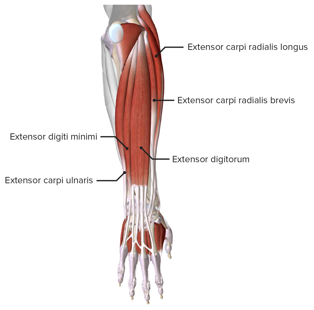
Extrinsic extensor muscles of the hand: superficial layer
Image by BioDigital, edited by Lecturio.| Muscle | Origin | Insertion | Innervation | Function |
|---|---|---|---|---|
| Abductor pollicis longus Abductor pollicis longus Forearm: Anatomy | Posterior surface of radius Radius The outer shorter of the two bones of the forearm, lying parallel to the ulna and partially revolving around it. Forearm: Anatomy and ulna Ulna The inner and longer bone of the forearm. Forearm: Anatomy, interosseous membrane Interosseous Membrane A sheet of fibrous connective tissue rich in collagen often linking two parallel bony structures forming a syndesmosis type joint. It provides longitudinal stability, tensile strength, and weight distribution/transfer and may allow limited movement in syndesmoses. Forearm: Anatomy | Base of 1st metacarpal | Posterior interosseous nerve (C7 and C8) from deep radial nerve Radial Nerve A major nerve of the upper extremity. In humans the fibers of the radial nerve originate in the lower cervical and upper thoracic spinal cord (usually C5 to T1), travel via the posterior cord of the brachial plexus, and supply motor innervation to extensor muscles of the arm and cutaneous sensory fibers to extensor regions of the arm and hand. Axilla and Brachial Plexus: Anatomy |
|
| Extensor pollicis longus Extensor pollicis longus Forearm: Anatomy | Posterior surface of ulna Ulna The inner and longer bone of the forearm. Forearm: Anatomy, interosseous membrane Interosseous Membrane A sheet of fibrous connective tissue rich in collagen often linking two parallel bony structures forming a syndesmosis type joint. It provides longitudinal stability, tensile strength, and weight distribution/transfer and may allow limited movement in syndesmoses. Forearm: Anatomy | Dorsal surface of distal phalanx of thumb |
|
|
| Extensor pollicis brevis Extensor pollicis brevis Forearm: Anatomy | Posterior surface of radius Radius The outer shorter of the two bones of the forearm, lying parallel to the ulna and partially revolving around it. Forearm: Anatomy, interosseous membrane Interosseous Membrane A sheet of fibrous connective tissue rich in collagen often linking two parallel bony structures forming a syndesmosis type joint. It provides longitudinal stability, tensile strength, and weight distribution/transfer and may allow limited movement in syndesmoses. Forearm: Anatomy | Dorsal surface of proximal phalanx of thumb | ||
| Extensor indicis Extensor indicis Forearm: Anatomy | Posterior surface of the ulna Ulna The inner and longer bone of the forearm. Forearm: Anatomy | Extensor expansion of 2nd finger |
|
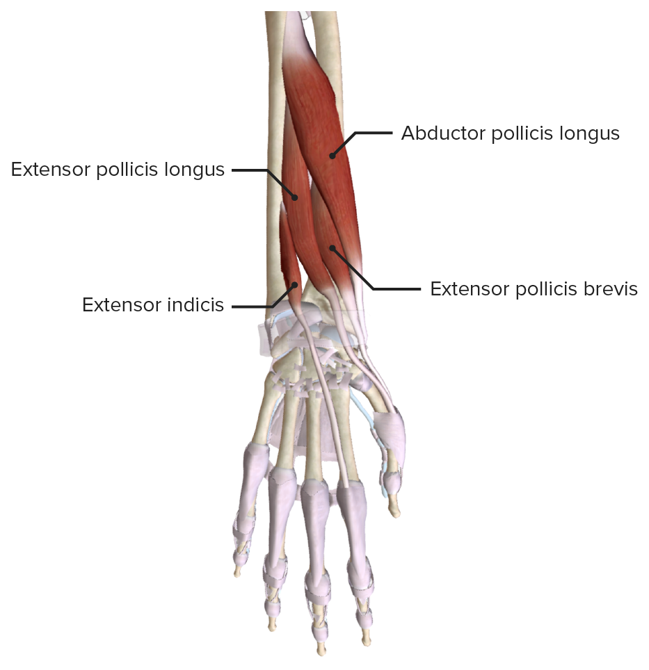
Extrinsic extensor muscles of the hand: deep layer
Image by BioDigital, edited by Lecturio.| Muscle | Origin | Insertion | Innervation | Function |
|---|---|---|---|---|
| Flexor carpi radialis Flexor carpi radialis Forearm: Anatomy | Medial epicondyle Medial epicondyle Arm: Anatomy of humerus Humerus Bone in humans and primates extending from the shoulder joint to the elbow joint. Arm: Anatomy | Base of 2nd–3rd metacarpals Metacarpals The five cylindrical bones of the metacarpus, articulating with the carpal bones proximally and the phalanges of fingers distally. Wrist Joint: Anatomy | Median nerve Median Nerve A major nerve of the upper extremity. In humans, the fibers of the median nerve originate in the lower cervical and upper thoracic spinal cord (usually C6 to T1), travel via the brachial plexus, and supply sensory and motor innervation to parts of the forearm and hand. Cubital Fossa: Anatomy (C7) | Flexes and abducts wrist |
| Palmaris longus Palmaris longus Forearm: Anatomy | Flexor retinaculum Flexor Retinaculum Ankle Joint: Anatomy and palmar aponeurosis | Flexes wrist weakly and tenses palmar aponeurosis | ||
| Flexor carpi ulnaris Flexor carpi ulnaris Forearm: Anatomy | Medial epicondyle Medial epicondyle Arm: Anatomy of the humerus Humerus Bone in humans and primates extending from the shoulder joint to the elbow joint. Arm: Anatomy, olecranon Olecranon A prominent projection of the ulna that articulates with the humerus and forms the outer protuberance of the elbow joint. Arm: Anatomy, and posterior ulna Ulna The inner and longer bone of the forearm. Forearm: Anatomy | Pisiform, hook of hamate, base of 5th metacarpal | Ulnar nerve Ulnar Nerve A major nerve of the upper extremity. In humans, the fibers of the ulnar nerve originate in the lower cervical and upper thoracic spinal cord (usually C7 to T1), travel via the medial cord of the brachial plexus, and supply sensory and motor innervation to parts of the hand and forearm. Axilla and Brachial Plexus: Anatomy (C8) | Flexes and adducts wrist |
| Flexor digitorum superficialis Flexor digitorum superficialis Forearm: Anatomy | Medial epicondyle Medial epicondyle Arm: Anatomy of the humerus Humerus Bone in humans and primates extending from the shoulder joint to the elbow joint. Arm: Anatomy, proximal shaft of the radius Radius The outer shorter of the two bones of the forearm, lying parallel to the ulna and partially revolving around it. Forearm: Anatomy | Middle phalanges of the medial 4 fingers | Median nerve Median Nerve A major nerve of the upper extremity. In humans, the fibers of the median nerve originate in the lower cervical and upper thoracic spinal cord (usually C6 to T1), travel via the brachial plexus, and supply sensory and motor innervation to parts of the forearm and hand. Cubital Fossa: Anatomy (C7) |
|
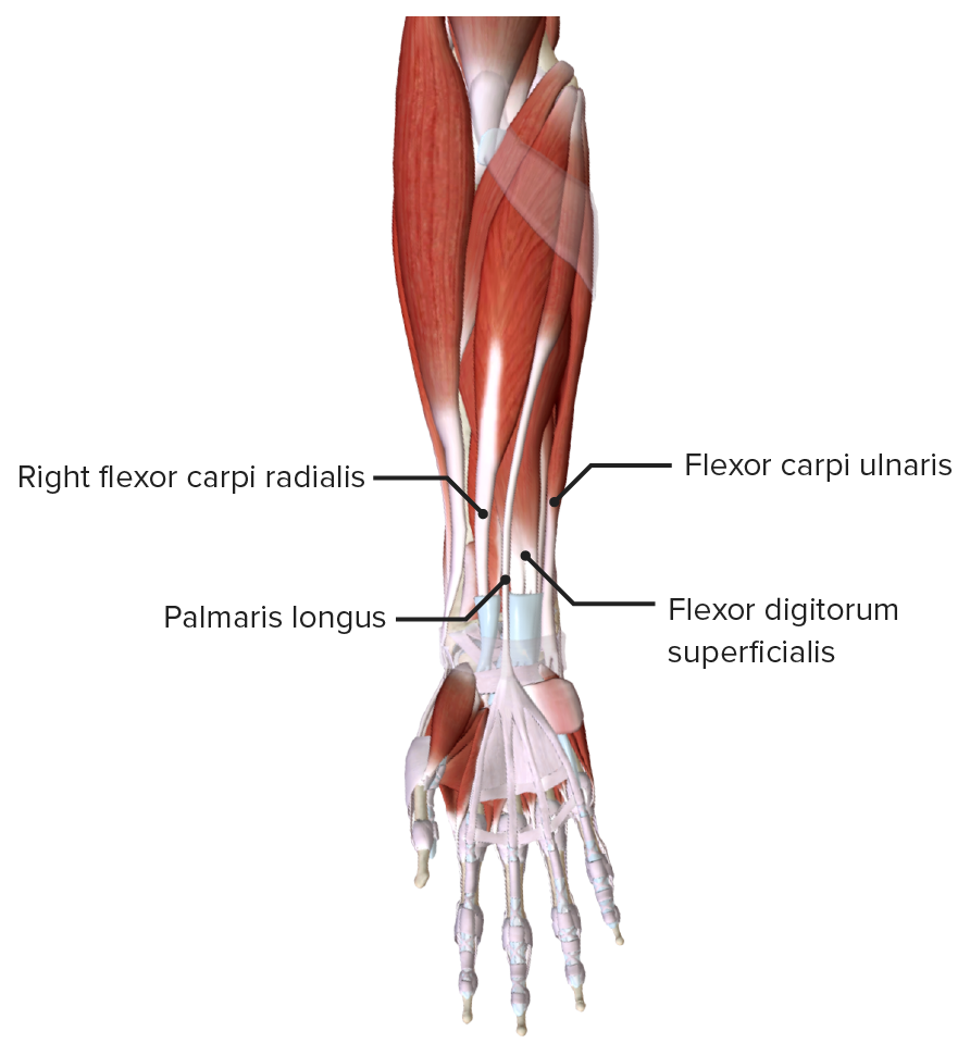
Extrinsic flexor muscles of the hand: superficial layer
Image by BioDigital, edited by Lecturio.| Muscle | Origin | Insertion | Innervation | Function |
|---|---|---|---|---|
| Flexor digitorum profundus Flexor digitorum profundus Forearm: Anatomy | Proximal end of the ulna Ulna The inner and longer bone of the forearm. Forearm: Anatomy (medial and anterior surfaces) and interosseous membrane Interosseous Membrane A sheet of fibrous connective tissue rich in collagen often linking two parallel bony structures forming a syndesmosis type joint. It provides longitudinal stability, tensile strength, and weight distribution/transfer and may allow limited movement in syndesmoses. Forearm: Anatomy | Distal phalanges of the 2nd–5th digits |
|
|
| Flexor pollicis longus Flexor pollicis longus Forearm: Anatomy | Shaft of radius Radius The outer shorter of the two bones of the forearm, lying parallel to the ulna and partially revolving around it. Forearm: Anatomy (anterior surface) and interosseous membrane Interosseous Membrane A sheet of fibrous connective tissue rich in collagen often linking two parallel bony structures forming a syndesmosis type joint. It provides longitudinal stability, tensile strength, and weight distribution/transfer and may allow limited movement in syndesmoses. Forearm: Anatomy | Distal phalanx of thumb | Anterior interosseous nerve Anterior Interosseous Nerve Supracondylar Fracture (branch of median) (C8) |
|
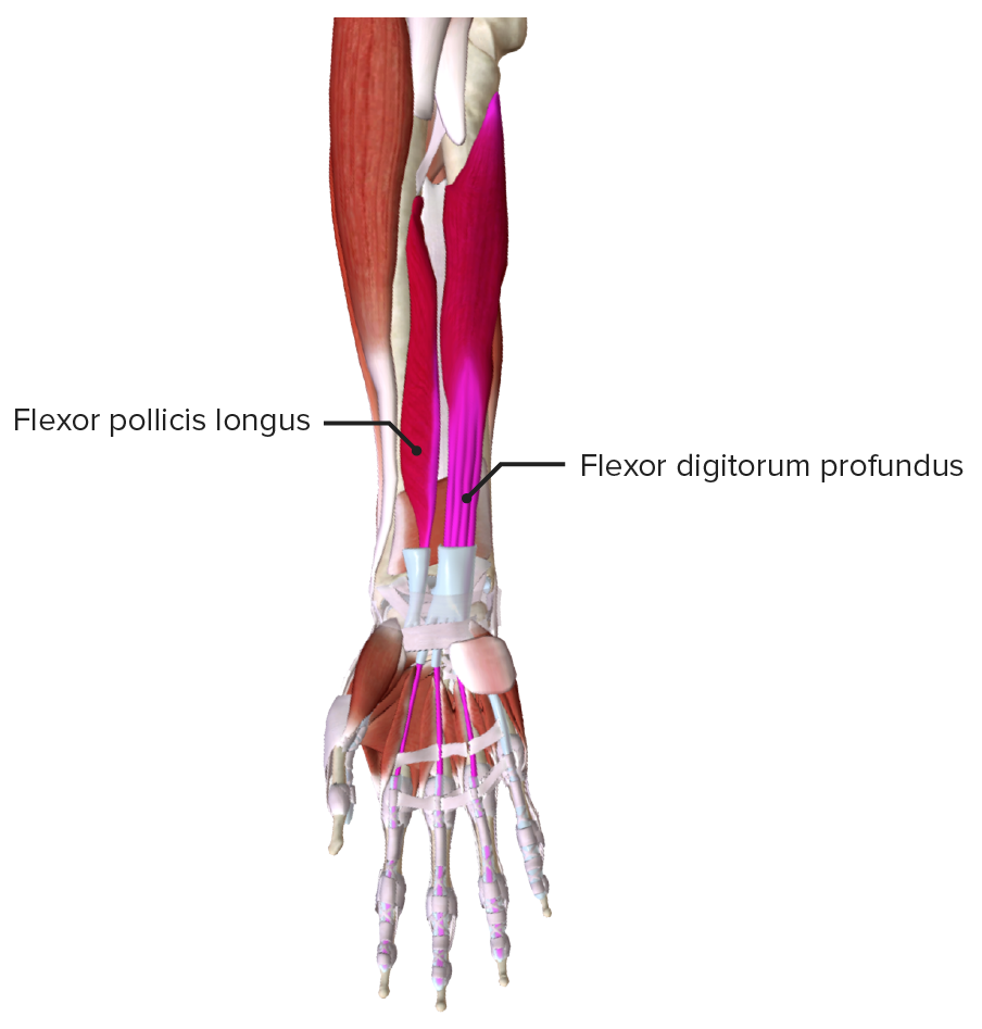
Extrinsic flexor muscles of the hand: deep layer
Image by BioDigital, edited by Lecturio.Also known as the extensor hood, dorsal expansion, dorsal hood, or dorsal aponeurosis of the hand and/or fingers. The extensor expansion comprises the tendons of the extensor muscles of the fingers and how these insert into the phalanges:
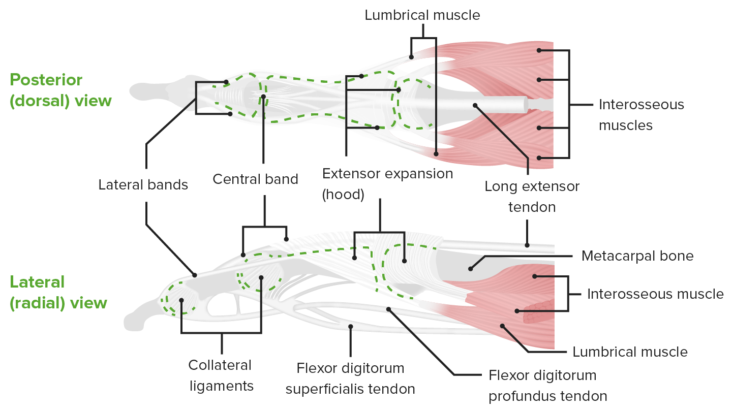
Schematic of a finger, featuring the extensor expansion or dorsal aponeurosis and insertion of the tendons of the flexor digitorum muscle
Image by Lecturio.Comprises the tendons of the flexor muscles of the fingers and how these insert into the phalanges, supported by a system of ligaments:
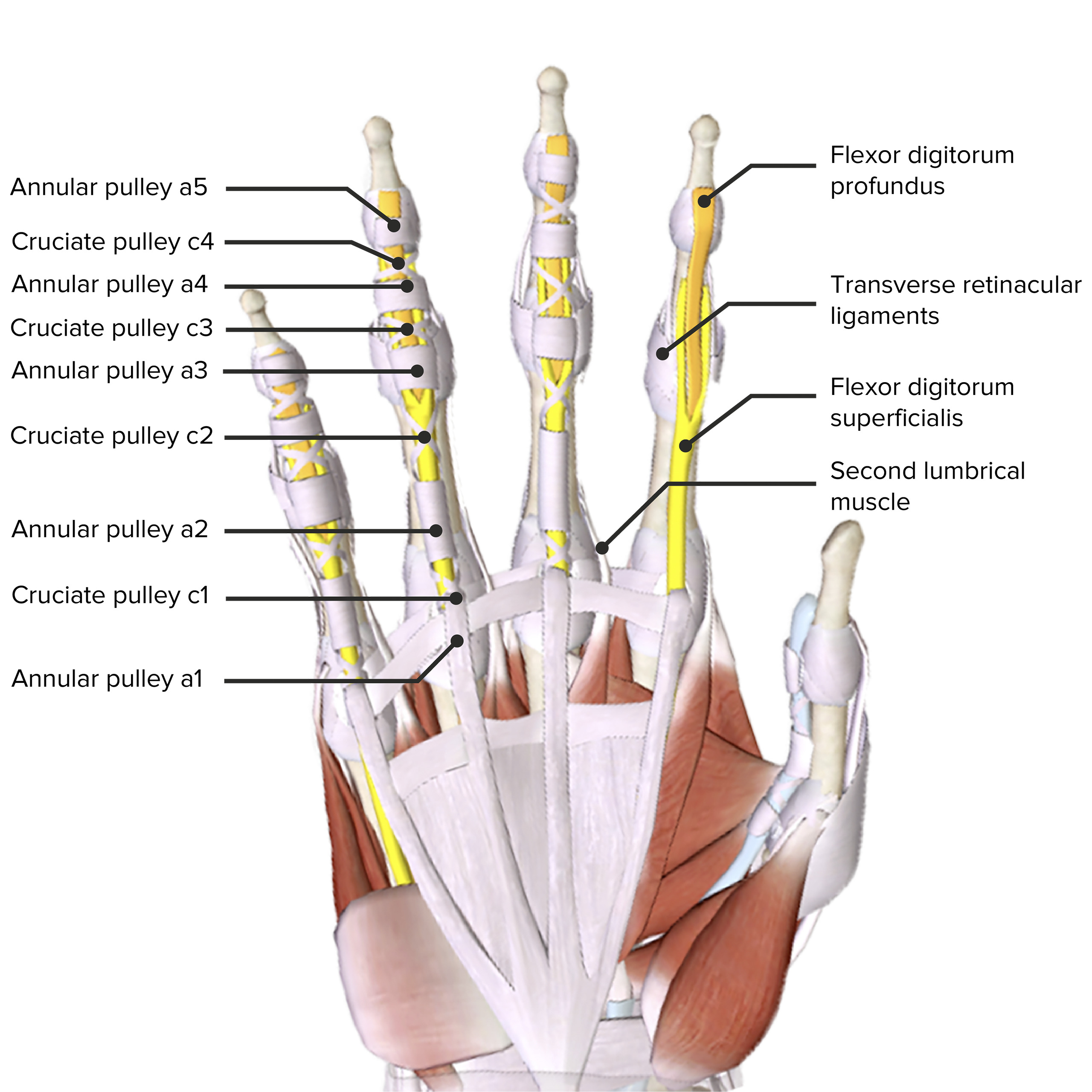
Palmar view of the hand, featuring pulleys:
A pulley is a thickening of flexor tendon sheath that stabilizes the tendon. Pulleys a1, a3, and a5 originate from palmar plates of the MCP, PIP, and DIP joints, respectively. Pulleys a2 and a4 originate from the proximal and middle phalanges, respectively. The most important pulleys, a2 and a4, biomechanically prevent bowstringing of flexor tendons. Pulley a1 tightness may cause a trigger finger.
MCP: metacarpophalangeal
PIP: proximal interphalangeal
DIP: distal interphalangeal
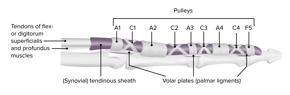
The fibro-osseous flexor digital sheath provides nutrition and biomechanical efficiency to the flexor tendons. Condensations of the synovial sheath form at strategic points along the digit to work in association with the transverse carpal ligament and the palmar aponeurosis pulley to maximize efficiency of joint rotation and force transmission.
Image by Lecturio.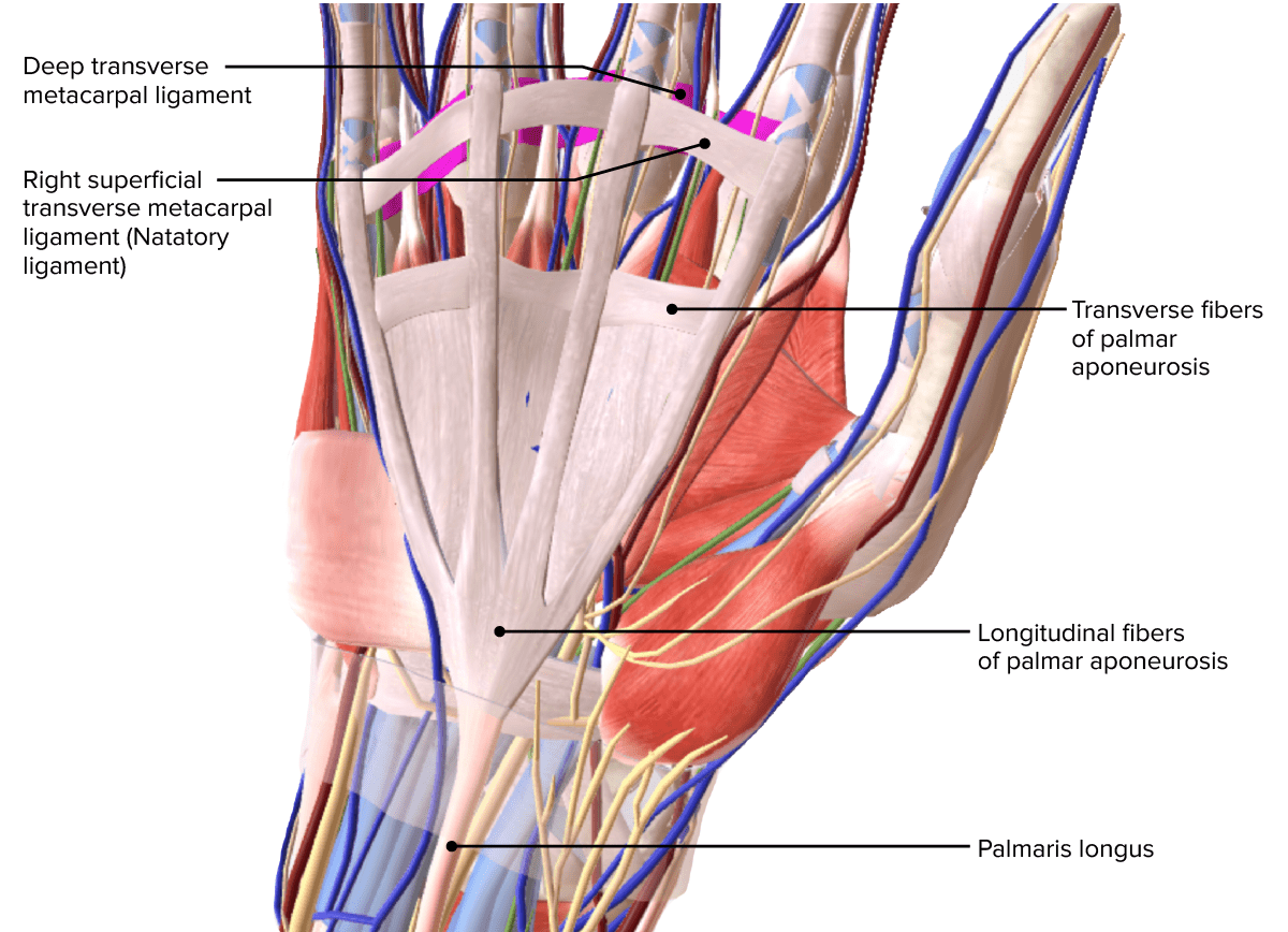
Palmar aponeurosis
Image by BioDigital, edited by Lecturio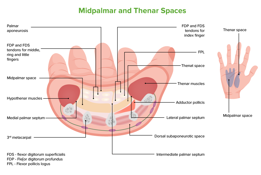
The medial margin of palmar aponeurosis gives off the medial palmar septum which attaches to the 5th metacarpal bone. The lateral margin of palmar aponeurosis gives off the lateral palmar septum (attaches to the 1st metacarpal bone) and the intermediate palmar septum (attaches to the 5th metacarpal bone).
Image by BioDigital, edited by Lecturio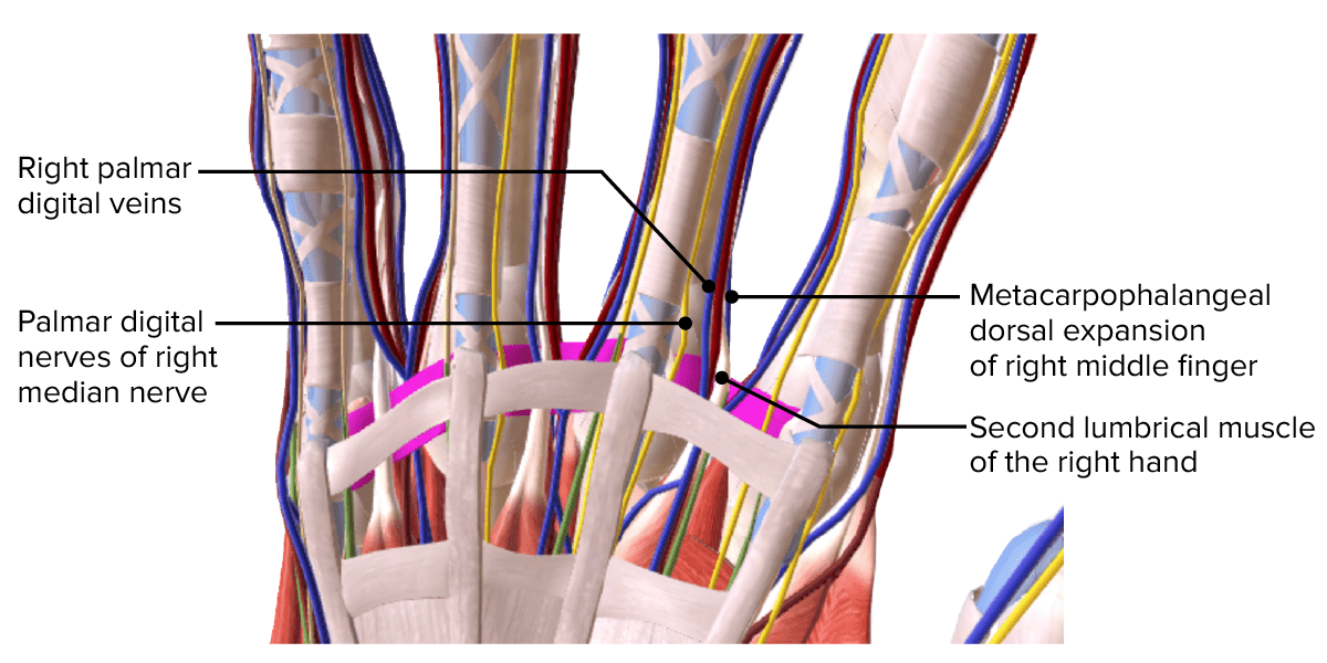
Detailed view of structures at the web space
Image by BioDigital, edited by LecturioThe arterial blood supply of the hand is provided by:
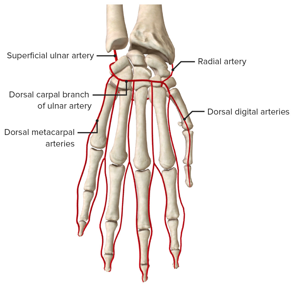
Blood supply of the dorsal aspect of the hand featuring the basal metacarpal arch
Image by BioDigital, edited by Lecturio.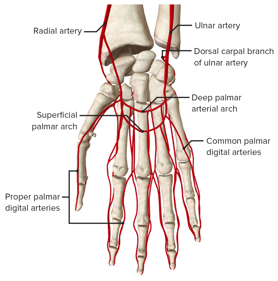
Blood supply to the palmar aspect of the hand featuring the superficial and deep palmar arches
Image by BioDigital, edited by Lecturio.The venous drainage of the hand is the origin of the veins Veins Veins are tubular collections of cells, which transport deoxygenated blood and waste from the capillary beds back to the heart. Veins are classified into 3 types: small veins/venules, medium veins, and large veins. Each type contains 3 primary layers: tunica intima, tunica media, and tunica adventitia. Veins: Histology of the upper extremity and begins in the dorsal and palmar venous network of the hand.

Venous drainage of the hand featuring the dorsal venous arch and digital veins
Image by BioDigital, edited by Lecturio.The innervation of the hand is primarily via the median and ulnar nerves, with the radial nerve Radial Nerve A major nerve of the upper extremity. In humans the fibers of the radial nerve originate in the lower cervical and upper thoracic spinal cord (usually C5 to T1), travel via the posterior cord of the brachial plexus, and supply motor innervation to extensor muscles of the arm and cutaneous sensory fibers to extensor regions of the arm and hand. Axilla and Brachial Plexus: Anatomy only contributing a small area of sensory Sensory Neurons which conduct nerve impulses to the central nervous system. Nervous System: Histology innervation on the radial dorsal aspect of the hand.
| Median nerve Median Nerve A major nerve of the upper extremity. In humans, the fibers of the median nerve originate in the lower cervical and upper thoracic spinal cord (usually C6 to T1), travel via the brachial plexus, and supply sensory and motor innervation to parts of the forearm and hand. Cubital Fossa: Anatomy |
|
|---|---|
| Ulnar nerve Ulnar Nerve A major nerve of the upper extremity. In humans, the fibers of the ulnar nerve originate in the lower cervical and upper thoracic spinal cord (usually C7 to T1), travel via the medial cord of the brachial plexus, and supply sensory and motor innervation to parts of the hand and forearm. Axilla and Brachial Plexus: Anatomy | Remaining muscles except those supplied by the median nerve Median Nerve A major nerve of the upper extremity. In humans, the fibers of the median nerve originate in the lower cervical and upper thoracic spinal cord (usually C6 to T1), travel via the brachial plexus, and supply sensory and motor innervation to parts of the forearm and hand. Cubital Fossa: Anatomy |
| Median nerve Median Nerve A major nerve of the upper extremity. In humans, the fibers of the median nerve originate in the lower cervical and upper thoracic spinal cord (usually C6 to T1), travel via the brachial plexus, and supply sensory and motor innervation to parts of the forearm and hand. Cubital Fossa: Anatomy |
|
|---|---|
| Ulnar nerve Ulnar Nerve A major nerve of the upper extremity. In humans, the fibers of the ulnar nerve originate in the lower cervical and upper thoracic spinal cord (usually C7 to T1), travel via the medial cord of the brachial plexus, and supply sensory and motor innervation to parts of the hand and forearm. Axilla and Brachial Plexus: Anatomy |
|
| Radial nerve Radial Nerve A major nerve of the upper extremity. In humans the fibers of the radial nerve originate in the lower cervical and upper thoracic spinal cord (usually C5 to T1), travel via the posterior cord of the brachial plexus, and supply motor innervation to extensor muscles of the arm and cutaneous sensory fibers to extensor regions of the arm and hand. Axilla and Brachial Plexus: Anatomy |
|
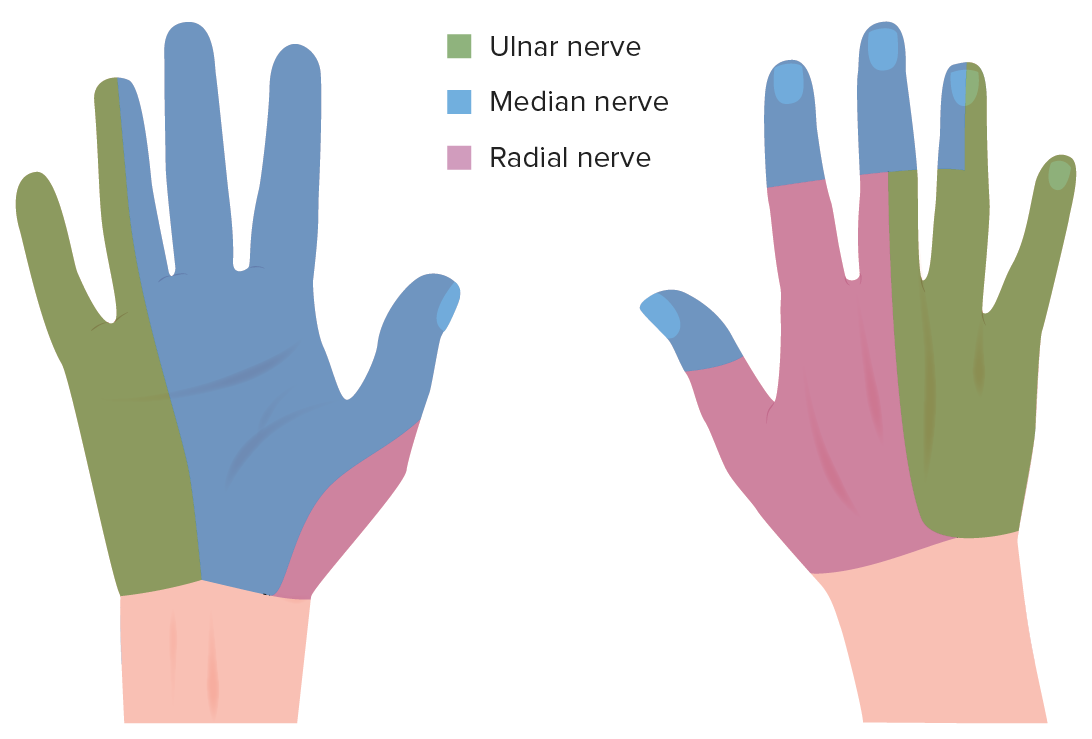
Sensory innervation of the hand
Image by Lecturio.The following are common problems associated with the hand: