The ankle is a hinged synovial joint Synovial joint Jaw and Temporomandibular Joint: Anatomy formed between the articular surfaces of the distal tibia Tibia The second longest bone of the skeleton. It is located on the medial side of the lower leg, articulating with the fibula laterally, the talus distally, and the femur proximally. Knee Joint: Anatomy, distal fibula Fibula The bone of the lower leg lateral to and smaller than the tibia. In proportion to its length, it is the most slender of the long bones. Leg: Anatomy, and talus. The ankle primarily allows plantar flexion Flexion Examination of the Upper Limbs and dorsiflexion of the foot Foot The foot is the terminal portion of the lower limb, whose primary function is to bear weight and facilitate locomotion. The foot comprises 26 bones, including the tarsal bones, metatarsal bones, and phalanges. The bones of the foot form longitudinal and transverse arches and are supported by various muscles, ligaments, and tendons. Foot: Anatomy. The subtalar joint and the other tarsal bones Tarsal Bones The seven bones which form the tarsus - namely, calcaneus; talus; cuboid, navicular, and the internal, middle, and external cuneiforms. Foot: Anatomy create many synergistic articulations, allowing for a wide range of motion Range of motion The distance and direction to which a bone joint can be extended. Range of motion is a function of the condition of the joints, muscles, and connective tissues involved. Joint flexibility can be improved through appropriate muscle strength exercises. Examination of the Upper Limbs (ROM)—plantar flexion Flexion Examination of the Upper Limbs, dorsiflexion, eversion Eversion Chronic Apophyseal Injury, inversion, abduction Abduction Examination of the Upper Limbs, and adduction Adduction Examination of the Upper Limbs. The movements are generated by large muscle groups that originate in the leg Leg The lower leg, or just "leg" in anatomical terms, is the part of the lower limb between the knee and the ankle joint. The bony structure is composed of the tibia and fibula bones, and the muscles of the leg are grouped into the anterior, lateral, and posterior compartments by extensions of fascia. Leg: Anatomy and insert as well as act upon the bones of the foot Foot The foot is the terminal portion of the lower limb, whose primary function is to bear weight and facilitate locomotion. The foot comprises 26 bones, including the tarsal bones, metatarsal bones, and phalanges. The bones of the foot form longitudinal and transverse arches and are supported by various muscles, ligaments, and tendons. Foot: Anatomy and tarsus.
Last updated: Dec 15, 2025
The ankle is a synovial joint Synovial joint Jaw and Temporomandibular Joint: Anatomy that consists of articulations between the tibia Tibia The second longest bone of the skeleton. It is located on the medial side of the lower leg, articulating with the fibula laterally, the talus distally, and the femur proximally. Knee Joint: Anatomy, fibula Fibula The bone of the lower leg lateral to and smaller than the tibia. In proportion to its length, it is the most slender of the long bones. Leg: Anatomy, and talus, which are reinforced by the medial, lateral, and syndesmotic ligament. Generally, the ankle joint Ankle joint The ankle is a hinged synovial joint formed between the articular surfaces of the distal tibia, distal fibula, and talus. The ankle primarily allows plantar flexion and dorsiflexion of the foot. has a shape that is wider superiorly and anteriorly to accommodate the wedge-shaped talus, which contributes to joint stability.
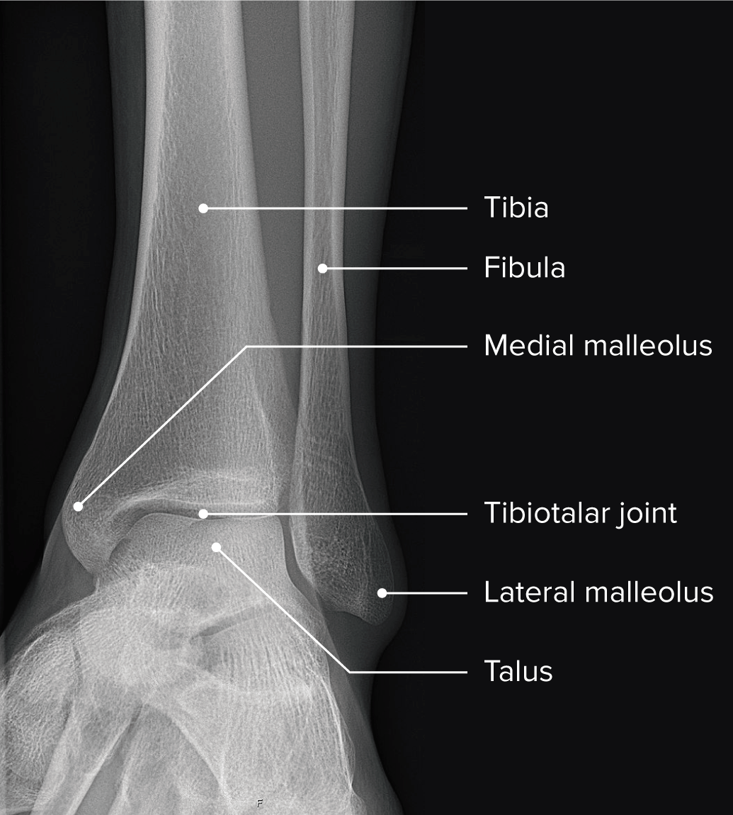
Radiograph of the ankle showing the normal anatomy of the ankle and the relationship of the bones and joints
Image by Lecturio.The tibia Tibia The second longest bone of the skeleton. It is located on the medial side of the lower leg, articulating with the fibula laterally, the talus distally, and the femur proximally. Knee Joint: Anatomy, fibula Fibula The bone of the lower leg lateral to and smaller than the tibia. In proportion to its length, it is the most slender of the long bones. Leg: Anatomy, and talus form the ankle joint Ankle joint The ankle is a hinged synovial joint formed between the articular surfaces of the distal tibia, distal fibula, and talus. The ankle primarily allows plantar flexion and dorsiflexion of the foot. and, along with the subtalar and talar joints, contribute to the coordinated movement of the ankle.
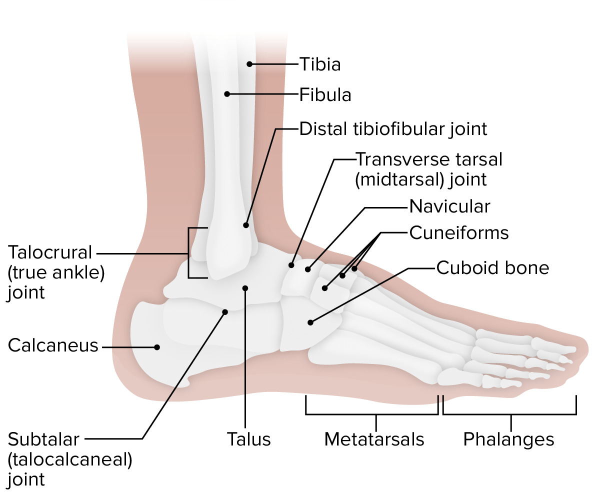
Lateral view of the ankle: Note the 4 different articulations that work synergistically to achieve the full range of motion of the ankle.
Image by Lecturio.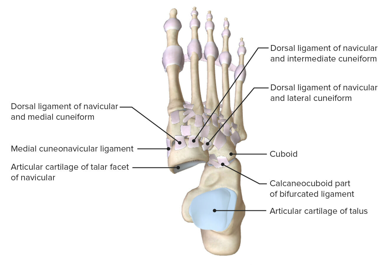
Superior view of the ankle with the talus removed, featuring the articular surfaces and supporting ligaments of the subtalar and transverse tarsal joints
Image by Lecturio.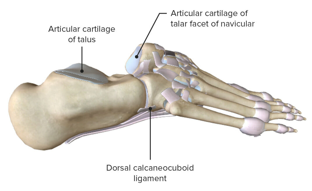
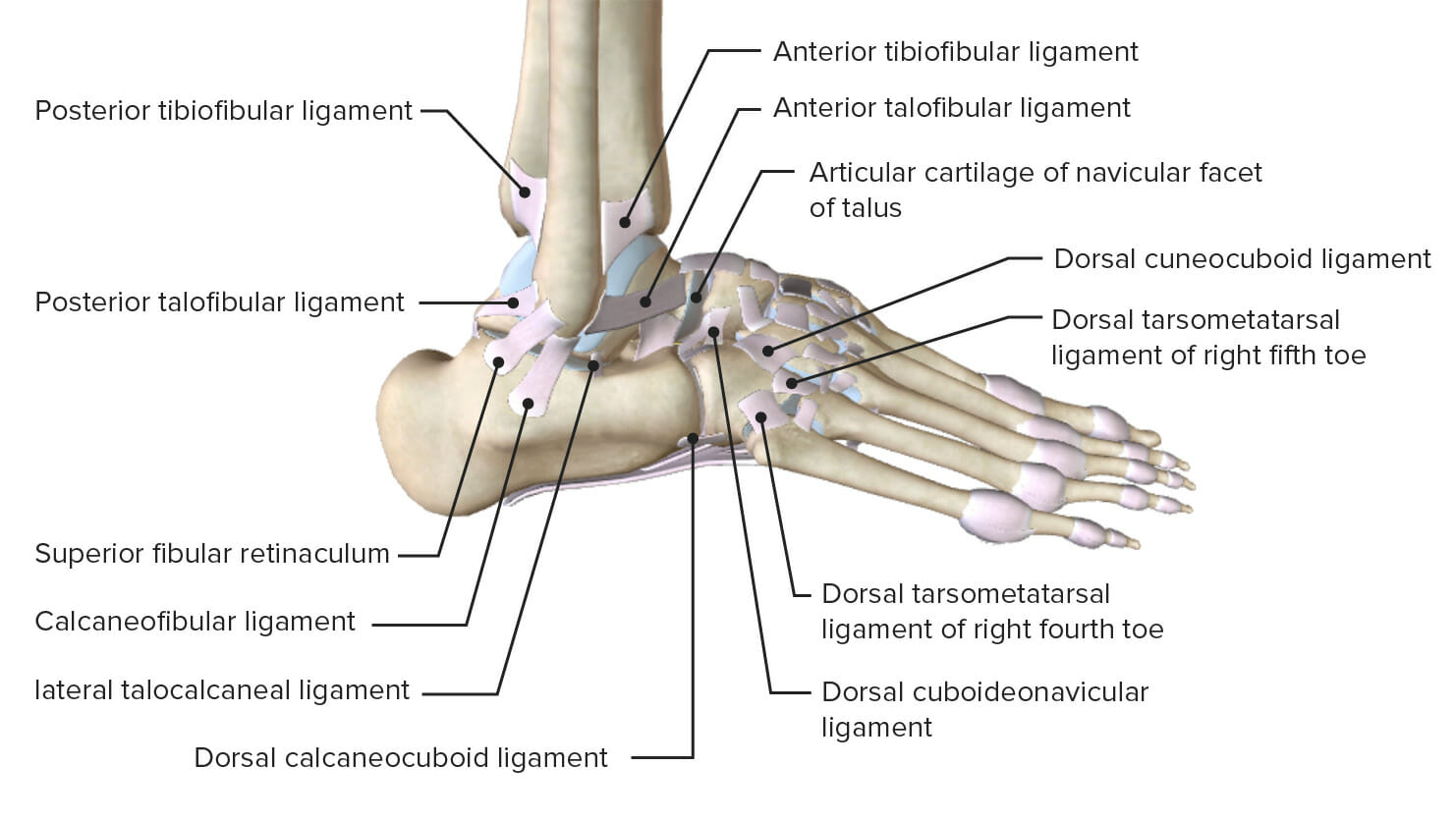
Lateral (right) views of the ankle with the talus removed, featuring the articular surfaces and supporting ligaments of the subtalar and transverse tarsal joints.
Image by Lecturio.Several major ligament complexes exist in the ankle to provide stability and support.
Lateral view of the ankle featuring the supporting ligaments of the talocrural and subtalar joints
Image by Lecturio.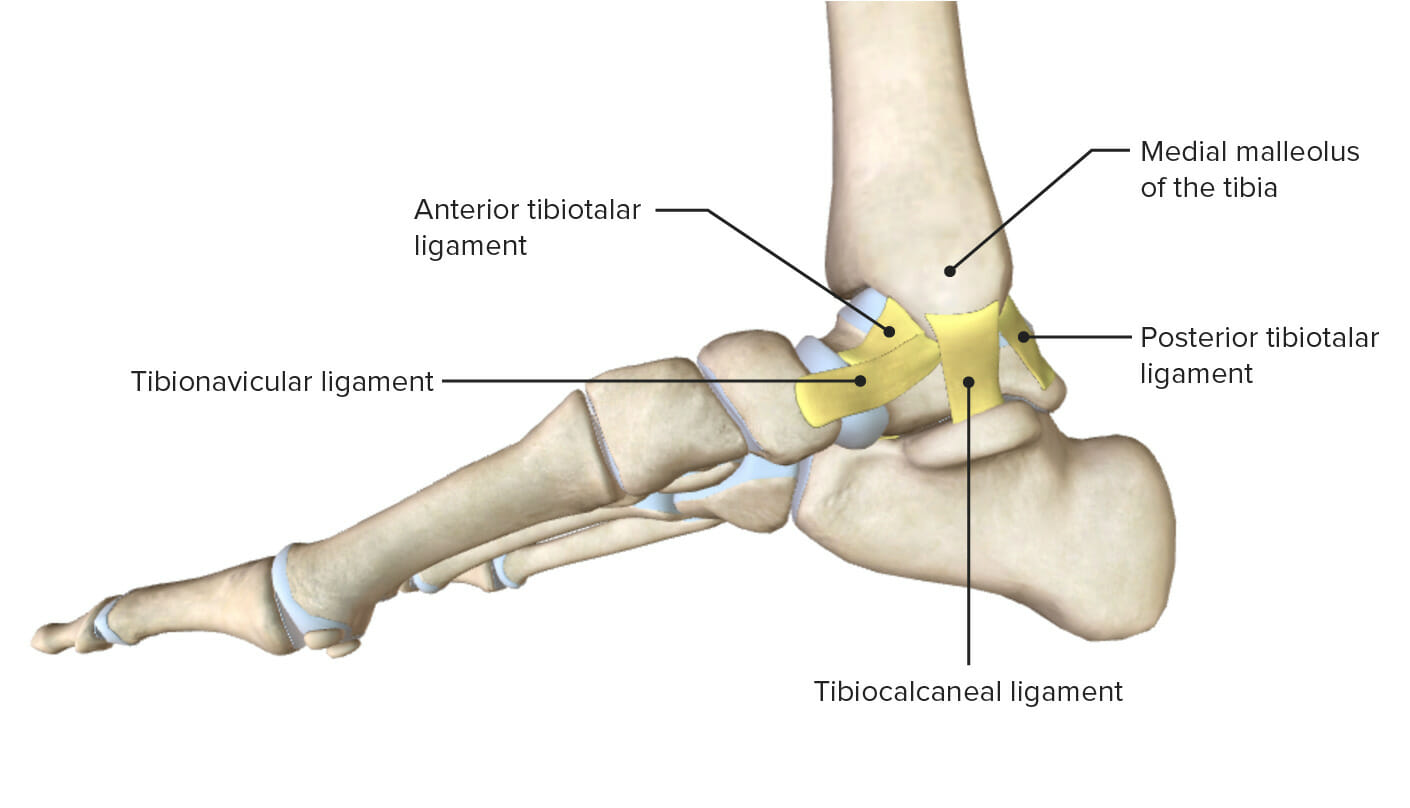
Medial view of the ankle highlighting the various ligaments of the medial ankle joint
Image by Lecturio.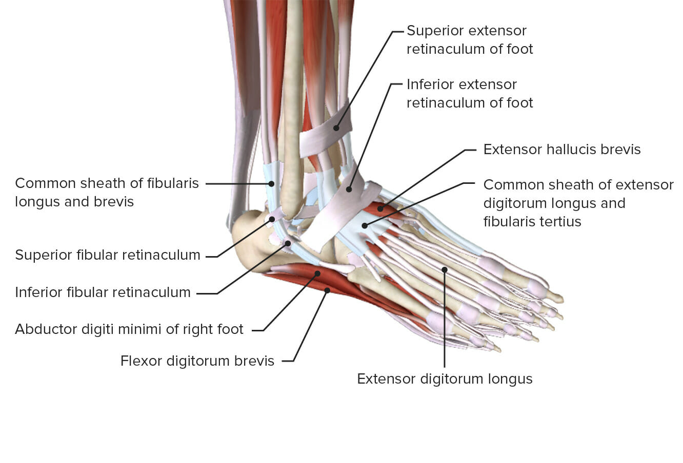
Oblique view of the ankle demonstrating the retinacula and various muscles
Image by Lecturio.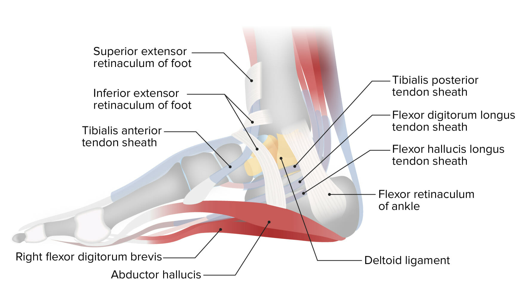
Lateral projection of the ankle showing the medial ankle retinacula
Image by Lecturio.The muscles of the leg Leg The lower leg, or just “leg” in anatomical terms, is the part of the lower limb between the knee and the ankle joint. The bony structure is composed of the tibia and fibula bones, and the muscles of the leg are grouped into the anterior, lateral, and posterior compartments by extensions of fascia. Leg: Anatomy insert into the bones of the ankle to create movement, and the range of motion Range of motion The distance and direction to which a bone joint can be extended. Range of motion is a function of the condition of the joints, muscles, and connective tissues involved. Joint flexibility can be improved through appropriate muscle strength exercises. Examination of the Upper Limbs (ROM) occurs in cardinal planes.
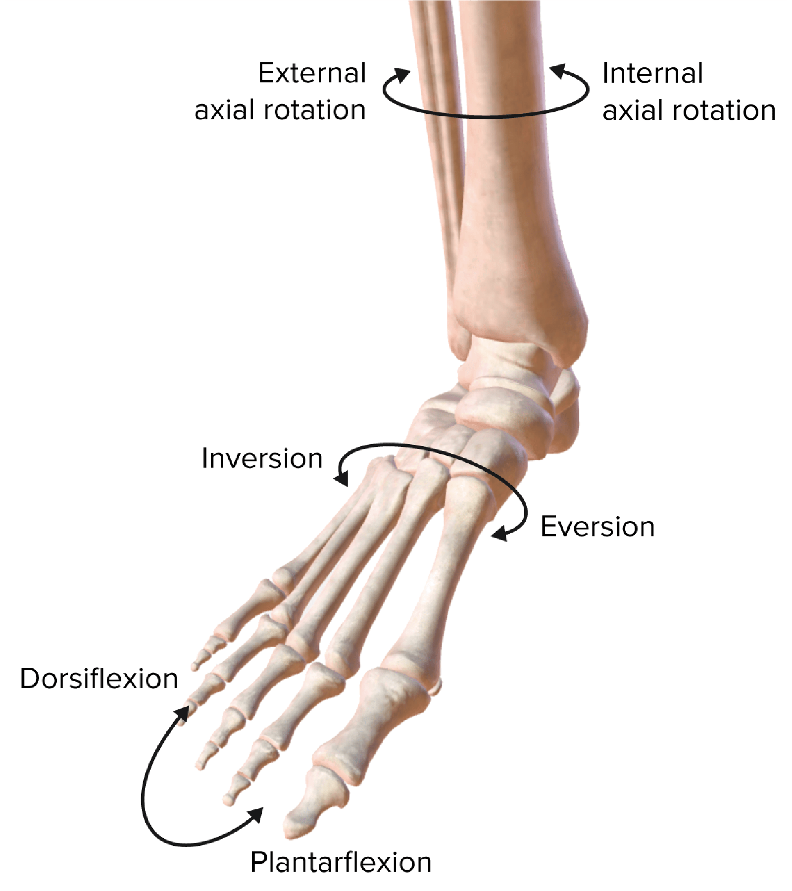
The movement planes of the ankle joint
Image by Lecturio.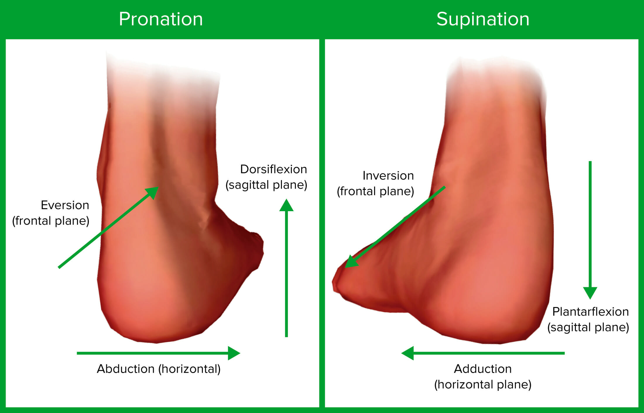
The various planes and movements of the ankle joint
Image by Lecturio.| Muscle | Origin | Insertion | Action | Innervation |
|---|---|---|---|---|
| Tibialis anterior Tibialis anterior Leg: Anatomy |
|
Interior surface of medial cuneiform | Dorsiflexion | Deep fibular (peroneal) nerve |
| Extensor hallucis longus Extensor hallucis longus Leg: Anatomy |
|
Distal phalanx big toe | Dorsiflexion | Deep fibular (peroneal) nerve |
| Extensor digitorum longus Extensor digitorum longus Leg: Anatomy |
|
Middle and distal phalanges Phalanges Bones that make up the skeleton of the fingers, consisting of two for the thumb, and three for each of the other fingers. Hand: Anatomy 2–5 | Dorsiflexion | Deep fibular (peroneal) nerve |
| Muscle | Origin | Insertion | Action | Innervation |
|---|---|---|---|---|
| Fibularis longus | Upper portion of lateral fibula Fibula The bone of the lower leg lateral to and smaller than the tibia. In proportion to its length, it is the most slender of the long bones. Leg: Anatomy | 1st metatarsal, medial cuneiform |
Plantar
flexion
Flexion
Examination of the Upper Limbs,
eversion Eversion Chronic Apophyseal Injury |
Superficial fibular |
| Fibularis brevis | Distal fibula Fibula The bone of the lower leg lateral to and smaller than the tibia. In proportion to its length, it is the most slender of the long bones. Leg: Anatomy shaft | Proximal end of 5th metatarsal | Plantar
flexion
Flexion
Examination of the Upper Limbs,
eversion Eversion Chronic Apophyseal Injury |
Superficial fibular |

Anterior view of the leg, featuring the muscles of the anterior compartment and their relationships with other muscles and with each other
Image by BioDigital, edited by Lecturio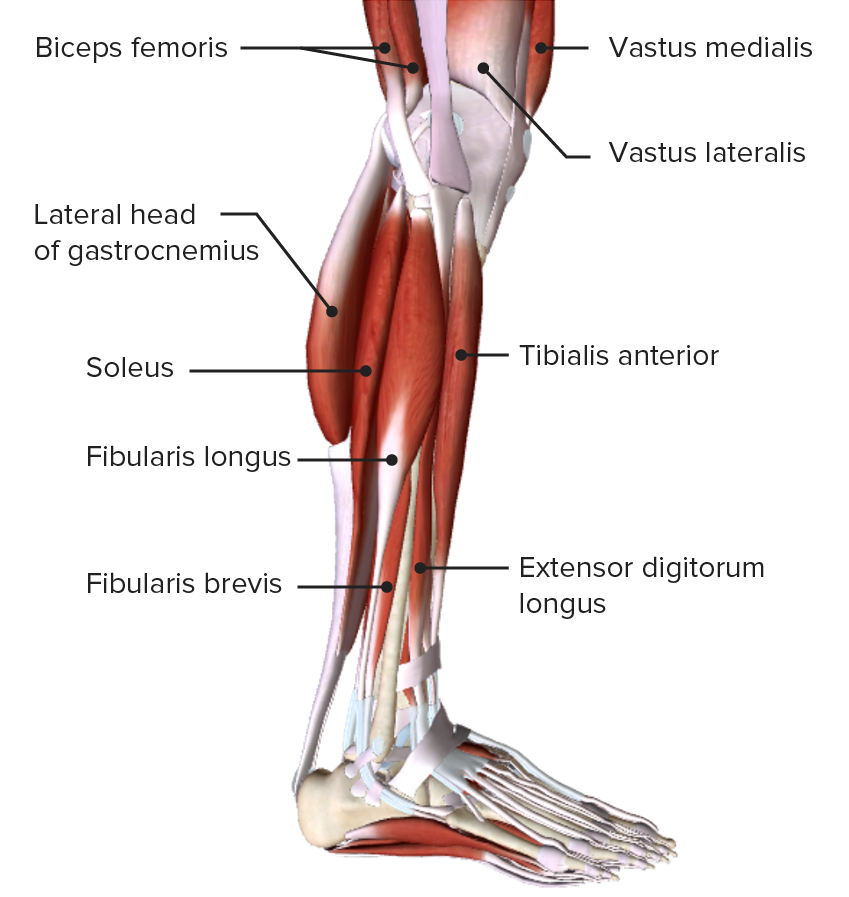
Lateral view of the leg, featuring the muscles of the lateral, or evertor, compartment and their relationships with other muscles and with each other
Image by BioDigital, edited by Lecturio| Muscle | Origin | Insertion | Action | Innervation |
|---|---|---|---|---|
| Gastrocnemius Gastrocnemius Leg: Anatomy | Medial and lateral condyles | Posterior calcaneus Calcaneus The largest of the tarsal bones which is situated at the lower and back part of the foot, forming the heel. Foot: Anatomy | Plantar flexion Flexion Examination of the Upper Limbs | Tibial nerve Tibial Nerve The medial terminal branch of the sciatic nerve. The tibial nerve fibers originate in lumbar and sacral spinal segments (L4 to S2). They supply motor and sensory innervation to parts of the calf and foot. Popliteal Fossa: Anatomy |
| Soleus Soleus Leg: Anatomy | Superior tibia Tibia The second longest bone of the skeleton. It is located on the medial side of the lower leg, articulating with the fibula laterally, the talus distally, and the femur proximally. Knee Joint: Anatomy, fibula Fibula The bone of the lower leg lateral to and smaller than the tibia. In proportion to its length, it is the most slender of the long bones. Leg: Anatomy, interosseous membrane Interosseous Membrane A sheet of fibrous connective tissue rich in collagen often linking two parallel bony structures forming a syndesmosis type joint. It provides longitudinal stability, tensile strength, and weight distribution/transfer and may allow limited movement in syndesmoses. Forearm: Anatomy | Posterior calcaneus Calcaneus The largest of the tarsal bones which is situated at the lower and back part of the foot, forming the heel. Foot: Anatomy | Plantar flexion Flexion Examination of the Upper Limbs | Tibial nerve Tibial Nerve The medial terminal branch of the sciatic nerve. The tibial nerve fibers originate in lumbar and sacral spinal segments (L4 to S2). They supply motor and sensory innervation to parts of the calf and foot. Popliteal Fossa: Anatomy |
| Plantaris Plantaris Leg: Anatomy | Posterior femur above lateral condyle | Calcaneus Calcaneus The largest of the tarsal bones which is situated at the lower and back part of the foot, forming the heel. Foot: Anatomy or calcaneus Calcaneus The largest of the tarsal bones which is situated at the lower and back part of the foot, forming the heel. Foot: Anatomy tendon | Plantar flexion Flexion Examination of the Upper Limbs | Tibial from anterior rami S1 S1 Heart Sounds– S2 S2 Heart Sounds |
| Tibialis posterior Tibialis posterior Leg: Anatomy | Superior tibia Tibia The second longest bone of the skeleton. It is located on the medial side of the lower leg, articulating with the fibula laterally, the talus distally, and the femur proximally. Knee Joint: Anatomy and fibula Fibula The bone of the lower leg lateral to and smaller than the tibia. In proportion to its length, it is the most slender of the long bones. Leg: Anatomy, interosseous membrane Interosseous Membrane A sheet of fibrous connective tissue rich in collagen often linking two parallel bony structures forming a syndesmosis type joint. It provides longitudinal stability, tensile strength, and weight distribution/transfer and may allow limited movement in syndesmoses. Forearm: Anatomy | Several tarsals and metatarsals 2–4 | Inversion and plantar flexion Flexion Examination of the Upper Limbs | Tibial nerve Tibial Nerve The medial terminal branch of the sciatic nerve. The tibial nerve fibers originate in lumbar and sacral spinal segments (L4 to S2). They supply motor and sensory innervation to parts of the calf and foot. Popliteal Fossa: Anatomy |
| Muscle | Origin | Insertion | Action | Innervation |
|---|---|---|---|---|
| Flexor digitorum longus Flexor digitorum longus Leg: Anatomy | Posterior tibia Tibia The second longest bone of the skeleton. It is located on the medial side of the lower leg, articulating with the fibula laterally, the talus distally, and the femur proximally. Knee Joint: Anatomy | Distal phalanges Phalanges Bones that make up the skeleton of the fingers, consisting of two for the thumb, and three for each of the other fingers. Hand: Anatomy 2-5 | Plantar flexion Flexion Examination of the Upper Limbs, inversion | Tibial nerve Tibial Nerve The medial terminal branch of the sciatic nerve. The tibial nerve fibers originate in lumbar and sacral spinal segments (L4 to S2). They supply motor and sensory innervation to parts of the calf and foot. Popliteal Fossa: Anatomy |
| Flexor hallucis longus Flexor hallucis longus Leg: Anatomy | Midshaft of fibula Fibula The bone of the lower leg lateral to and smaller than the tibia. In proportion to its length, it is the most slender of the long bones. Leg: Anatomy, interosseous membrane Interosseous Membrane A sheet of fibrous connective tissue rich in collagen often linking two parallel bony structures forming a syndesmosis type joint. It provides longitudinal stability, tensile strength, and weight distribution/transfer and may allow limited movement in syndesmoses. Forearm: Anatomy | Distal phalanx of big toe | Plantar flexion Flexion Examination of the Upper Limbs, inversion | Tibial nerve Tibial Nerve The medial terminal branch of the sciatic nerve. The tibial nerve fibers originate in lumbar and sacral spinal segments (L4 to S2). They supply motor and sensory innervation to parts of the calf and foot. Popliteal Fossa: Anatomy |
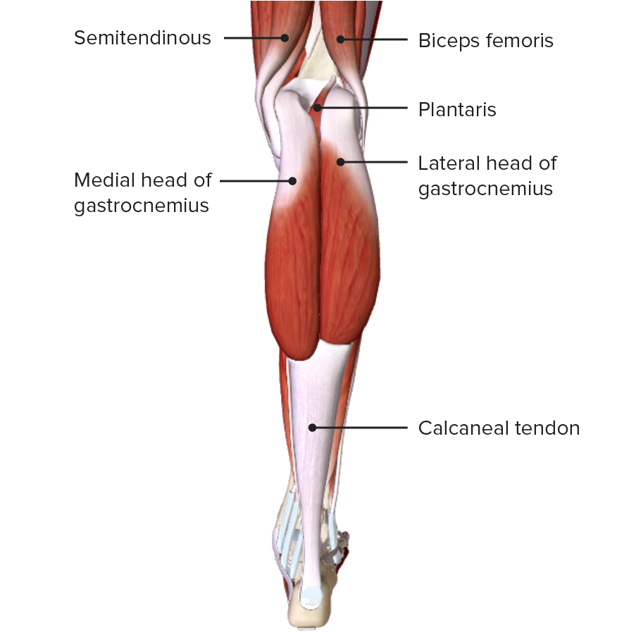
Posterior view of the leg, featuring the gastrocnemius muscles of the superficial layer of the posterior compartment
Image by BioDigital, edited by Lecturio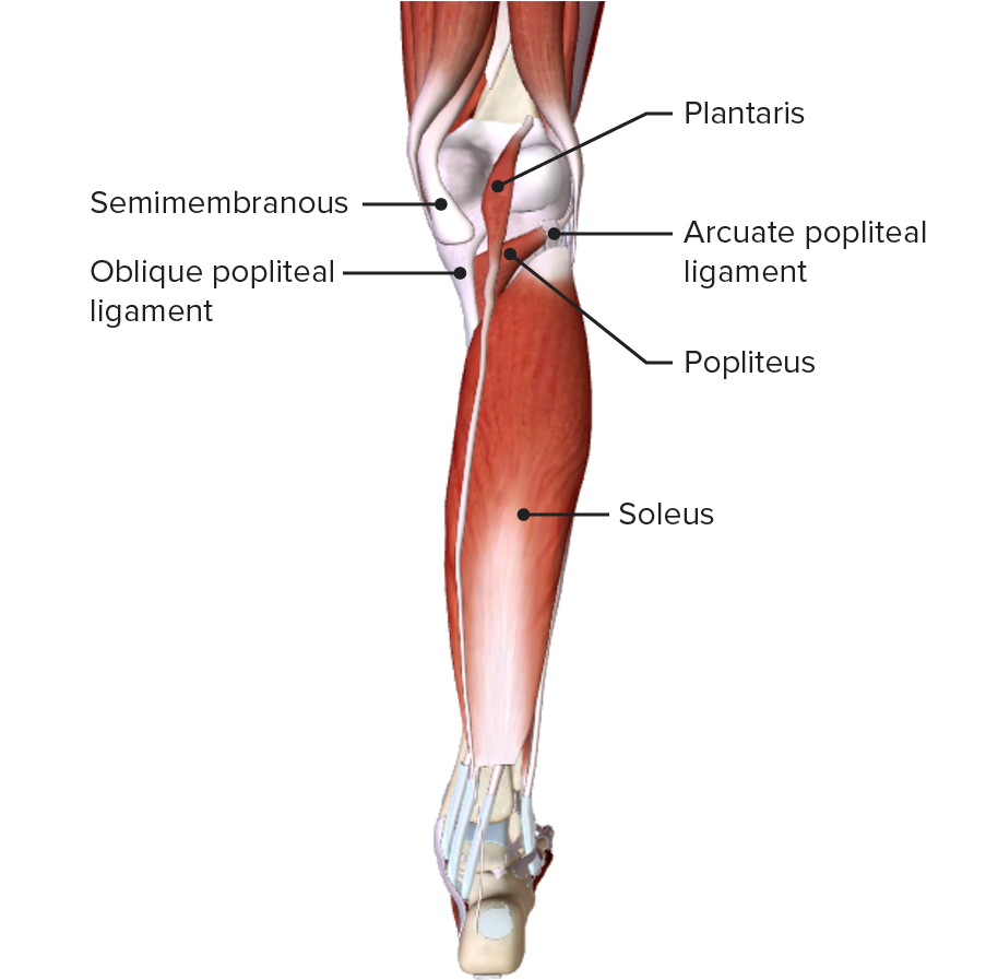
Posterior view of the leg, featuring the plantaris and soleus muscles of the superficial layer of the posterior compartment
Image by BioDigital, edited by Lecturio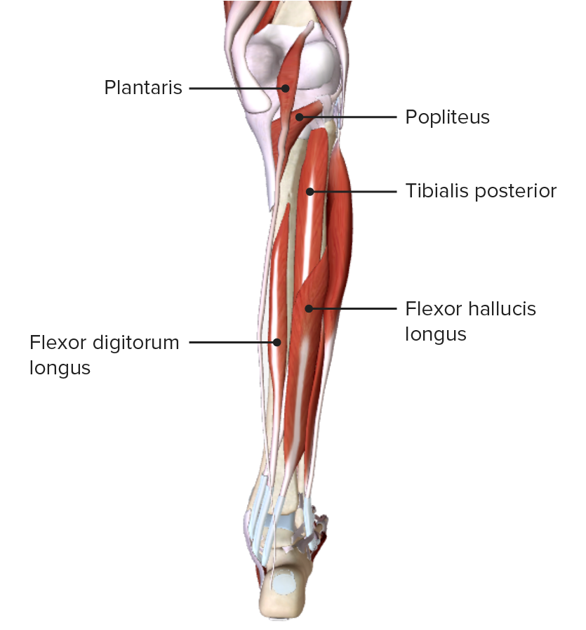
Posterior view of the leg, featuring the muscles of the deep layer of the posterior compartment and their spatial relationships with other muscles and with each other
Image by BioDigital, edited by Lecturio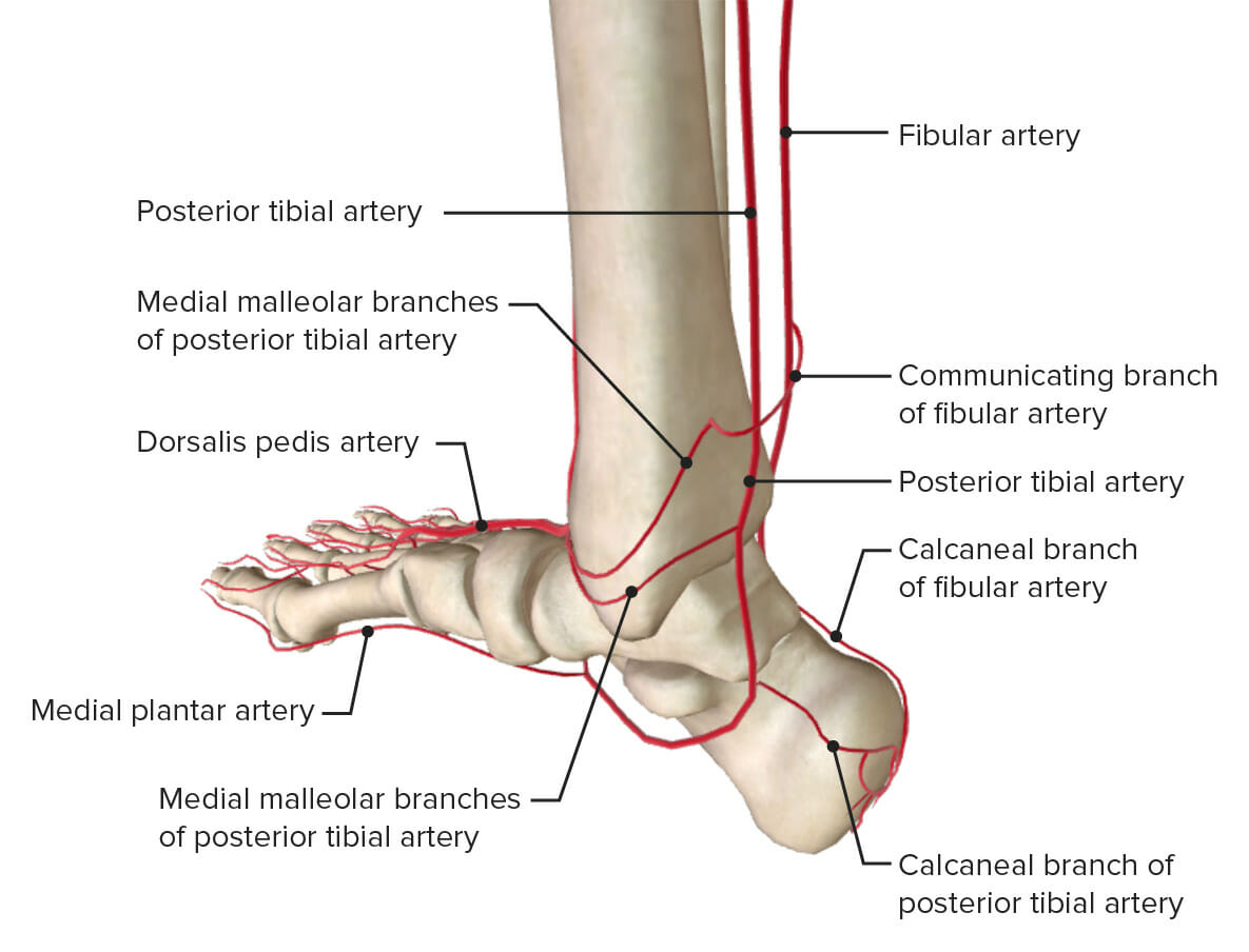
The arterial supply to the ankle: Note that the anterior tibial artery is shown as its continuation, the dorsalis pedis artery.
Image by Lecturio.The nerve supply of the ankle is provided by the roots from spinal cord Spinal cord The spinal cord is the major conduction pathway connecting the brain to the body; it is part of the CNS. In cross section, the spinal cord is divided into an H-shaped area of gray matter (consisting of synapsing neuronal cell bodies) and a surrounding area of white matter (consisting of ascending and descending tracts of myelinated axons). Spinal Cord: Anatomy level L4 to S2 S2 Heart Sounds. The following nerves also supply branches to the ankle joint Ankle joint The ankle is a hinged synovial joint formed between the articular surfaces of the distal tibia, distal fibula, and talus. The ankle primarily allows plantar flexion and dorsiflexion of the foot. :

Anterior view of the nerves of the ankle joint
Image by BioDigital, edited by Lecturio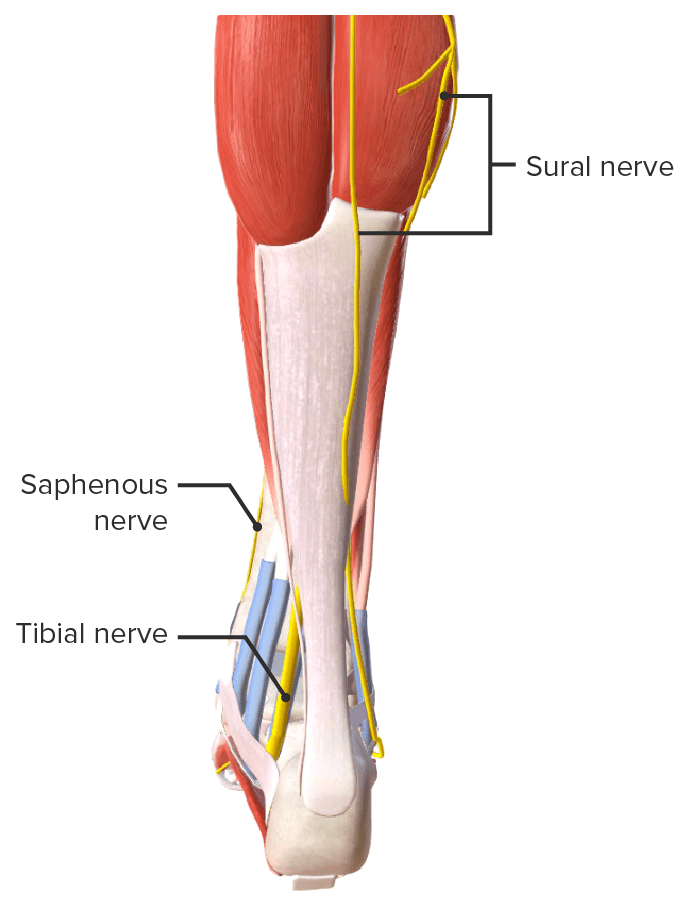
Posterior view of the nerves of the ankle joint
Image by BioDigital, edited by Lecturio