Cartilage is a type of connective tissue Connective tissue Connective tissues originate from embryonic mesenchyme and are present throughout the body except inside the brain and spinal cord. The main function of connective tissues is to provide structural support to organs. Connective tissues consist of cells and an extracellular matrix. Connective Tissue: Histology derived from embryonic mesenchyme that is responsible for structural support, resilience, and the smoothness of physical actions. Perichondrium ( connective tissue Connective tissue Connective tissues originate from embryonic mesenchyme and are present throughout the body except inside the brain and spinal cord. The main function of connective tissues is to provide structural support to organs. Connective tissues consist of cells and an extracellular matrix. Connective Tissue: Histology membrane surrounding most cartilage) compensates for the absence of vasculature in cartilage by providing nutrition and support. The abundant ground substance Ground substance Connective Tissue: Histology contains large amounts of chondroitin sulfate Chondroitin sulfate Derivatives of chondroitin which have a sulfate moiety esterified to the galactosamine moiety of chondroitin. Chondroitin sulfate a, or chondroitin 4-sulfate, and chondroitin sulfate c, or chondroitin 6-sulfate, have the sulfate esterified in the 4- and 6-positions, respectively. Chondroitin sulfate B is a misnomer and this compound is not a true chondroitin sulfate. Connective Tissue: Histology, hyaluronic acid Hyaluronic acid A natural high-viscosity mucopolysaccharide with alternating beta (1-3) glucuronide and beta (1-4) glucosaminidase bonds. It is found in the umbilical cord, in vitreous body and in synovial fluid. A high urinary level is found in progeria. Connective Tissue: Histology, and water (80% of cartilage is water). All types of cartilage contain type II collagen Collagen A polypeptide substance comprising about one third of the total protein in mammalian organisms. It is the main constituent of skin; connective tissue; and the organic substance of bones (bone and bones) and teeth (tooth). Connective Tissue: Histology produced by chondrocytes. Elastic Elastic Connective Tissue: Histology cartilage additionally contains elastic Elastic Connective Tissue: Histology fibers, whereas fibrocartilage also contains dense connective tissue Dense connective tissue Connective Tissue: Histology ( type 1 Type 1 Spinal Muscular Atrophy collagen Collagen A polypeptide substance comprising about one third of the total protein in mammalian organisms. It is the main constituent of skin; connective tissue; and the organic substance of bones (bone and bones) and teeth (tooth). Connective Tissue: Histology).
Last updated: Dec 15, 2025
Cartilage is a type of connective tissue Connective tissue Connective tissues originate from embryonic mesenchyme and are present throughout the body except inside the brain and spinal cord. The main function of connective tissues is to provide structural support to organs. Connective tissues consist of cells and an extracellular matrix. Connective Tissue: Histology that forms structural components of the human skeleton and provides support to various organs.
Chondrocytes:
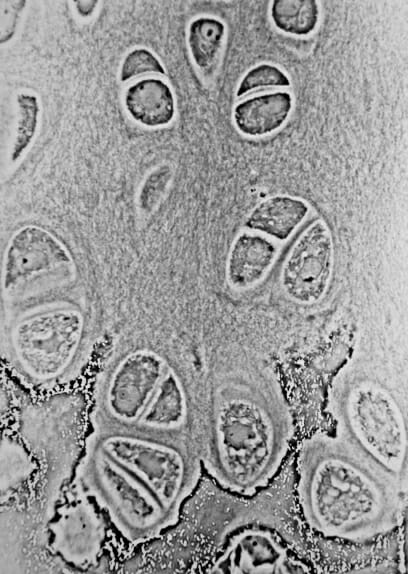
Light micrograph of an epiphyseal plate showing chondrocytes
Image: “Cartilage” by Robert M. Hunt. License: Public DomainChondroblasts:
Extracellular matrix Extracellular matrix A meshwork-like substance found within the extracellular space and in association with the basement membrane of the cell surface. It promotes cellular proliferation and provides a supporting structure to which cells or cell lysates in culture dishes adhere. Hypertrophic and Keloid Scars:
There are 3 main types of cartilage tissue:
| Hyaline cartilage | Elastic Elastic Connective Tissue: Histology (yellow) cartilage | Fibrocartilage | |
|---|---|---|---|
| Composition of extracellular matrix Extracellular matrix A meshwork-like substance found within the extracellular space and in association with the basement membrane of the cell surface. It promotes cellular proliferation and provides a supporting structure to which cells or cell lysates in culture dishes adhere. Hypertrophic and Keloid Scars |
|
|
|
| Major cells |
|
|
|
| Arrangement of chondrocytes | Isolated or in small isogenous groups | Usually in small isogenous groups | Isolated or in isogenous groups arranged axially |
| Presence of perichondrium | Yes (except at epiphysis Epiphysis The head of a long bone that is separated from the shaft by the epiphyseal plate until bone growth stops. At that time, the plate disappears and the head and shaft are united. Bones: Structure and Types and articular cartilage) | Yes | No |
| Locations |
|
|
|
| Functions |
|
|
|
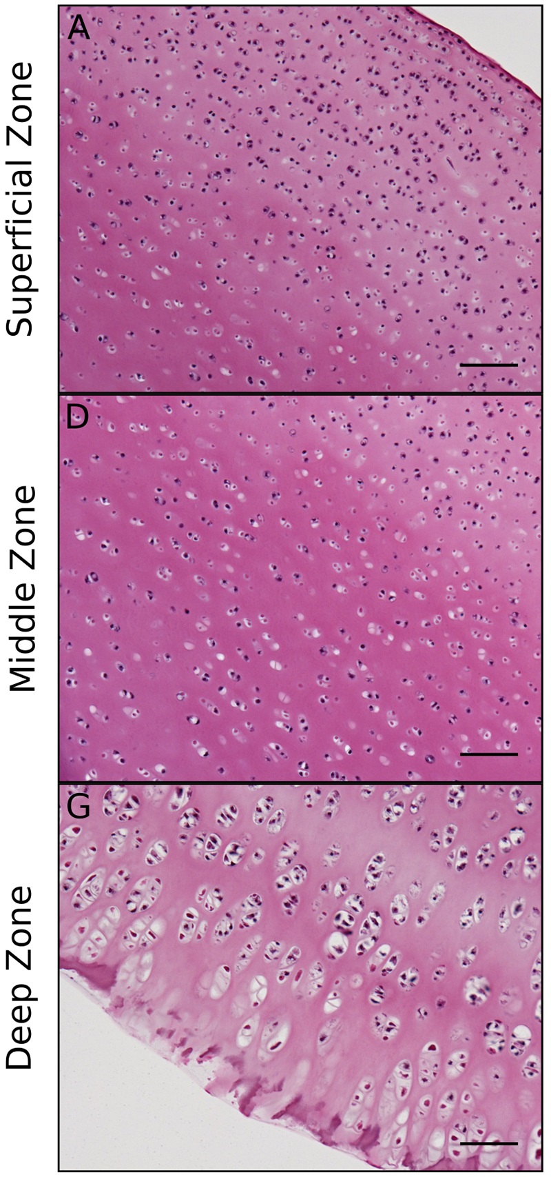
Histology of articular cartilage zones:
The superficial zone consists mostly of collagen II fibers aligned parallel to the articular surface to resist shear forces, whereas the deep zone consists of the same fibers aligned perpendicularly to the bone interface to absorb compressive loads.
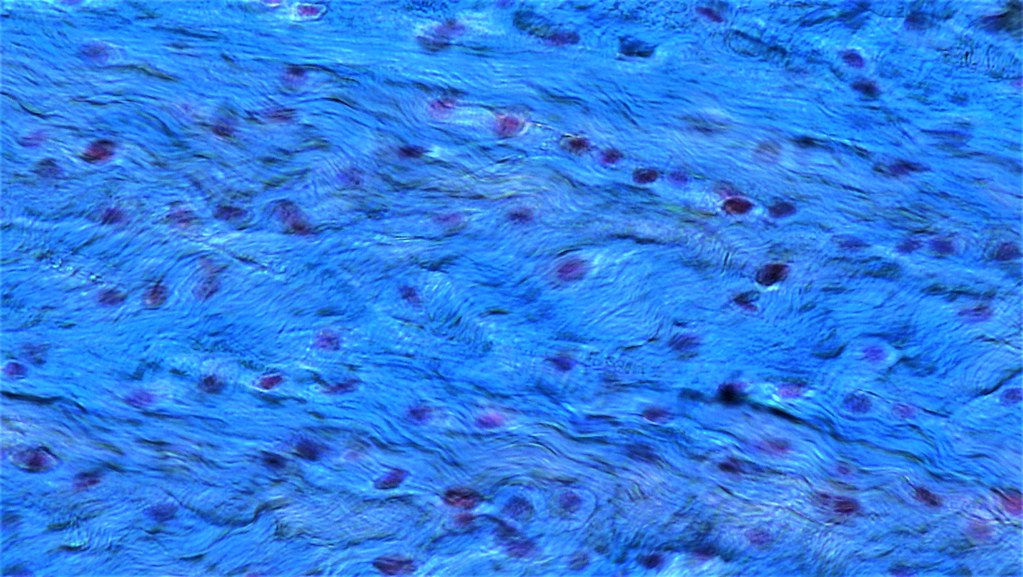
Fibrocartilage:
Light microscopy shows fibrocartilage, composed of chondrocytes and dense connective tissue.
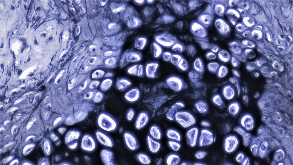
Elastic cartilage:
Composed of chondrocytes and abundant elastic fibers, which gives the cartilage yellow color