Achieve Mastery of Medical Concepts

Study for medical school and boards with Lecturio

Osteochondritis Dissecans

Osteochondritis dissecans ( OCD OCD Obsessive-compulsive disorder (OCD) is a condition characterized by obsessions (recurring and intrusive thoughts, urges, or images) and/or compulsions (repetitive actions the person is compelled to perform) that are time-consuming and associated with functional impairment. Obsessive-compulsive Disorder (OCD)) is an orthopedic disorder characterized by the detachment of a focal segment of subchondral bone Bone Bone is a compact type of hardened connective tissue composed of bone cells, membranes, an extracellular mineralized matrix, and central bone marrow. The 2 primary types of bone are compact and spongy. Bones: Structure and Types and cartilage Cartilage Cartilage is a type of connective tissue derived from embryonic mesenchyme that is responsible for structural support, resilience, and the smoothness of physical actions. Perichondrium (connective tissue membrane surrounding cartilage) compensates for the absence of vasculature in cartilage by providing nutrition and support. Cartilage: Histology as a result of focal aseptic necrosis Aseptic Necrosis Septic Arthritis. This can occur at any age, but it is most commonly seen in adolescents who participate in competitive sports. Patients Patients Individuals participating in the health care system for the purpose of receiving therapeutic, diagnostic, or preventive procedures. Clinician–Patient Relationship can be asymptomatic or may present with joint pain Pain An unpleasant sensation induced by noxious stimuli which are detected by nerve endings of nociceptive neurons. Pain: Types and Pathways, stiffness, and swelling Swelling Inflammation that is worse with activity. The diagnosis can be made with imaging. Management depends on the severity, but can include restricted weight-bearing activity, physical therapy Physical Therapy Becker Muscular Dystrophy, or surgery.

Last updated: Dec 5, 2022

Editorial responsibility: Stanley Oiseth, Lindsay Jones, Evelin Maza

Overview

Definition

Osteochondritis dissecans ( OCD OCD Obsessive-compulsive disorder (OCD) is a condition characterized by obsessions (recurring and intrusive thoughts, urges, or images) and/or compulsions (repetitive actions the person is compelled to perform) that are time-consuming and associated with functional impairment. Obsessive-compulsive Disorder (OCD)) is a disorder characterized by aseptic necrosis Aseptic Necrosis Septic Arthritis of subchondral bone Bone Bone is a compact type of hardened connective tissue composed of bone cells, membranes, an extracellular mineralized matrix, and central bone marrow. The 2 primary types of bone are compact and spongy. Bones: Structure and Types that results in focal detachment and displacement Displacement The process by which an emotional or behavioral response that is appropriate for one situation appears in another situation for which it is inappropriate. Defense Mechanisms of bone Bone Bone is a compact type of hardened connective tissue composed of bone cells, membranes, an extracellular mineralized matrix, and central bone marrow. The 2 primary types of bone are compact and spongy. Bones: Structure and Types and cartilage Cartilage Cartilage is a type of connective tissue derived from embryonic mesenchyme that is responsible for structural support, resilience, and the smoothness of physical actions. Perichondrium (connective tissue membrane surrounding cartilage) compensates for the absence of vasculature in cartilage by providing nutrition and support. Cartilage: Histology into the joint space. The term osteochondritis is a misnomer, as histologic analysis reveals a lack of inflammatory cells.

Epidemiology

  • Prevalence Prevalence The total number of cases of a given disease in a specified population at a designated time. It is differentiated from incidence, which refers to the number of new cases in the population at a given time. Measures of Disease Frequency: 15–29 per 100,000 patients Patients Individuals participating in the health care system for the purpose of receiving therapeutic, diagnostic, or preventive procedures. Clinician–Patient Relationship
  • Peak age: 12–19 years
  • Male > female
  • Higher incidence Incidence The number of new cases of a given disease during a given period in a specified population. It also is used for the rate at which new events occur in a defined population. It is differentiated from prevalence, which refers to all cases in the population at a given time. Measures of Disease Frequency in young athletes

Etiology and Pathophysiology

Etiology

The exact etiology is unknown. Many theories have been proposed, and OCD OCD Obsessive-compulsive disorder (OCD) is a condition characterized by obsessions (recurring and intrusive thoughts, urges, or images) and/or compulsions (repetitive actions the person is compelled to perform) that are time-consuming and associated with functional impairment. Obsessive-compulsive Disorder (OCD) is likely multifactorial.

  • Trauma:
  • Ischemia Ischemia A hypoperfusion of the blood through an organ or tissue caused by a pathologic constriction or obstruction of its blood vessels, or an absence of blood circulation. Ischemic Cell Damage
  • Genetic predisposition

Pathophysiology

Pathogenesis of osteochondritis dissecans:

  • Repetitive microtrauma Microtrauma Small injuries caused by external force applied to the body including bones, muscles, nerves and tendons. Osgood-Schlatter Disease → injury to the subchondral bone Bone Bone is a compact type of hardened connective tissue composed of bone cells, membranes, an extracellular mineralized matrix, and central bone marrow. The 2 primary types of bone are compact and spongy. Bones: Structure and Types fragmentation Fragmentation Chronic Apophyseal Injury
  • A defect develops between the osteochondral lesion and the underlying bone Bone Bone is a compact type of hardened connective tissue composed of bone cells, membranes, an extracellular mineralized matrix, and central bone marrow. The 2 primary types of bone are compact and spongy. Bones: Structure and Types.
  • Results in hypovascularity → osteonecrosis of the lesion
  • Involvement of the overlying cartilage Cartilage Cartilage is a type of connective tissue derived from embryonic mesenchyme that is responsible for structural support, resilience, and the smoothness of physical actions. Perichondrium (connective tissue membrane surrounding cartilage) compensates for the absence of vasculature in cartilage by providing nutrition and support. Cartilage: Histology → instability of the fragment
  • Subchondral bone Bone Bone is a compact type of hardened connective tissue composed of bone cells, membranes, an extracellular mineralized matrix, and central bone marrow. The 2 primary types of bone are compact and spongy. Bones: Structure and Types and cartilage Cartilage Cartilage is a type of connective tissue derived from embryonic mesenchyme that is responsible for structural support, resilience, and the smoothness of physical actions. Perichondrium (connective tissue membrane surrounding cartilage) compensates for the absence of vasculature in cartilage by providing nutrition and support. Cartilage: Histology may become partially or totally separated.

Stages of progression in osteochondritis dissecans:

  • Stage I: small area of compression Compression Blunt Chest Trauma of the subchondral bone Bone Bone is a compact type of hardened connective tissue composed of bone cells, membranes, an extracellular mineralized matrix, and central bone marrow. The 2 primary types of bone are compact and spongy. Bones: Structure and Types
  • Stage II: partial detachment of an osteochondral fragment
  • Stage III:
    • Complete detachment of a fragment
    • Remains within the underlying crater bed
  • Stage IV:
    • Complete detachment of a fragment
    • Displaced from crater bed (loose body)
Osteochondritis dissecans 4 stages

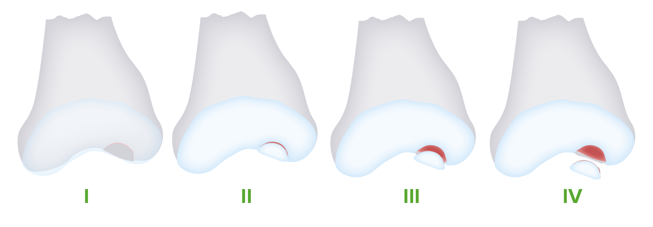
Osteochondritis dissecans progresses through 4 stages. With continued injury, the lesion fragments and becomes separated from the underlying bone, eventually becoming displaced in the joint space (loose body).

Image by Lecturio.

Clinical Presentation

Symptoms

Some patients Patients Individuals participating in the health care system for the purpose of receiving therapeutic, diagnostic, or preventive procedures. Clinician–Patient Relationship may be asymptomatic, with OCD OCD Obsessive-compulsive disorder (OCD) is a condition characterized by obsessions (recurring and intrusive thoughts, urges, or images) and/or compulsions (repetitive actions the person is compelled to perform) that are time-consuming and associated with functional impairment. Obsessive-compulsive Disorder (OCD) found incidentally on imaging. However, symptomatic individuals may experience:

  • Chronic pain Chronic pain Aching sensation that persists for more than a few months. It may or may not be associated with trauma or disease, and may persist after the initial injury has healed. Its localization, character, and timing are more vague than with acute pain. Pain Management in the affected joint:
    • Increases with activity
    • Alleviated at rest
  • Progressive symptoms, such as:
    • Joint stiffness
    • Intermittent swelling Swelling Inflammation
    • Joint locking or catching (may signal a loose body)
    • Joint instability
  • Most commonly affected joints:
    • Knee (75% of cases)
    • Elbow (6% of cases)
    • Ankle (4% of cases)

Physical examination

  • Localized tenderness to palpation Palpation Application of fingers with light pressure to the surface of the body to determine consistency of parts beneath in physical diagnosis; includes palpation for determining the outlines of organs. Dermatologic Examination at the site of the lesion, particularly:
  • Painful range of motion Range of motion The distance and direction to which a bone joint can be extended. Range of motion is a function of the condition of the joints, muscles, and connective tissues involved. Joint flexibility can be improved through appropriate muscle strength exercises. Examination of the Upper Limbs
  • Range of motion Range of motion The distance and direction to which a bone joint can be extended. Range of motion is a function of the condition of the joints, muscles, and connective tissues involved. Joint flexibility can be improved through appropriate muscle strength exercises. Examination of the Upper Limbs:
    • Usually normal if the knee is affected
    • Often diminished in an affected elbow
  • Joint effusion Joint Effusion Septic Arthritis
  • Crepitus Crepitus Osteoarthritis
  • Wilson’s test:
  • Abnormal gait Gait Manner or style of walking. Neurological Examination:
    • May be seen in some patients Patients Individuals participating in the health care system for the purpose of receiving therapeutic, diagnostic, or preventive procedures. Clinician–Patient Relationship with knee involvement
    • Affected leg Leg The lower leg, or just “leg” in anatomical terms, is the part of the lower limb between the knee and the ankle joint. The bony structure is composed of the tibia and fibula bones, and the muscles of the leg are grouped into the anterior, lateral, and posterior compartments by extensions of fascia. Leg: Anatomy is externally rotated with ambulation.
Wilson's test

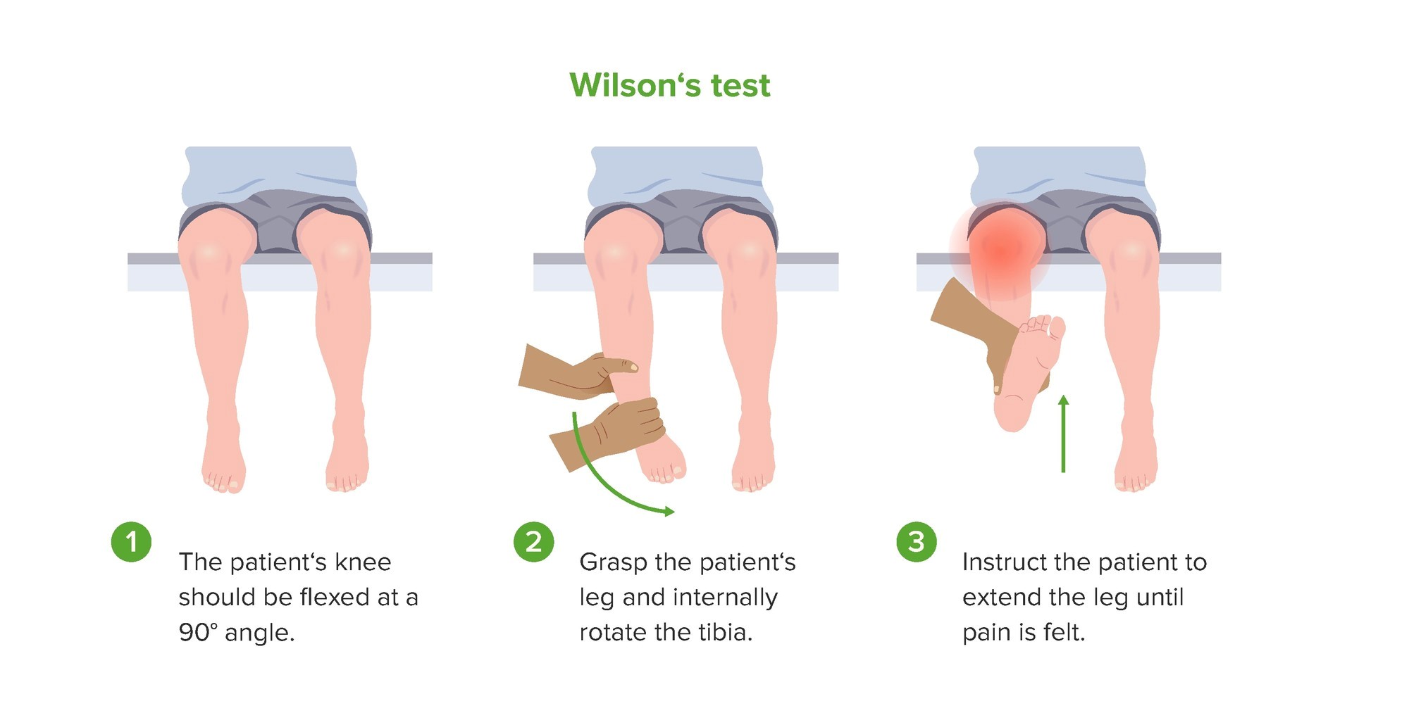
Demonstration of Wilson’s test, which may be positive in patients with osteochondritis dissecans of the knee.

Image by Lecturio.

Diagnosis

A diagnosis of OCD OCD Obsessive-compulsive disorder (OCD) is a condition characterized by obsessions (recurring and intrusive thoughts, urges, or images) and/or compulsions (repetitive actions the person is compelled to perform) that are time-consuming and associated with functional impairment. Obsessive-compulsive Disorder (OCD) should be suspected in adolescents with a characteristic presentation of joint pain Pain An unpleasant sensation induced by noxious stimuli which are detected by nerve endings of nociceptive neurons. Pain: Types and Pathways and involvement in repetitive activities and sports.

X-ray X-ray Penetrating electromagnetic radiation emitted when the inner orbital electrons of an atom are excited and release radiant energy. X-ray wavelengths range from 1 pm to 10 nm. Hard x-rays are the higher energy, shorter wavelength x-rays. Soft x-rays or grenz rays are less energetic and longer in wavelength. The short wavelength end of the x-ray spectrum overlaps the gamma rays wavelength range. The distinction between gamma rays and x-rays is based on their radiation source. Pulmonary Function Tests

X-rays X-rays X-rays are high-energy particles of electromagnetic radiation used in the medical field for the generation of anatomical images. X-rays are projected through the body of a patient and onto a film, and this technique is called conventional or projectional radiography. X-rays of the affected joint are usually diagnostic and are the initial testing method of choice. However, these images can be normal in the early disease stages.

Knee:

  • Findings:
    • Subcortical, crescent-shaped lucency is often first detected.
    • Subchondral bony fragment
  • Frequent lesion locations:
    • Medial condyle (77%)
    • Lateral condyle (17%)
    • Patella Patella The flat, triangular bone situated at the anterior part of the knee. Knee Joint: Anatomy (7%)
Osteochondritis dissecans knee x-ray

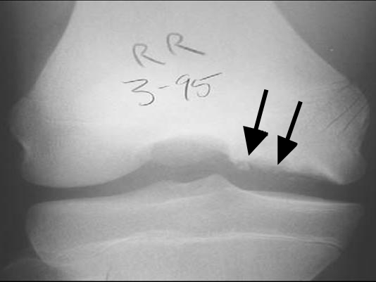
Knee X-ray of a patient with osteochondritis dissecans:
Note the fragmentation of the bone and the radiolucency underneath (arrows).

Image: “Osteochondritis dissecans diagram” by Kristin M Houghton. License: CC BY 2.0

Elbow:

Osteochondritis dissecans elbow x-ray

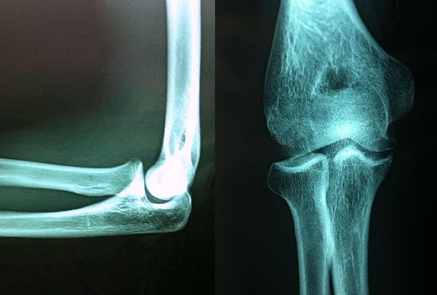
X-ray images from a 16-year-old athlete with osteochondritis dissecans of the elbow.
Shown is a radiolucent focus of the capitellum over the radial head.

Image: “Osteochondritis dissecans of the capitellum in adolescents: report of a case and review of the literature” by Erraji M, Kharraji A, Abbassi N, Najib A, Yacoubi H. License: CC BY 2.0

Ankle:

  • Normal in ⅓ of cases
  • Cup-shaped lesion on the talus Talus The second largest of the tarsal bones. It articulates with the tibia and fibula to form the ankle joint. Ankle Joint: Anatomy
Osteochondritis dissecans ankle x-ray

X-ray images of an ankle showing osteochondritis dissecans of the superomedial talus.
Note the radiolucency and fragmentation (arrows).

Image: “CT and projectional radiography of osteochondritis dissecans – annotated” by Mikael Häggström. License: CC0 1.0, cropped by Lecturio.

MRI

  • Should be considered in symptomatic patients Patients Individuals participating in the health care system for the purpose of receiving therapeutic, diagnostic, or preventive procedures. Clinician–Patient Relationship with normal X-rays X-rays X-rays are high-energy particles of electromagnetic radiation used in the medical field for the generation of anatomical images. X-rays are projected through the body of a patient and onto a film, and this technique is called conventional or projectional radiography. X-rays
  • More sensitive in detecting subchondral changes and can be used in staging Staging Methods which attempt to express in replicable terms the extent of the neoplasm in the patient. Grading, Staging, and Metastasis
  • Gadolinium Gadolinium An element of the rare earth family of metals. It has the atomic symbol gd, atomic number 64, and atomic weight 157. 25. Its oxide is used in the control rods of some nuclear reactors. Magnetic Resonance Imaging (MRI) can be used to evaluate the blood supply to the area.
  • Frequently used in preoperative planning
Osteochondritis dissecans knee mri

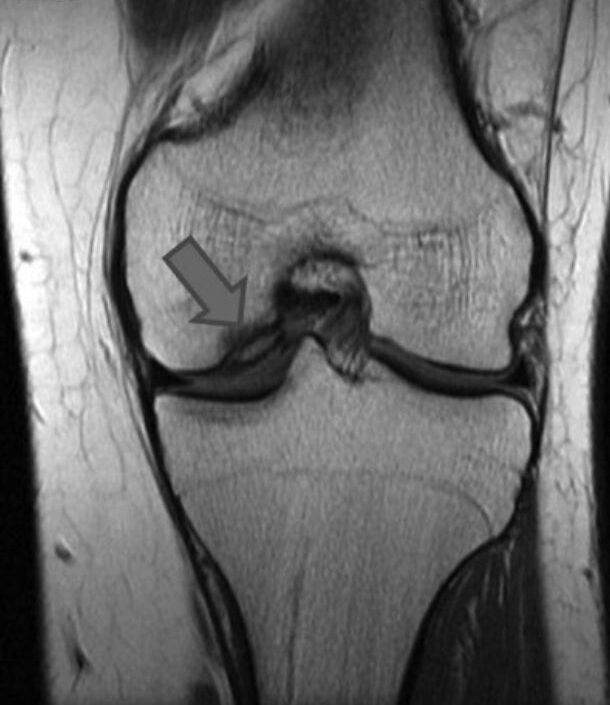
MRI of a knee showing an area of osteochondritis dissecans affecting the medial femoral condyle

Image: “The present state of treatments for articular cartilage defects in the knee” by Perera JR, Gikas PD, Bentley G. License: CC BY 3.0, cropped by Lecturio.

Management

The main goal of management is to achieve healing of the osteochondral lesion and return the affected joint to full function. Specialty referrals for sports medicine or orthopedic surgery may be needed.

Conservative management

  • Limit Limit A value (e.g., pressure or time) that should not be exceeded and which is specified by the operator to protect the lung Invasive Mechanical Ventilation weight-bearing and activity:
    • Critically important to prevent progression
    • Return to activity may occur when:
      • Pain-free
      • Active full range of motion Range of motion The distance and direction to which a bone joint can be extended. Range of motion is a function of the condition of the joints, muscles, and connective tissues involved. Joint flexibility can be improved through appropriate muscle strength exercises. Examination of the Upper Limbs improves
      • Evidence of healing
  • Joint immobilization Immobilization Delirium
  • Physical therapy Physical Therapy Becker Muscular Dystrophy
  • Indications for conservative management:
  • Asymptomatic patients Patients Individuals participating in the health care system for the purpose of receiving therapeutic, diagnostic, or preventive procedures. Clinician–Patient Relationship with incidental X-ray X-ray Penetrating electromagnetic radiation emitted when the inner orbital electrons of an atom are excited and release radiant energy. X-ray wavelengths range from 1 pm to 10 nm. Hard x-rays are the higher energy, shorter wavelength x-rays. Soft x-rays or grenz rays are less energetic and longer in wavelength. The short wavelength end of the x-ray spectrum overlaps the gamma rays wavelength range. The distinction between gamma rays and x-rays is based on their radiation source. Pulmonary Function Tests findings of osteochondral lesions:
    • Repeat X-rays X-rays X-rays are high-energy particles of electromagnetic radiation used in the medical field for the generation of anatomical images. X-rays are projected through the body of a patient and onto a film, and this technique is called conventional or projectional radiography. X-rays every 6 months.
    • Follow the patient until skeletal maturity or until the lesion has healed.

Surgical management

  • Indications:
    • Signs of joint instability
    • Expanding lesion on x-rays X-rays X-rays are high-energy particles of electromagnetic radiation used in the medical field for the generation of anatomical images. X-rays are projected through the body of a patient and onto a film, and this technique is called conventional or projectional radiography. X-rays
    • Failed nonoperative treatment
    • Intra-articular loose body
  • Techniques:
    • Arthroscopic subchondral drilling 
    • Fixation of unstable lesion
    • Arthroscopic excision, curettage Curettage A scraping, usually of the interior of a cavity or tract, for removal of new growth or other abnormal tissue, or to obtain material for tissue diagnosis. It is performed with a curet (curette), a spoon-shaped instrument designed for that purpose. Benign Bone Tumors, and drilling
    • Autologous chondral transplantation 
  • Choice depends on skeletal maturity, stage of the disease, and size of the lesion.

Prognosis Prognosis A prediction of the probable outcome of a disease based on a individual’s condition and the usual course of the disease as seen in similar situations. Non-Hodgkin Lymphomas

  • Most patients Patients Individuals participating in the health care system for the purpose of receiving therapeutic, diagnostic, or preventive procedures. Clinician–Patient Relationship with stable lesions will have spontaneous healing with conservative measures.
  • Success rates for unstable lesions varies depending on the severity and surgical technique: 30%–100%
  • Patients Patients Individuals participating in the health care system for the purpose of receiving therapeutic, diagnostic, or preventive procedures. Clinician–Patient Relationship have an increased risk of developing osteoarthritis Osteoarthritis Osteoarthritis (OA) is the most common form of arthritis, and is due to cartilage destruction and changes of the subchondral bone. The risk of developing this disorder increases with age, obesity, and repetitive joint use or trauma. Patients develop gradual joint pain, stiffness lasting < 30 minutes, and decreased range of motion. Osteoarthritis of the joint.
  • Factors linked with an improved prognosis Prognosis A prediction of the probable outcome of a disease based on a individual’s condition and the usual course of the disease as seen in similar situations. Non-Hodgkin Lymphomas:
    • Younger age
    • Intact articular cartilage Cartilage Cartilage is a type of connective tissue derived from embryonic mesenchyme that is responsible for structural support, resilience, and the smoothness of physical actions. Perichondrium (connective tissue membrane surrounding cartilage) compensates for the absence of vasculature in cartilage by providing nutrition and support. Cartilage: Histology
    • Smaller osteochondral lesions
    • Stable lesions

Differential Diagnosis

  • Meniscal tear: an acute injury often seen in younger patients Patients Individuals participating in the health care system for the purpose of receiving therapeutic, diagnostic, or preventive procedures. Clinician–Patient Relationship: The presentation may include a tearing or popping sensation during the injury, knee pain Pain An unpleasant sensation induced by noxious stimuli which are detected by nerve endings of nociceptive neurons. Pain: Types and Pathways, decreased range of motion Range of motion The distance and direction to which a bone joint can be extended. Range of motion is a function of the condition of the joints, muscles, and connective tissues involved. Joint flexibility can be improved through appropriate muscle strength exercises. Examination of the Upper Limbs, locking or catching, and joint effusion Joint Effusion Septic Arthritis. The diagnosis is based on the physical examination and imaging. Management includes conservative measures and surgery for severe injury.
  • Plica syndrome: irritation and inflammation Inflammation Inflammation is a complex set of responses to infection and injury involving leukocytes as the principal cellular mediators in the body’s defense against pathogenic organisms. Inflammation is also seen as a response to tissue injury in the process of wound healing. The 5 cardinal signs of inflammation are pain, heat, redness, swelling, and loss of function. Inflammation of the medial synovial component of the knee, which can occur with repetitive trauma: Patients Patients Individuals participating in the health care system for the purpose of receiving therapeutic, diagnostic, or preventive procedures. Clinician–Patient Relationship may experience knee pain Pain An unpleasant sensation induced by noxious stimuli which are detected by nerve endings of nociceptive neurons. Pain: Types and Pathways, often of the medial aspect of the joint, with a catching or popping sensation. A definitive diagnosis is provided by arthroscopy Arthroscopy Endoscopic examination, therapy and surgery of the joint. Knee Ligament Injuries. Management consists of conservative measures, physical therapy Physical Therapy Becker Muscular Dystrophy, intra-articular steroid injections, and arthroscopic surgery for refractory cases.
  • Patellofemoral syndrome: most common cause of anterior knee pain Pain An unpleasant sensation induced by noxious stimuli which are detected by nerve endings of nociceptive neurons. Pain: Types and Pathways, which may result from malalignment of the leg Leg The lower leg, or just “leg” in anatomical terms, is the part of the lower limb between the knee and the ankle joint. The bony structure is composed of the tibia and fibula bones, and the muscles of the leg are grouped into the anterior, lateral, and posterior compartments by extensions of fascia. Leg: Anatomy, muscular imbalance, overactivity, or trauma: Patellofemoral syndrome produces dull and generalized anterior knee pain Pain An unpleasant sensation induced by noxious stimuli which are detected by nerve endings of nociceptive neurons. Pain: Types and Pathways that is exacerbated by activity or prolonged sitting. Patellofemoral syndrome is a diagnosis of exclusion, and other intra-articular etiologies should be ruled out. Conservative measures and physical therapy Physical Therapy Becker Muscular Dystrophy are used in management.
  • Little League elbow Little league elbow medial epicondyle apophysitis Chronic Apophyseal Injury: overuse injury Overuse Injury Osgood-Schlatter Disease that results in apophysitis Apophysitis Osgood-Schlatter Disease of the medial epicondyle Medial epicondyle Arm: Anatomy of the elbow: This condition is often associated with repetitive throwing, commonly in preadolescent and adolescent baseball pitchers. Patients Patients Individuals participating in the health care system for the purpose of receiving therapeutic, diagnostic, or preventive procedures. Clinician–Patient Relationship develop elbow pain Pain An unpleasant sensation induced by noxious stimuli which are detected by nerve endings of nociceptive neurons. Pain: Types and Pathways, particularly of the medial epicondyle Medial epicondyle Arm: Anatomy. The diagnosis is clinical, and management includes conservative measures and physical therapy Physical Therapy Becker Muscular Dystrophy.
  • Panner disease: osteochondrosis of the humeral capitellum epiphysis Epiphysis The head of a long bone that is separated from the shaft by the epiphyseal plate until bone growth stops. At that time, the plate disappears and the head and shaft are united. Bones: Structure and Types consisting of atypical ossification Ossification The process of bone formation. Histogenesis of bone including ossification. Bones: Development and Ossification, necrosis Necrosis The death of cells in an organ or tissue due to disease, injury or failure of the blood supply. Ischemic Cell Damage, and regeneration Regeneration The physiological renewal, repair, or replacement of tissue. Wound Healing: In contrast to OCD OCD Obsessive-compulsive disorder (OCD) is a condition characterized by obsessions (recurring and intrusive thoughts, urges, or images) and/or compulsions (repetitive actions the person is compelled to perform) that are time-consuming and associated with functional impairment. Obsessive-compulsive Disorder (OCD), this disorder is seen at younger ages and does not progress to a loose body. Patients Patients Individuals participating in the health care system for the purpose of receiving therapeutic, diagnostic, or preventive procedures. Clinician–Patient Relationship present with elbow pain Pain An unpleasant sensation induced by noxious stimuli which are detected by nerve endings of nociceptive neurons. Pain: Types and Pathways, stiffness, and limited range of motion Range of motion The distance and direction to which a bone joint can be extended. Range of motion is a function of the condition of the joints, muscles, and connective tissues involved. Joint flexibility can be improved through appropriate muscle strength exercises. Examination of the Upper Limbs. The diagnosis is made with imaging, and management includes conservative measures.
  • Medial epicondylitis: also known as “ golfer’s elbow Golfer’S Elbow Examination of the Upper Limbs”: This disorder is caused by valgus force on the elbow resulting in inflammation Inflammation Inflammation is a complex set of responses to infection and injury involving leukocytes as the principal cellular mediators in the body’s defense against pathogenic organisms. Inflammation is also seen as a response to tissue injury in the process of wound healing. The 5 cardinal signs of inflammation are pain, heat, redness, swelling, and loss of function. Inflammation of the wrist flexor group tendon at its insertion on the medial epicondyle Medial epicondyle Arm: Anatomy. Patients Patients Individuals participating in the health care system for the purpose of receiving therapeutic, diagnostic, or preventive procedures. Clinician–Patient Relationship will have medial elbow pain Pain An unpleasant sensation induced by noxious stimuli which are detected by nerve endings of nociceptive neurons. Pain: Types and Pathways and tenderness. Pain Pain An unpleasant sensation induced by noxious stimuli which are detected by nerve endings of nociceptive neurons. Pain: Types and Pathways will be provoked by flexion Flexion Examination of the Upper Limbs of the wrist and pronation Pronation Applies to movements of the forearm in turning the palm backward or downward. When referring to the foot, a combination of eversion and abduction movements in the tarsal and metatarsal joints (turning the foot up and in toward the midline of the body). Examination of the Upper Limbs against resistance Resistance Physiologically, the opposition to flow of air caused by the forces of friction. As a part of pulmonary function testing, it is the ratio of driving pressure to the rate of air flow. Ventilation: Mechanics of Breathing. The diagnosis is clinical, and management involves conservative measures and physical therapy Physical Therapy Becker Muscular Dystrophy.
  • Lateral epicondylitis: also known as “ tennis elbow Tennis elbow A condition characterized by pain in or near the lateral humeral epicondyle or in the forearm extensor muscle mass as a result of unusual strain. It occurs due repetitive stresses on the elbow from activities such as tennis playing. Examination of the Upper Limbs”: This condition results from inflammation Inflammation Inflammation is a complex set of responses to infection and injury involving leukocytes as the principal cellular mediators in the body’s defense against pathogenic organisms. Inflammation is also seen as a response to tissue injury in the process of wound healing. The 5 cardinal signs of inflammation are pain, heat, redness, swelling, and loss of function. Inflammation of the extensor tendons of the forearm Forearm The forearm is the region of the upper limb between the elbow and the wrist. The term “forearm” is used in anatomy to distinguish this area from the arm, a term that is commonly used to describe the entire upper limb. The forearm consists of 2 long bones (the radius and the ulna), the interosseous membrane, and multiple arteries, nerves, and muscles. Forearm: Anatomy at their insertion on the lateral epicondyle Lateral epicondyle Arm: Anatomy. Patients Patients Individuals participating in the health care system for the purpose of receiving therapeutic, diagnostic, or preventive procedures. Clinician–Patient Relationship will have lateral elbow pain Pain An unpleasant sensation induced by noxious stimuli which are detected by nerve endings of nociceptive neurons. Pain: Types and Pathways that can radiate to the forearm Forearm The forearm is the region of the upper limb between the elbow and the wrist. The term “forearm” is used in anatomy to distinguish this area from the arm, a term that is commonly used to describe the entire upper limb. The forearm consists of 2 long bones (the radius and the ulna), the interosseous membrane, and multiple arteries, nerves, and muscles. Forearm: Anatomy. Pain Pain An unpleasant sensation induced by noxious stimuli which are detected by nerve endings of nociceptive neurons. Pain: Types and Pathways will be provoked by wrist extension Extension Examination of the Upper Limbs with resistance Resistance Physiologically, the opposition to flow of air caused by the forces of friction. As a part of pulmonary function testing, it is the ratio of driving pressure to the rate of air flow. Ventilation: Mechanics of Breathing. The diagnosis is clinical, and management involves conservative measures and physical therapy Physical Therapy Becker Muscular Dystrophy.
  • Ankle sprain: injury of the ankle, most often from inversion of the foot Foot The foot is the terminal portion of the lower limb, whose primary function is to bear weight and facilitate locomotion. The foot comprises 26 bones, including the tarsal bones, metatarsal bones, and phalanges. The bones of the foot form longitudinal and transverse arches and are supported by various muscles, ligaments, and tendons. Foot: Anatomy: Patients Patients Individuals participating in the health care system for the purpose of receiving therapeutic, diagnostic, or preventive procedures. Clinician–Patient Relationship will have pain Pain An unpleasant sensation induced by noxious stimuli which are detected by nerve endings of nociceptive neurons. Pain: Types and Pathways, tenderness, and swelling Swelling Inflammation of the anterolateral ankle (most common location). The diagnosis is clinical. Imaging may be done to exclude other causes of ankle pain Pain An unpleasant sensation induced by noxious stimuli which are detected by nerve endings of nociceptive neurons. Pain: Types and Pathways. Management includes conservative measures and physical therapy Physical Therapy Becker Muscular Dystrophy.

References

  1. Pallin, D. J. (2018). Knee and lower leg. In Walls, R. M., et al. (Eds.). Rosen’s emergency medicine: Concepts and clinical practice. pp. 614‒633.e2. Retrieved March 15, 2021, from https://www.clinicalkey.es/#!/content/3-s2.0-B9780323354790000507
  2. O’Keefe, K. P. (2020). Hip and knee pain. In Tintinalli, J. E., et al. (Eds.). Tintinalli’s emergency medicine: A comprehensive study guide. New York, NY: McGraw-Hill Education. https://accessmedicine.mhmedical.com/content.aspx?aid=1167030636
  3. Ferri, F., Ferri, F. (2020). Ferri’s clinical advisor 2020: 5 books in 1. Philadelphia: Elsevier.
  4. Park, N.H., Kim, H.S., Yi, S.Y., Min, B.C. (2013). Multiple osteochondritis dissecans of knee joint in a patient with Wilson disease, focusing on magnetic resonance findings. Knee Surg Relat Res 25(4):225‒229. https://doi.org/10.5792/ksrr.2013.25.4.225
  5. Cooper, G., Warren, R. (2018). Osteochondritis dissecans. In DeBerardino, T.M. (Ed.). Medscape. Retrieved March 15, 2021, from https://emedicine.medscape.com/article/1253074-overview
  6. Wood, D., Davis, D.D., Carter, K.R. (2021). Osteochondritis dissecans. StatPearls. Retrieved March 15, 2021, from https://www.ncbi.nlm.nih.gov/books/NBK526091/
  7. Hergenroeder, A.C., Harvey, B.S. (2019). Osteochondritis dissecans (OCD): Clinical manifestations and diagnosis. In Wiley, J.F. (Ed.). UpToDate. Retrieved March 15, 2021, from https://www.uptodate.com/contents/osteochondritis-dissecans-ocd-clinical-manifestations-and-diagnosis
  8. Hergenroeder, A.C., Harvey, B.S. (2019). Management of osteochondritis dissecans (OCD). In Wiley, J.F. (Ed.). UpToDate. Retrieved March 15, 2021, from https://www.uptodate.com/contents/management-of-osteochondritis-dissecans-ocd

Create your free account or log in to continue reading!

Sign up now and get free access to Lecturio with concept pages, medical videos, and questions for your medical education.

User Reviews

Details