Advertisement

Advertisement

Advertisement

Advertisement

Wound Healing

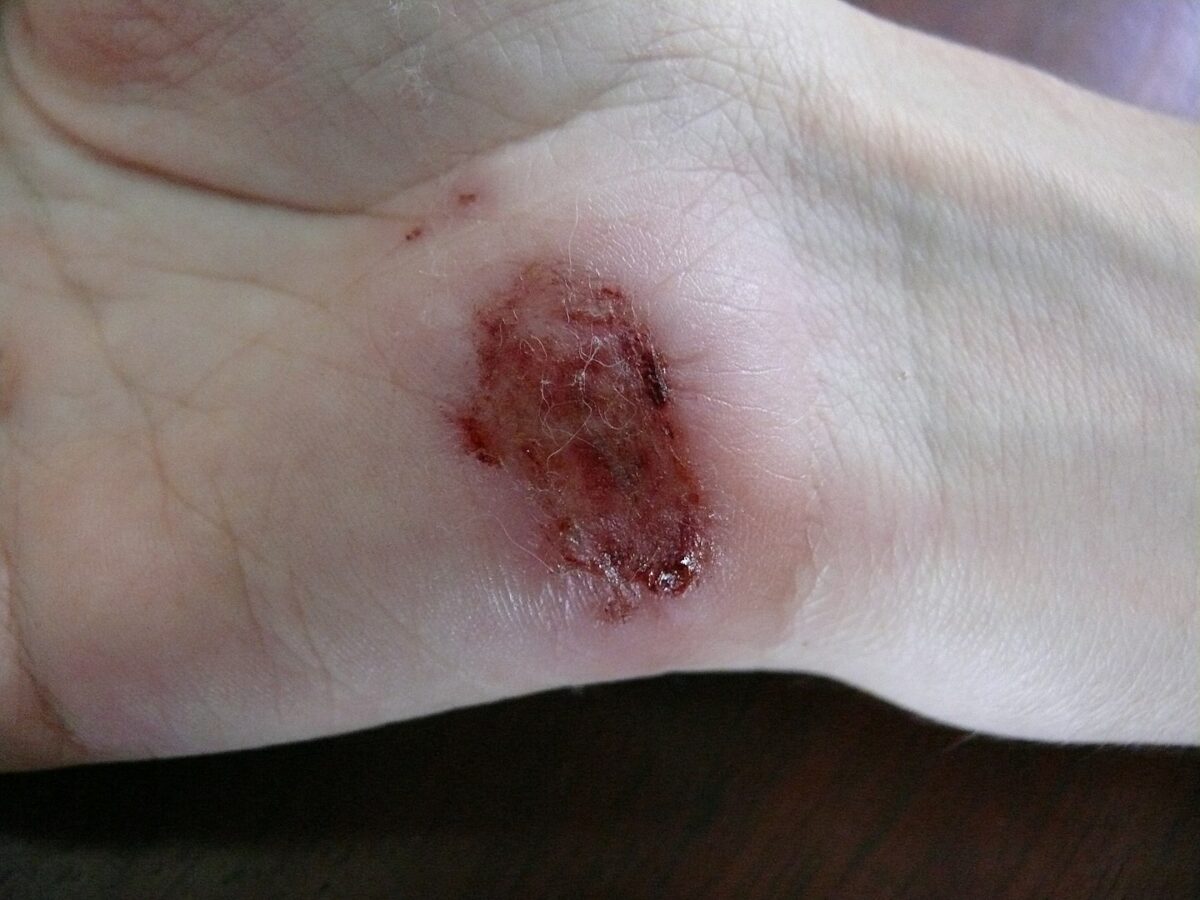
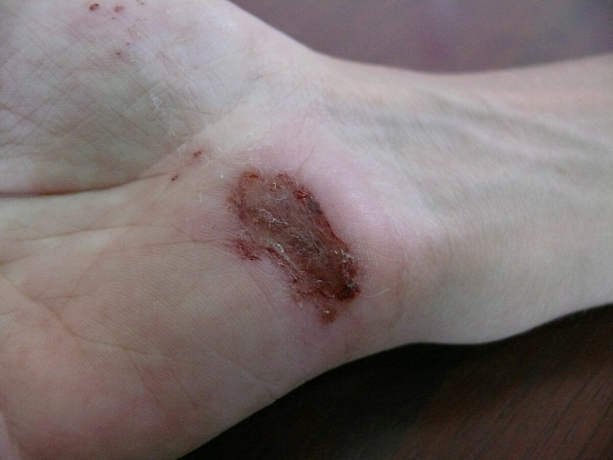
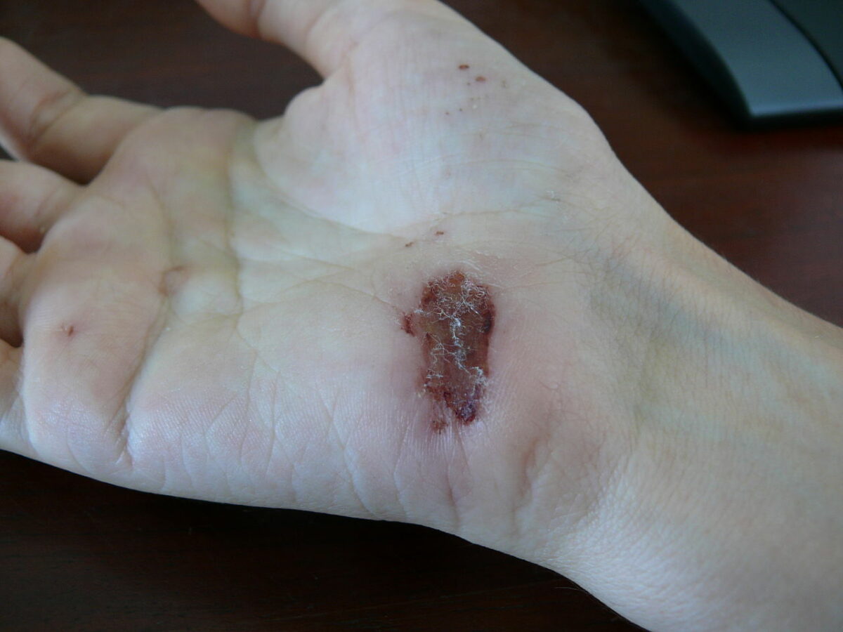
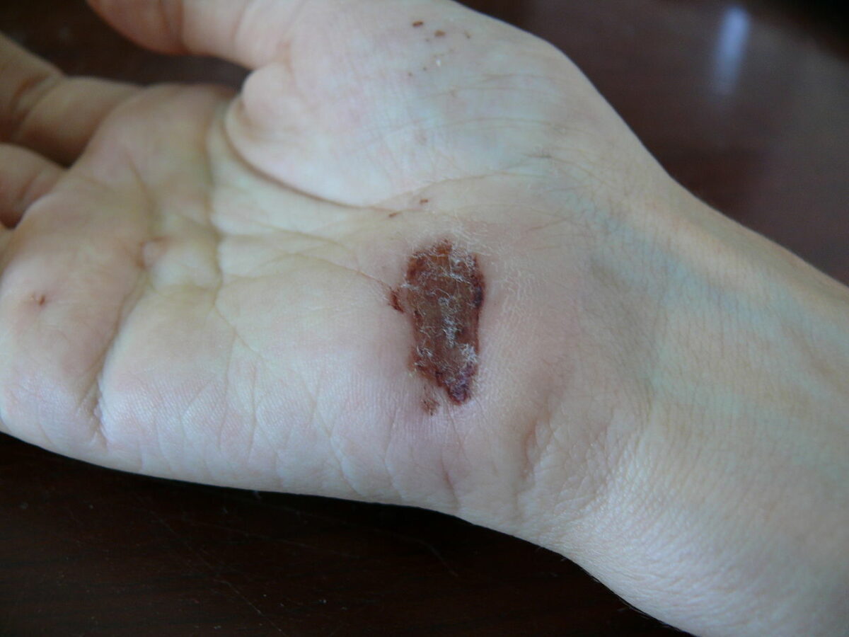
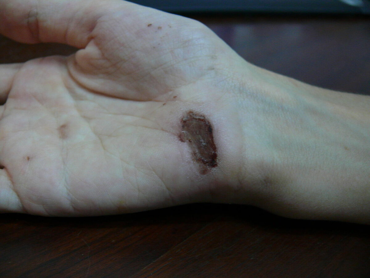
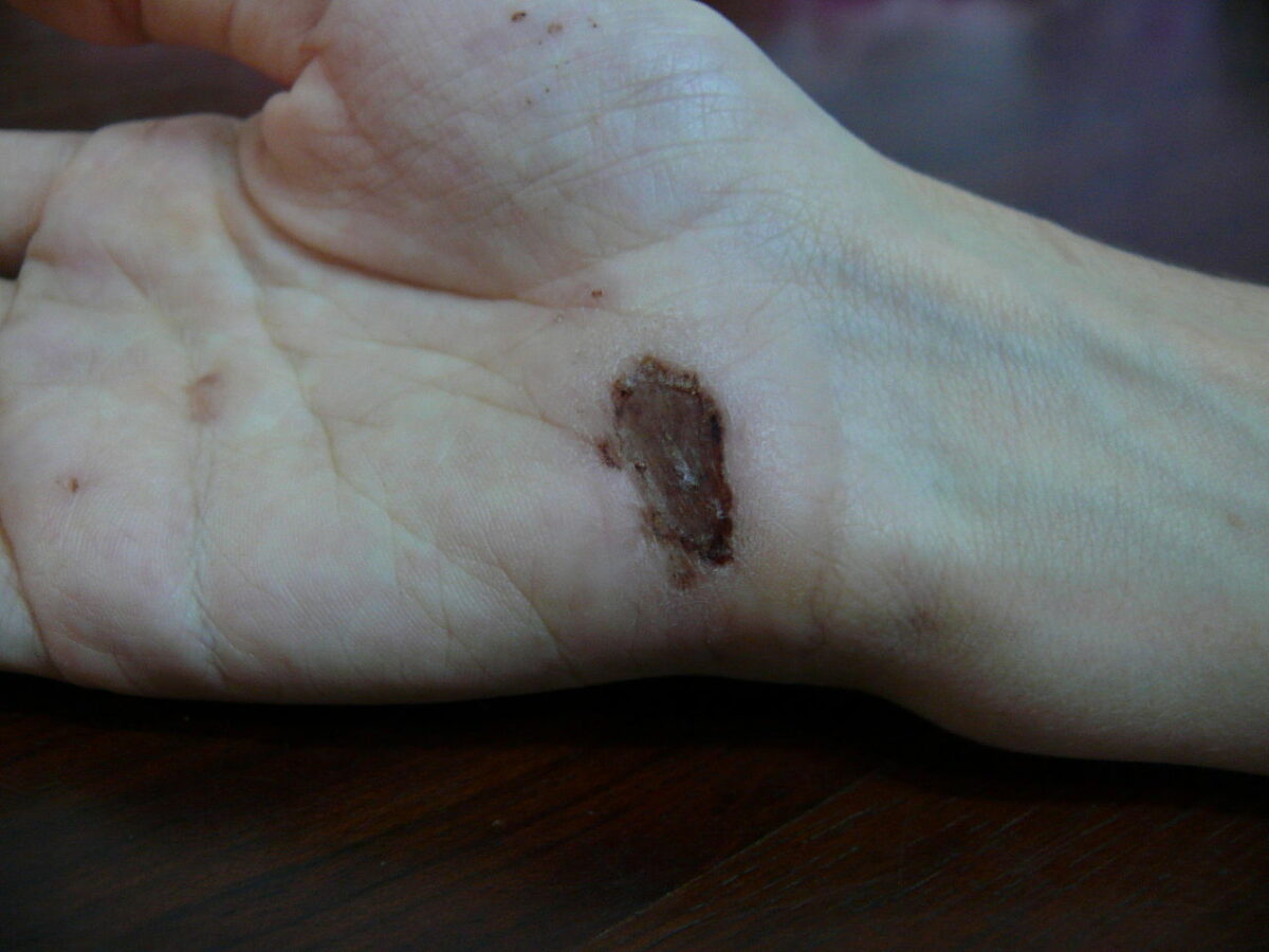
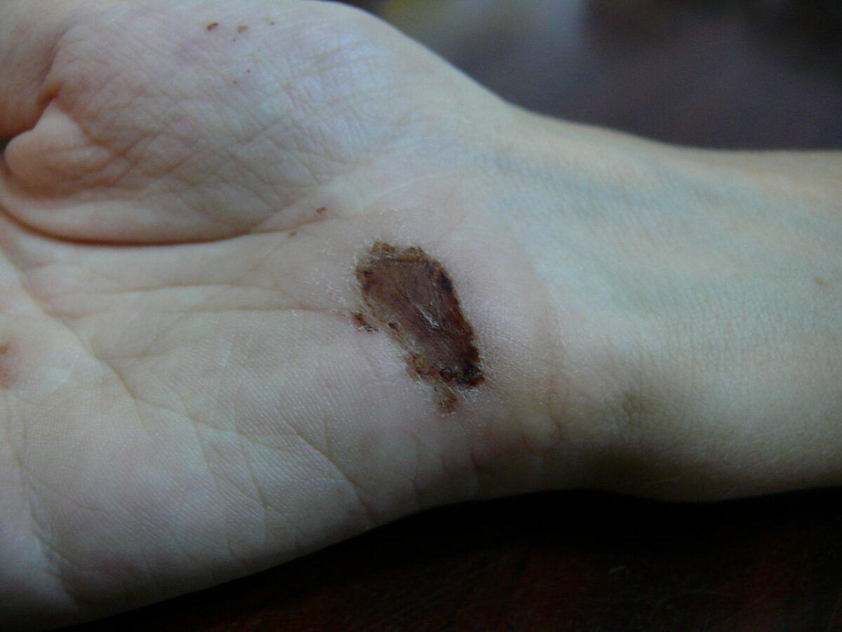
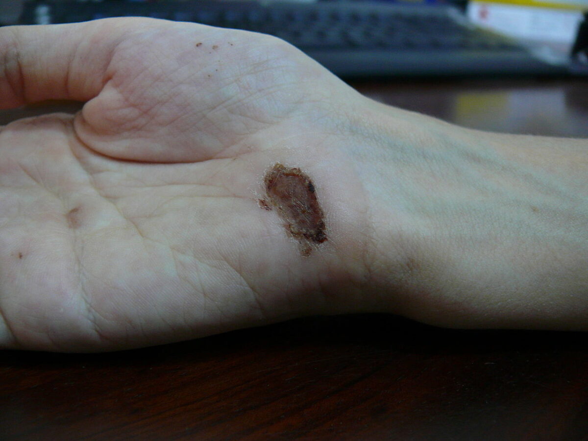
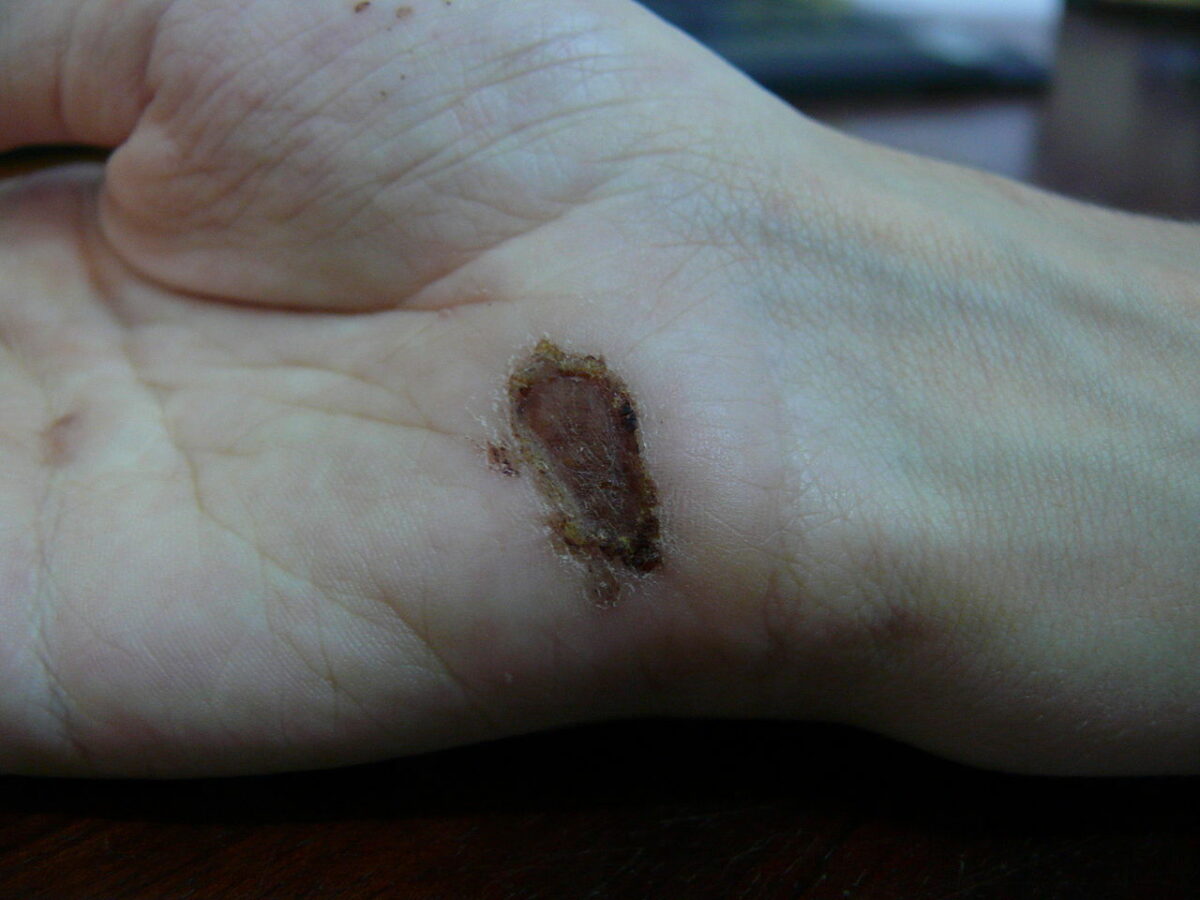
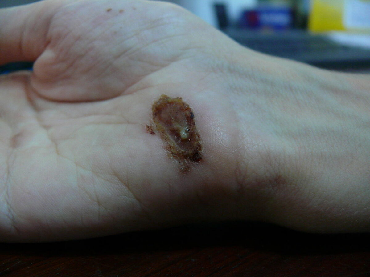
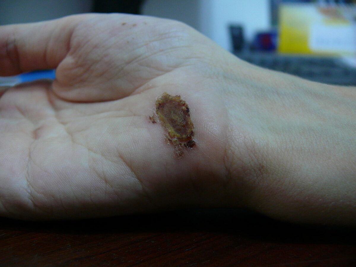
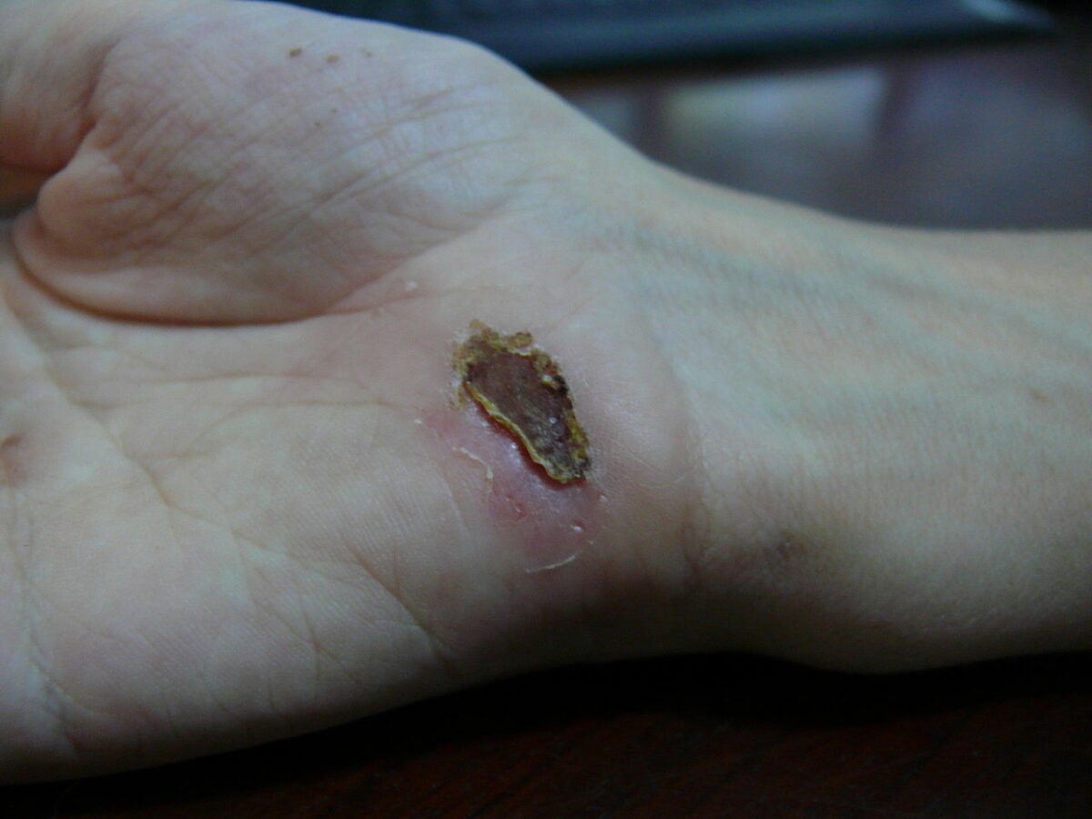
Wound healing is a physiological process involving tissue repair in response to injury. It involves a complex interaction of various cell types, cytokines Cytokines Non-antibody proteins secreted by inflammatory leukocytes and some non-leukocytic cells, that act as intercellular mediators. They differ from classical hormones in that they are produced by a number of tissue or cell types rather than by specialized glands. They generally act locally in a paracrine or autocrine rather than endocrine manner. Adaptive Immune Response, and inflammatory mediators. The overall process protects underlying structures from further damage. Wound healing stages include hemostasis Hemostasis Hemostasis refers to the innate, stepwise body processes that occur following vessel injury, resulting in clot formation and cessation of bleeding. Hemostasis occurs in 2 phases, namely, primary and secondary. Primary hemostasis involves forming a plug that stops the bleeding temporarily. Secondary hemostasis involves the activation of the coagulation cascade. Hemostasis, inflammation Inflammation Inflammation is a complex set of responses to infection and injury involving leukocytes as the principal cellular mediators in the body's defense against pathogenic organisms. Inflammation is also seen as a response to tissue injury in the process of wound healing. The 5 cardinal signs of inflammation are pain, heat, redness, swelling, and loss of function. Inflammation, granulation, and remodeling. Certain conditions such as diabetes Diabetes Diabetes mellitus (DM) is a metabolic disease characterized by hyperglycemia and dysfunction of the regulation of glucose metabolism by insulin. Type 1 DM is diagnosed mostly in children and young adults as the result of autoimmune destruction of β cells in the pancreas and the resulting lack of insulin. Type 2 DM has a significant association with obesity and is characterized by insulin resistance. Diabetes Mellitus, peripheral vascular disease, and malnutrition Malnutrition Malnutrition is a clinical state caused by an imbalance or deficiency of calories and/or micronutrients and macronutrients. The 2 main manifestations of acute severe malnutrition are marasmus (total caloric insufficiency) and kwashiorkor (protein malnutrition with characteristic edema). Malnutrition in children in resource-limited countries can limit Limit A value (e.g., pressure or time) that should not be exceeded and which is specified by the operator to protect the lung Invasive Mechanical Ventilation the body’s ability to heal itself, thus resulting in complications.

Last updated: May 17, 2024

Editorial responsibility: Stanley Oiseth, Lindsay Jones, Evelin Maza

Advertisement

Advertisement

Advertisement

Advertisement

Advertisement

Advertisement

Stages of Wound Repair

There are 3 main stages of wound healing:

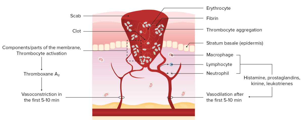
  1. Exudative stage (day 1–4)
    • Hemostasis Hemostasis Hemostasis refers to the innate, stepwise body processes that occur following vessel injury, resulting in clot formation and cessation of bleeding. Hemostasis occurs in 2 phases, namely, primary and secondary. Primary hemostasis involves forming a plug that stops the bleeding temporarily. Secondary hemostasis involves the activation of the coagulation cascade. Hemostasis: limits blood loss from the wound via vasoconstriction Vasoconstriction The physiological narrowing of blood vessels by contraction of the vascular smooth muscle. Vascular Resistance, Flow, and Mean Arterial Pressure. There is a release of inflammatory mediators (growth factors, chemokines Chemokines Class of pro-inflammatory cytokines that have the ability to attract and activate leukocytes. They can be divided into at least three structural branches: c; cc; and cxc; according to variations in a shared cysteine motif. Adaptive Cell-mediated Immunity, and cytokines Cytokines Non-antibody proteins secreted by inflammatory leukocytes and some non-leukocytic cells, that act as intercellular mediators. They differ from classical hormones in that they are produced by a number of tissue or cell types rather than by specialized glands. They generally act locally in a paracrine or autocrine rather than endocrine manner. Adaptive Immune Response) via the activation of platelets Platelets Platelets are small cell fragments involved in hemostasis. Thrombopoiesis takes place primarily in the bone marrow through a series of cell differentiation and is influenced by several cytokines. Platelets are formed after fragmentation of the megakaryocyte cytoplasm. Platelets: Histology
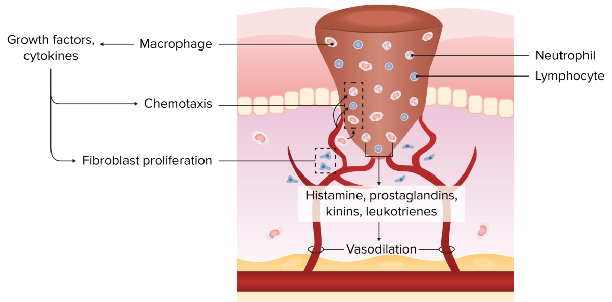
    • Inflammation Inflammation Inflammation is a complex set of responses to infection and injury involving leukocytes as the principal cellular mediators in the body’s defense against pathogenic organisms. Inflammation is also seen as a response to tissue injury in the process of wound healing. The 5 cardinal signs of inflammation are pain, heat, redness, swelling, and loss of function. Inflammation: clears cellular debris through phagocytosis Phagocytosis The engulfing and degradation of microorganisms; other cells that are dead, dying, or pathogenic; and foreign particles by phagocytic cells (phagocytes). Innate Immunity: Phagocytes and Antigen Presentation. There is a migration of inflammatory cells that facilitate tissue regeneration (via growth factors).
  2. Proliferative stage (day 2–16)
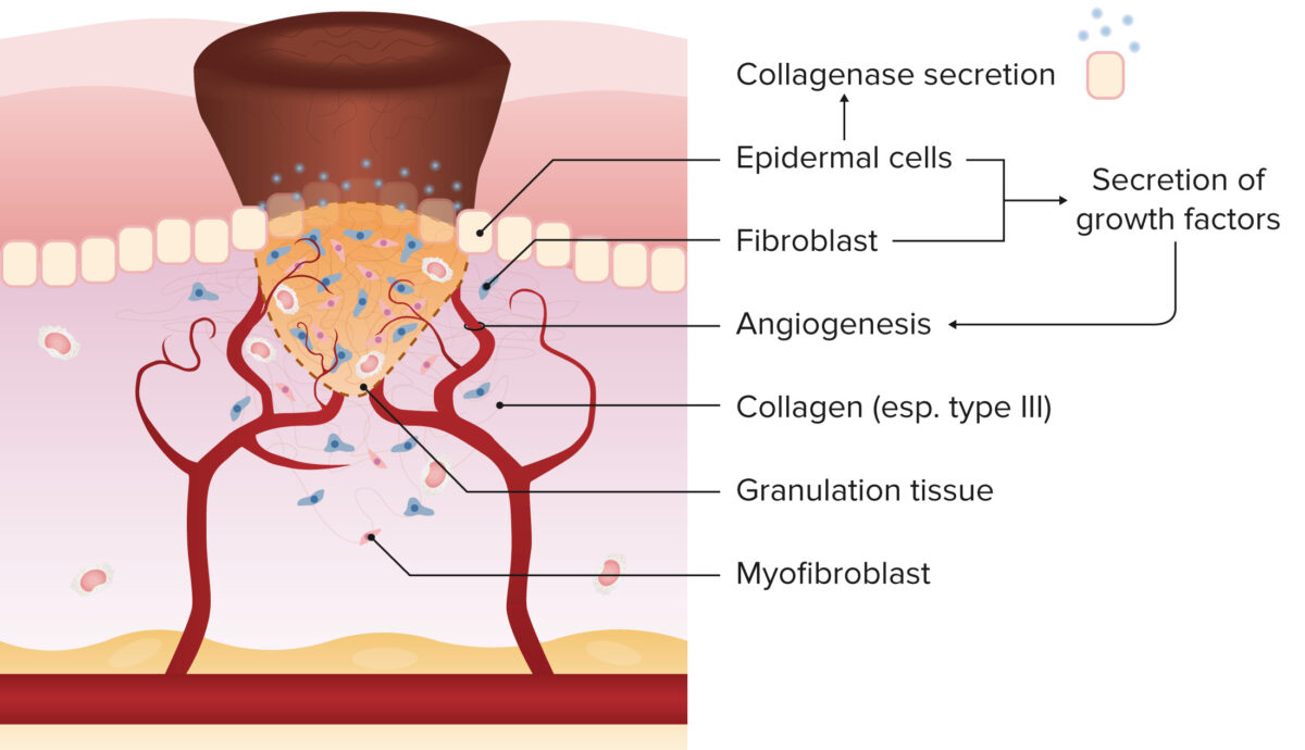
    • Granulation: lays the framework for new connective tissue Connective tissue Connective tissues originate from embryonic mesenchyme and are present throughout the body except inside the brain and spinal cord. The main function of connective tissues is to provide structural support to organs. Connective tissues consist of cells and an extracellular matrix. Connective Tissue: Histology and vasculature
      • Fibroblast growth factor Fibroblast growth factor A family of small polypeptide growth factors that share several common features including a strong affinity for heparin, and a central barrel-shaped core region of 140 amino acids that is highly homologous between family members. Although originally studied as proteins that stimulate the growth of fibroblasts this distinction is no longer a requirement for membership in the fibroblast growth factor family. X-linked Hypophosphatemic Rickets and vascular endothelial growth factor promote cellular proliferation and angiogenesis Angiogenesis Bartonella, respectively.
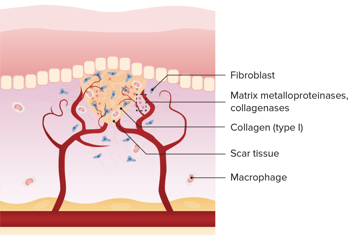
  3. Remodeling stage (day 5–25): modifies the wound contents to increase its tensile strength
    • May last years after an injury
    • Apoptosis Apoptosis A regulated cell death mechanism characterized by distinctive morphologic changes in the nucleus and cytoplasm, including the endonucleolytic cleavage of genomic DNA, at regularly spaced, internucleosomal sites, I.e., DNA fragmentation. It is genetically-programmed and serves as a balance to mitosis in regulating the size of animal tissues and in mediating pathologic processes associated with tumor growth. Ischemic Cell Damage of excess cells 
    • Degradation and alignment of collagen Collagen A polypeptide substance comprising about one third of the total protein in mammalian organisms. It is the main constituent of skin; connective tissue; and the organic substance of bones (bone and bones) and teeth (tooth). Connective Tissue: Histology fibers
Wound healing timeline

Timeline of wound healing

Image: “Timeline of Wound Healing” by Wikimedia Commons. License: Public Domain Files
Timeline and steps of wound healing
Day 1
Day 2
  • Squamous cells seal off the wound
  • Macrophages Macrophages The relatively long-lived phagocytic cell of mammalian tissues that are derived from blood monocytes. Main types are peritoneal macrophages; alveolar macrophages; histiocytes; kupffer cells of the liver; and osteoclasts. They may further differentiate within chronic inflammatory lesions to epithelioid cells or may fuse to form foreign body giant cells or langhans giant cells. Innate Immunity: Phagocytes and Antigen Presentation migrate into the wound
Day 3
  • Granulation tissue begins to form
  • Initial deposition of type III collagen Collagen A polypeptide substance comprising about one third of the total protein in mammalian organisms. It is the main constituent of skin; connective tissue; and the organic substance of bones (bone and bones) and teeth (tooth). Connective Tissue: Histology
  • Macrophages Macrophages The relatively long-lived phagocytic cell of mammalian tissues that are derived from blood monocytes. Main types are peritoneal macrophages; alveolar macrophages; histiocytes; kupffer cells of the liver; and osteoclasts. They may further differentiate within chronic inflammatory lesions to epithelioid cells or may fuse to form foreign body giant cells or langhans giant cells. Innate Immunity: Phagocytes and Antigen Presentation replace neutrophils Neutrophils Granular leukocytes having a nucleus with three to five lobes connected by slender threads of chromatin, and cytoplasm containing fine inconspicuous granules and stainable by neutral dyes. Innate Immunity: Phagocytes and Antigen Presentation
Days 4-6
  • Peak granulation tissue formation
  • Fibronectin Fibronectin Glycoproteins found on the surfaces of cells, particularly in fibrillar structures. The proteins are lost or reduced when these cells undergo viral or chemical transformation. They are highly susceptible to proteolysis and are substrates for activated blood coagulation factor VIII. The forms present in plasma are called cold-insoluble globulins. Connective Tissue: Histology (key glycoprotein; used by cancer cells to metastasize)
Week 2
  • Tensile strength ~10%
Month 1
  • Remodeling of the wound (collagenase/lysyl oxidase Oxidase Neisseria)
  • Tensile strength approximately 80% in 3 months (never reaches 100% tensile strength after injury)

Types of Wound Healing

Regeneration vs. reparation

  • Regeneration ( epithelialization Epithelialization Anal Fistula) is the process of returning the site of injury to its original state.
    • This is seen in re-epithelialization after having minor lacerations.
  • Reparation (or tissue repair, a form of wound healing) is the process of generating a scar Scar Dermatologic Examination or less functional tissue with a different form and/or composition of the original tissue.
    • Does not restore complete functionality

Three types of reparation

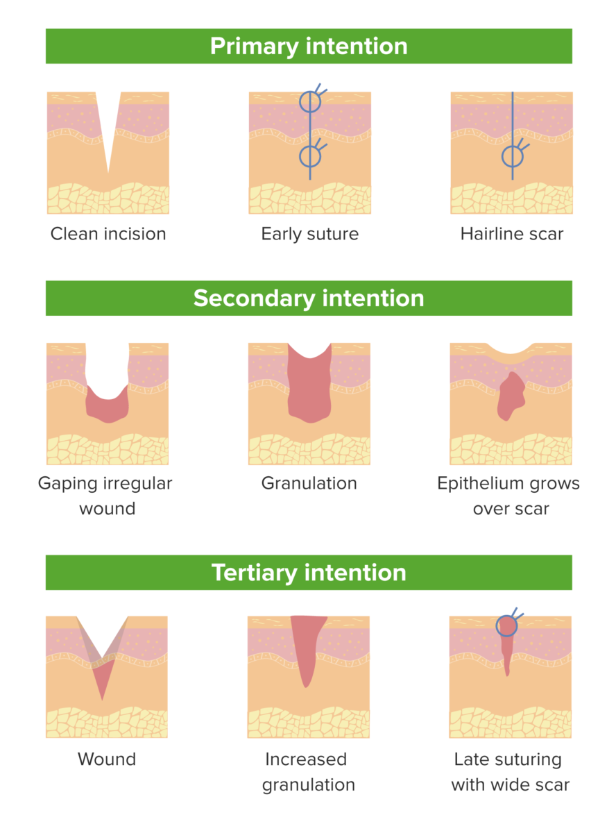
  1. Primary intention: when the tissue surface edges have been approximated
    • Small defect with little risk of complications and/or infection
  2. Secondary intention: when there are significant tissue losses and the wound surface cannot be brought together (e.g., lacerations, burns Burns A burn is a type of injury to the skin and deeper tissues caused by exposure to heat, electricity, chemicals, friction, or radiation. Burns are classified according to their depth as superficial (1st-degree), partial-thickness (2nd-degree), full-thickness (3rd-degree), and 4th-degree burns. Burns, and ulcers)
    • Granulation tissue (consisting of connective tissue Connective tissue Connective tissues originate from embryonic mesenchyme and are present throughout the body except inside the brain and spinal cord. The main function of connective tissues is to provide structural support to organs. Connective tissues consist of cells and an extracellular matrix. Connective Tissue: Histology cells and ingrowing young blood vessels) is needed to close the defect → scar Scar Dermatologic Examination formation occurs with a higher risk of infection 
  3. Tertiary or delayed primary intention: when there is a need to delay the closure of a wound (due to contamination risk, poor circulation Circulation The movement of the blood as it is pumped through the cardiovascular system. ABCDE Assessment, etc ETC The electron transport chain (ETC) sends electrons through a series of proteins, which generate an electrochemical proton gradient that produces energy in the form of adenosine triphosphate (ATP). Electron Transport Chain (ETC).)
Wound healing

Processes of primary, secondary, and tertiary wound healing

Image by Lecturio.
Complications and Impaired Wound Healing

Wound healing complications

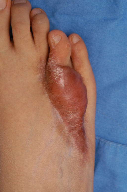
  • Hypertrophic scars
    • Confined to wound borders
    • Common in young individuals and particularly prevalent in dark-skinned individuals
    • Common after burns Burns A burn is a type of injury to the skin and deeper tissues caused by exposure to heat, electricity, chemicals, friction, or radiation. Burns are classified according to their depth as superficial (1st-degree), partial-thickness (2nd-degree), full-thickness (3rd-degree), and 4th-degree burns. Burns
    • Occur when there is excess immature collagen Collagen A polypeptide substance comprising about one third of the total protein in mammalian organisms. It is the main constituent of skin; connective tissue; and the organic substance of bones (bone and bones) and teeth (tooth). Connective Tissue: Histology production during the reparative phase of wound healing
    • Often do not require treatment and resolve spontaneously
    • Increased proportion of type III collagen Collagen A polypeptide substance comprising about one third of the total protein in mammalian organisms. It is the main constituent of skin; connective tissue; and the organic substance of bones (bone and bones) and teeth (tooth). Connective Tissue: Histology compared with type I
  • Keloids
    • Extends beyond wound borders
    • More common in dark-skinned individuals
    • Seen in patients Patients Individuals participating in the health care system for the purpose of receiving therapeutic, diagnostic, or preventive procedures. Clinician–Patient Relationship who participate in contact sports (e.g., boxing, football) and in those with piercings
    • Occur when excessive collagen Collagen A polypeptide substance comprising about one third of the total protein in mammalian organisms. It is the main constituent of skin; connective tissue; and the organic substance of bones (bone and bones) and teeth (tooth). Connective Tissue: Histology production occurs during the reparative phase of wound healing
    • Especially prevalent on the ear lobe and sternum Sternum A long, narrow, and flat bone commonly known as breastbone occurring in the midsection of the anterior thoracic segment or chest region, which stabilizes the rib cage and serves as the point of origin for several muscles that move the arms, head, and neck. Chest Wall: Anatomy
    • Remain years after injury and may need intervention (excision, steroid injections, radiotherapy, etc ETC The electron transport chain (ETC) sends electrons through a series of proteins, which generate an electrochemical proton gradient that produces energy in the form of adenosine triphosphate (ATP). Electron Transport Chain (ETC).)
  • Hyper/ hypopigmentation Hypopigmentation A condition caused by a deficiency or a loss of melanin pigmentation in the epidermis, also known as hypomelanosis. Hypopigmentation can be localized or generalized, and may result from genetic defects, trauma, inflammation, or infections. Malassezia Fungi
    • Postinflammatory hyperpigmentation Postinflammatory Hyperpigmentation Acne Vulgaris
      • Temporary pigmentation following injury or inflammation Inflammation Inflammation is a complex set of responses to infection and injury involving leukocytes as the principal cellular mediators in the body’s defense against pathogenic organisms. Inflammation is also seen as a response to tissue injury in the process of wound healing. The 5 cardinal signs of inflammation are pain, heat, redness, swelling, and loss of function. Inflammation of the skin Skin The skin, also referred to as the integumentary system, is the largest organ of the body. The skin is primarily composed of the epidermis (outer layer) and dermis (deep layer). The epidermis is primarily composed of keratinocytes that undergo rapid turnover, while the dermis contains dense layers of connective tissue. Skin: Structure and Functions
      • More common in dark-skinned individuals
      • More severe injuries result in postinflammatory hypopigmentation Hypopigmentation A condition caused by a deficiency or a loss of melanin pigmentation in the epidermis, also known as hypomelanosis. Hypopigmentation can be localized or generalized, and may result from genetic defects, trauma, inflammation, or infections. Malassezia Fungi that is usually permanent.
      • Pathogenesis: Inflammation Inflammation Inflammation is a complex set of responses to infection and injury involving leukocytes as the principal cellular mediators in the body’s defense against pathogenic organisms. Inflammation is also seen as a response to tissue injury in the process of wound healing. The 5 cardinal signs of inflammation are pain, heat, redness, swelling, and loss of function. Inflammation and injury stimulate melanocytes Melanocytes Mammalian pigment cells that produce melanins, pigments found mainly in the epidermis, but also in the eyes and the hair, by a process called melanogenesis. Coloration can be altered by the number of melanocytes or the amount of pigment produced and stored in the organelles called melanosomes. The large non-mammalian melanin-containing cells are called melanophores. Skin: Structure and Functions to increase melanin Melanin Insoluble polymers of tyrosine derivatives found in and causing darkness in skin (skin pigmentation), hair, and feathers providing protection against sunburn induced by sunlight. Carotenes contribute yellow and red coloration. Seborrheic Keratosis synthesis Synthesis Polymerase Chain Reaction (PCR) and transfer pigment to keratinocytes Keratinocytes Epidermal cells which synthesize keratin and undergo characteristic changes as they move upward from the basal layers of the epidermis to the cornified (horny) layer of the skin. Successive stages of differentiation of the keratinocytes forming the epidermal layers are basal cell, spinous or prickle cell, and the granular cell. Skin: Structure and Functions.
      • Clinical features:
        • Located at site of original disease after healed
        • May become darker if exposed to sunlight
    • Hyperpigmentation Hyperpigmentation Excessive pigmentation of the skin, usually as a result of increased epidermal or dermal melanin pigmentation, hypermelanosis. Hyperpigmentation can be localized or generalized. The condition may arise from exposure to light, chemicals or other substances, or from a primary metabolic imbalance. Malassezia Fungi
      • Can be seen with skin Skin The skin, also referred to as the integumentary system, is the largest organ of the body. The skin is primarily composed of the epidermis (outer layer) and dermis (deep layer). The epidermis is primarily composed of keratinocytes that undergo rapid turnover, while the dermis contains dense layers of connective tissue. Skin: Structure and Functions grafts after burns Burns A burn is a type of injury to the skin and deeper tissues caused by exposure to heat, electricity, chemicals, friction, or radiation. Burns are classified according to their depth as superficial (1st-degree), partial-thickness (2nd-degree), full-thickness (3rd-degree), and 4th-degree burns. Burns
      • Pigmentation disorders are common among dark-skinned burn patients Patients Individuals participating in the health care system for the purpose of receiving therapeutic, diagnostic, or preventive procedures. Clinician–Patient Relationship, in particular Africans and Asians.
        • Characterized by hyperpigmentation Hyperpigmentation Excessive pigmentation of the skin, usually as a result of increased epidermal or dermal melanin pigmentation, hypermelanosis. Hyperpigmentation can be localized or generalized. The condition may arise from exposure to light, chemicals or other substances, or from a primary metabolic imbalance. Malassezia Fungi in darker-skinned individuals and hypopigmentation Hypopigmentation A condition caused by a deficiency or a loss of melanin pigmentation in the epidermis, also known as hypomelanosis. Hypopigmentation can be localized or generalized, and may result from genetic defects, trauma, inflammation, or infections. Malassezia Fungi in lighter-skinned individuals
        • This is often due to genetic as well as environmental factors.
    • Hypopigmentation Hypopigmentation A condition caused by a deficiency or a loss of melanin pigmentation in the epidermis, also known as hypomelanosis. Hypopigmentation can be localized or generalized, and may result from genetic defects, trauma, inflammation, or infections. Malassezia Fungi
      • Common with deep wounds when melanocytes Melanocytes Mammalian pigment cells that produce melanins, pigments found mainly in the epidermis, but also in the eyes and the hair, by a process called melanogenesis. Coloration can be altered by the number of melanocytes or the amount of pigment produced and stored in the organelles called melanosomes. The large non-mammalian melanin-containing cells are called melanophores. Skin: Structure and Functions are the only remaining cell in the wound
      • Melanocytes Melanocytes Mammalian pigment cells that produce melanins, pigments found mainly in the epidermis, but also in the eyes and the hair, by a process called melanogenesis. Coloration can be altered by the number of melanocytes or the amount of pigment produced and stored in the organelles called melanosomes. The large non-mammalian melanin-containing cells are called melanophores. Skin: Structure and Functions act as stem cells to heal the wound (being converted into epithelial cells) and therefore cannot act in their role of providing pigmentation to the skin Skin The skin, also referred to as the integumentary system, is the largest organ of the body. The skin is primarily composed of the epidermis (outer layer) and dermis (deep layer). The epidermis is primarily composed of keratinocytes that undergo rapid turnover, while the dermis contains dense layers of connective tissue. Skin: Structure and Functions.
  • Incisional hernias Incisional hernias Protrusion of tissue at or near the site of an incision from a previous surgery. Abdominal Hernias: increased risk in obese patients Patients Individuals participating in the health care system for the purpose of receiving therapeutic, diagnostic, or preventive procedures. Clinician–Patient Relationship and patients Patients Individuals participating in the health care system for the purpose of receiving therapeutic, diagnostic, or preventive procedures. Clinician–Patient Relationship with chronic obstructive pulmonary disease Pulmonary disease Diseases involving the respiratory system. Blastomyces/Blastomycosis
  • Wound dehiscence: increased risk in patients Patients Individuals participating in the health care system for the purpose of receiving therapeutic, diagnostic, or preventive procedures. Clinician–Patient Relationship taking corticosteroids Corticosteroids Chorioretinitis or other medications that inhibit phases of wound healing
  • Contractures: in burn wounds and wounds across joints

Patient-related factors negatively affecting wound healing

  • Infection: Bacteria Bacteria Bacteria are prokaryotic single-celled microorganisms that are metabolically active and divide by binary fission. Some of these organisms play a significant role in the pathogenesis of diseases. Bacteriology degrade the building blocks of wound healing,
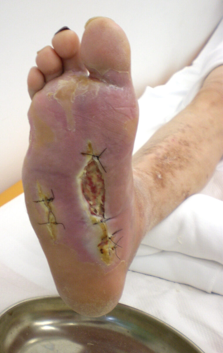
  • Diabetes Diabetes Diabetes mellitus (DM) is a metabolic disease characterized by hyperglycemia and dysfunction of the regulation of glucose metabolism by insulin. Type 1 DM is diagnosed mostly in children and young adults as the result of autoimmune destruction of β cells in the pancreas and the resulting lack of insulin. Type 2 DM has a significant association with obesity and is characterized by insulin resistance. Diabetes Mellitus: Decreased blood flow Blood flow Blood flow refers to the movement of a certain volume of blood through the vasculature over a given unit of time (e.g., mL per minute). Vascular Resistance, Flow, and Mean Arterial Pressure and excess of serum glucose Glucose A primary source of energy for living organisms. It is naturally occurring and is found in fruits and other parts of plants in its free state. It is used therapeutically in fluid and nutrient replacement. Lactose Intolerance prevents the migration of inflammatory cells into the wound.
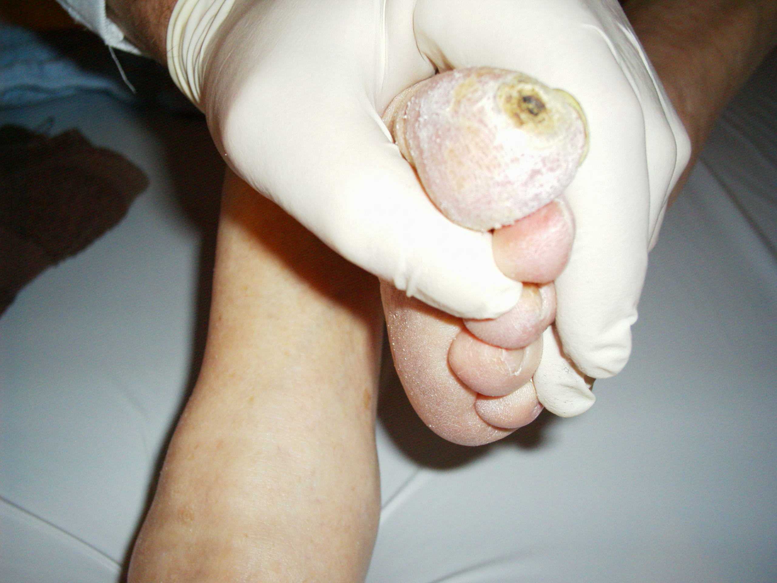
    • Diabetic foot Foot The foot is the terminal portion of the lower limb, whose primary function is to bear weight and facilitate locomotion. The foot comprises 26 bones, including the tarsal bones, metatarsal bones, and phalanges. The bones of the foot form longitudinal and transverse arches and are supported by various muscles, ligaments, and tendons. Foot: Anatomy syndrome is often a primary manifestation of problems with wound healing in diabetics: ulceration Ulceration Corneal Abrasions, Erosion, and Ulcers of the foot Foot The foot is the terminal portion of the lower limb, whose primary function is to bear weight and facilitate locomotion. The foot comprises 26 bones, including the tarsal bones, metatarsal bones, and phalanges. The bones of the foot form longitudinal and transverse arches and are supported by various muscles, ligaments, and tendons. Foot: Anatomy (distally from the ankle and including the ankle) is associated with neuropathy Neuropathy Leprosy and different degrees of ischemia Ischemia A hypoperfusion of the blood through an organ or tissue caused by a pathologic constriction or obstruction of its blood vessels, or an absence of blood circulation. Ischemic Cell Damage and infection.
  • Malnutrition Malnutrition Malnutrition is a clinical state caused by an imbalance or deficiency of calories and/or micronutrients and macronutrients. The 2 main manifestations of acute severe malnutrition are marasmus (total caloric insufficiency) and kwashiorkor (protein malnutrition with characteristic edema). Malnutrition in children in resource-limited countries: Insufficient protein leads to a relative immunodeficient state.
  • Vitamin C Vitamin C A six carbon compound related to glucose. It is found naturally in citrus fruits and many vegetables. Ascorbic acid is an essential nutrient in human diets, and necessary to maintain connective tissue and bone. Its biologically active form, vitamin C, functions as a reducing agent and coenzyme in several metabolic pathways. Vitamin C is considered an antioxidant. Water-soluble Vitamins and their Deficiencies (cofactor for hydroxylases) deficiency → impedes collagen Collagen A polypeptide substance comprising about one third of the total protein in mammalian organisms. It is the main constituent of skin; connective tissue; and the organic substance of bones (bone and bones) and teeth (tooth). Connective Tissue: Histology cross-linking
  • Zinc Zinc A metallic element of atomic number 30 and atomic weight 65. 38. It is a necessary trace element in the diet, forming an essential part of many enzymes, and playing an important role in protein synthesis and in cell division. Zinc deficiency is associated with anemia, short stature, hypogonadism, impaired wound healing, and geophagia. It is known by the symbol zn. Trace Elements (cofactor for collagenase) and copper Copper A heavy metal trace element with the atomic symbol cu, atomic number 29, and atomic weight 63. 55. Trace Elements (cofactor for lysyl oxidase Oxidase Neisseria) deficiencies: impede remodeling and maturation of collagen Collagen A polypeptide substance comprising about one third of the total protein in mammalian organisms. It is the main constituent of skin; connective tissue; and the organic substance of bones (bone and bones) and teeth (tooth). Connective Tissue: Histology
  • Use of corticosteroids Corticosteroids Chorioretinitis/ glucocorticoids Glucocorticoids Glucocorticoids are a class within the corticosteroid family. Glucocorticoids are chemically and functionally similar to endogenous cortisol. There are a wide array of indications, which primarily benefit from the antiinflammatory and immunosuppressive effects of this class of drugs. Glucocorticoids: inhibits the inflammatory phase of wound healing
  • Genetic factors: connective tissue Connective tissue Connective tissues originate from embryonic mesenchyme and are present throughout the body except inside the brain and spinal cord. The main function of connective tissues is to provide structural support to organs. Connective tissues consist of cells and an extracellular matrix. Connective Tissue: Histology disorders ( Ehlers-Danlos syndrome Ehlers-Danlos syndrome Ehlers-Danlos syndrome (EDS) is a heterogeneous group of inherited connective tissue disorders that are characterized by hyperextensible skin, hypermobile joints, and fragility of the skin and connective tissue. Ehlers-Danlos Syndrome: various mutations in collagen Collagen A polypeptide substance comprising about one third of the total protein in mammalian organisms. It is the main constituent of skin; connective tissue; and the organic substance of bones (bone and bones) and teeth (tooth). Connective Tissue: Histology and collagen-binding proteins Proteins Linear polypeptides that are synthesized on ribosomes and may be further modified, crosslinked, cleaved, or assembled into complex proteins with several subunits. The specific sequence of amino acids determines the shape the polypeptide will take, during protein folding, and the function of the protein. Energy Homeostasis, most common is type III collagen Collagen A polypeptide substance comprising about one third of the total protein in mammalian organisms. It is the main constituent of skin; connective tissue; and the organic substance of bones (bone and bones) and teeth (tooth). Connective Tissue: Histology; Marfan syndrome Marfan syndrome Marfan syndrome is a genetic condition with autosomal dominant inheritance. Marfan syndrome affects the elasticity of connective tissues throughout the body, most notably in the cardiovascular, ocular, and musculoskeletal systems. Marfan Syndrome: fibrillin-1 Fibrillin-1 A fibrillin (fbn1) that functions as a structural support protein for microfibrils. It also regulates the maturation of osteoblasts by controlling the availability and concentration of tgf-beta and bone morphogenetic proteins. Mutations in the fbn1 gene are associated with marfan syndrome. Marfan Syndrome gene mutation Gene Mutation Myotonic Dystrophies)
  • Color of skin Skin The skin, also referred to as the integumentary system, is the largest organ of the body. The skin is primarily composed of the epidermis (outer layer) and dermis (deep layer). The epidermis is primarily composed of keratinocytes that undergo rapid turnover, while the dermis contains dense layers of connective tissue. Skin: Structure and Functions: Darker-skinned individuals tend to be more prone to keloids.
    • Post-injury pigmentation disorders can occur, leading to either hyper- or hypopigmentation Hypopigmentation A condition caused by a deficiency or a loss of melanin pigmentation in the epidermis, also known as hypomelanosis. Hypopigmentation can be localized or generalized, and may result from genetic defects, trauma, inflammation, or infections. Malassezia Fungi.

Clinical Relevance

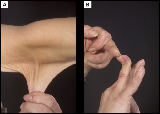
  • Ehlers-Danlos syndrome Ehlers-Danlos syndrome Ehlers-Danlos syndrome (EDS) is a heterogeneous group of inherited connective tissue disorders that are characterized by hyperextensible skin, hypermobile joints, and fragility of the skin and connective tissue. Ehlers-Danlos Syndrome is a deficiency of type III or V collagen Collagen A polypeptide substance comprising about one third of the total protein in mammalian organisms. It is the main constituent of skin; connective tissue; and the organic substance of bones (bone and bones) and teeth (tooth). Connective Tissue: Histology. Patients Patients Individuals participating in the health care system for the purpose of receiving therapeutic, diagnostic, or preventive procedures. Clinician–Patient Relationship with this condition will often present with hyperextensible skin Skin The skin, also referred to as the integumentary system, is the largest organ of the body. The skin is primarily composed of the epidermis (outer layer) and dermis (deep layer). The epidermis is primarily composed of keratinocytes that undergo rapid turnover, while the dermis contains dense layers of connective tissue. Skin: Structure and Functions.
  • Osteogenesis imperfecta Osteogenesis imperfecta Osteogenesis imperfecta (OI), or “brittle bone disease,” is a rare genetic connective tissue disorder characterized by severe bone fragility. Although OI is considered a single disease, OI includes over 16 genotypes and clinical phenotypes with differing symptom severity. Osteogenesis Imperfecta is a deficiency of type I collagen Type I Collagen Ehlers-Danlos Syndrome. Patients Patients Individuals participating in the health care system for the purpose of receiving therapeutic, diagnostic, or preventive procedures. Clinician–Patient Relationship with this condition often present with multiple fractures and blue sclera Sclera The white, opaque, fibrous, outer tunic of the eyeball, covering it entirely excepting the segment covered anteriorly by the cornea. It is essentially avascular but contains apertures for vessels, lymphatics, and nerves. Eye: Anatomy.

Create your free account or log in to continue reading!

Sign up now and get free access to Lecturio with concept pages, medical videos, and questions for your medical education.

User Reviews

It's Deal Week! 💸 Save 30% on all plans!

Details