Vascular Resistance, Flow, and Mean Arterial Pressure
Vascular Resistance, Flow, and Mean Arterial Pressure
Blood flows through the heart, arteriesArteriesArteries are tubular collections of cells that transport oxygenated blood and nutrients from the heart to the tissues of the body. The blood passes through the arteries in order of decreasing luminal diameter, starting in the largest artery (the aorta) and ending in the small arterioles. Arteries are classified into 3 types: large elastic arteries, medium muscular arteries, and small arteries and arterioles. Arteries: Histology, capillariesCapillariesCapillaries are the primary structures in the circulatory system that allow the exchange of gas, nutrients, and other materials between the blood and the extracellular fluid (ECF). Capillaries are the smallest of the blood vessels. Because a capillary diameter is so small, only 1 RBC may pass through at a time.Capillaries: Histology, and veinsVeinsVeins are tubular collections of cells, which transport deoxygenated blood and waste from the capillary beds back to the heart. Veins are classified into 3 types: small veins/venules, medium veins, and large veins. Each type contains 3 primary layers: tunica intima, tunica media, and tunica adventitia. Veins: Histology in a closed, continuous circuit. Flow is the movement of volume per unit of time. Flow is affected by the pressure gradient and the resistanceResistancePhysiologically, the opposition to flow of air caused by the forces of friction. As a part of pulmonary function testing, it is the ratio of driving pressure to the rate of air flow.Ventilation: Mechanics of Breathing fluid encounters between 2 points. Vascular resistanceResistancePhysiologically, the opposition to flow of air caused by the forces of friction. As a part of pulmonary function testing, it is the ratio of driving pressure to the rate of air flow.Ventilation: Mechanics of Breathing is the opposition to flow, which is caused primarily by blood friction against vessel walls. Vascular resistanceResistancePhysiologically, the opposition to flow of air caused by the forces of friction. As a part of pulmonary function testing, it is the ratio of driving pressure to the rate of air flow.Ventilation: Mechanics of Breathing is directly related to the diameter of the vessel (smaller vessels have higher resistanceResistancePhysiologically, the opposition to flow of air caused by the forces of friction. As a part of pulmonary function testing, it is the ratio of driving pressure to the rate of air flow.Ventilation: Mechanics of Breathing). Mean arterial pressure (MAP) is the average systemic arterial pressure and is directly related to cardiac outputCardiac outputThe volume of blood passing through the heart per unit of time. It is usually expressed as liters (volume) per minute so as not to be confused with stroke volume (volume per beat).Cardiac Mechanics (CO) and systemic vascular resistanceResistancePhysiologically, the opposition to flow of air caused by the forces of friction. As a part of pulmonary function testing, it is the ratio of driving pressure to the rate of air flow.Ventilation: Mechanics of Breathing (SVR). The SVR and MAP are affected by the vascular anatomy as well as a number of local and neurohumoral factors.
Blood flows through the heart, arteriesArteriesArteries are tubular collections of cells that transport oxygenated blood and nutrients from the heart to the tissues of the body. The blood passes through the arteries in order of decreasing luminal diameter, starting in the largest artery (the aorta) and ending in the small arterioles. Arteries are classified into 3 types: large elastic arteries, medium muscular arteries, and small arteries and arterioles. Arteries: Histology, capillariesCapillariesCapillaries are the primary structures in the circulatory system that allow the exchange of gas, nutrients, and other materials between the blood and the extracellular fluid (ECF). Capillaries are the smallest of the blood vessels. Because a capillary diameter is so small, only 1 RBC may pass through at a time.Capillaries: Histology, and veinsVeinsVeins are tubular collections of cells, which transport deoxygenated blood and waste from the capillary beds back to the heart. Veins are classified into 3 types: small veins/venules, medium veins, and large veins. Each type contains 3 primary layers: tunica intima, tunica media, and tunica adventitia. Veins: Histology in a closed, continuous circuit.
Blood flow refers to the movement of a certain volume of blood through the vasculature over a given unit of time (e.g., mL per minute).
Hemodynamics refer to the physical principles governing blood flow, which are:
The pressure gradient between 1 point and another
ResistanceResistancePhysiologically, the opposition to flow of air caused by the forces of friction. As a part of pulmonary function testing, it is the ratio of driving pressure to the rate of air flow.Ventilation: Mechanics of Breathing of the vessel
Note: This animation does not have sound.
Ohm’s law
Ohm’s law is an important basic formula in physics. A derivation of Ohm’s law can be used to calculate blood flow.
Ohm’s law:I = V / R:
Current (I) = flow of charged particles
Voltage (V) = the difference in concentration of charged particles at 2 different points
ResistanceResistancePhysiologically, the opposition to flow of air caused by the forces of friction. As a part of pulmonary function testing, it is the ratio of driving pressure to the rate of air flow.Ventilation: Mechanics of Breathing (R) = opposition to current flow
The current (flow of electrons) in a closed system is directly proportional to voltage and inversely proportional to resistanceResistancePhysiologically, the opposition to flow of air caused by the forces of friction. As a part of pulmonary function testing, it is the ratio of driving pressure to the rate of air flow.Ventilation: Mechanics of Breathing within the system.
$$I= \frac{V}{R}$$
Ohm’s law applied to the cardiovascular system: F = ΔP / R:
Flow (F) = blood flow through a vessel
Pressure gradient (ΔP) = change in pressure between 2 different points (i.e., ΔP = P1 ‒ P2)
ResistanceResistancePhysiologically, the opposition to flow of air caused by the forces of friction. As a part of pulmonary function testing, it is the ratio of driving pressure to the rate of air flow.Ventilation: Mechanics of Breathing (R) = opposition to blood flow
$$F= \frac{\Delta P}{R}$$
Flow
Flow: the volume of fluid passing a point per unit of time:
Caused by a ΔP between 2 points (there is no flow without a ΔP)
2 types of flow: laminar and turbulent
Laminar flow:
Smooth-walled vessels allow for smooth flow through the tube
Flow is fastest in the center of the vessel (less friction) and slowest against the walls (more friction).
Results in cylindrical “layers” of different flow rates
Characteristic of healthy vessels
Turbulent flow:
Irregular swirling or turnover of fluid in the vessel
Results in ↑ contact with vessel walls → ↑ friction → ↑ resistanceResistancePhysiologically, the opposition to flow of air caused by the forces of friction. As a part of pulmonary function testing, it is the ratio of driving pressure to the rate of air flow.Ventilation: Mechanics of Breathing
→ ↓ Flow at a given ΔP (compared to laminar flow)
Occurs when there is:
Too much pressure for a given vessel
An occlusion in the vessel
An atherosclerotic vessel causes turbulent flow.
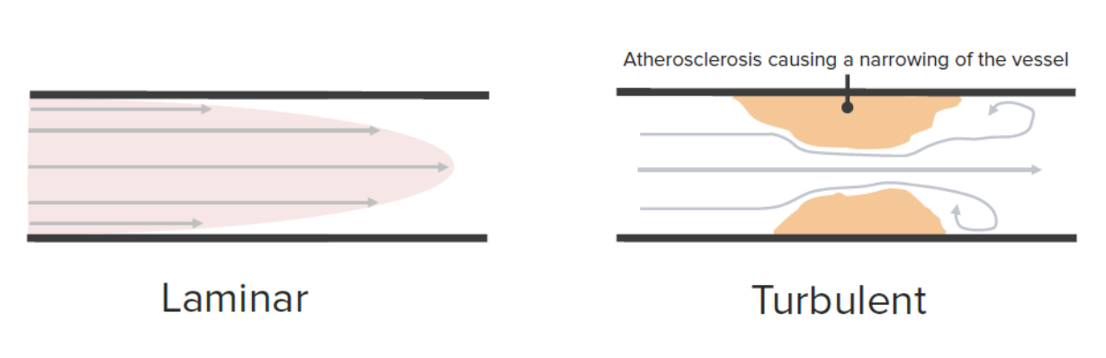
Laminar versus turbulent flow:
In smooth-walled vessels, blood moves by laminar flow. The blood moves the fastest in the center of the vessel where there is the least resistance. In atherosclerotic vessels with uneven walls, blood flow is turbulent.
Image by Lecturio.
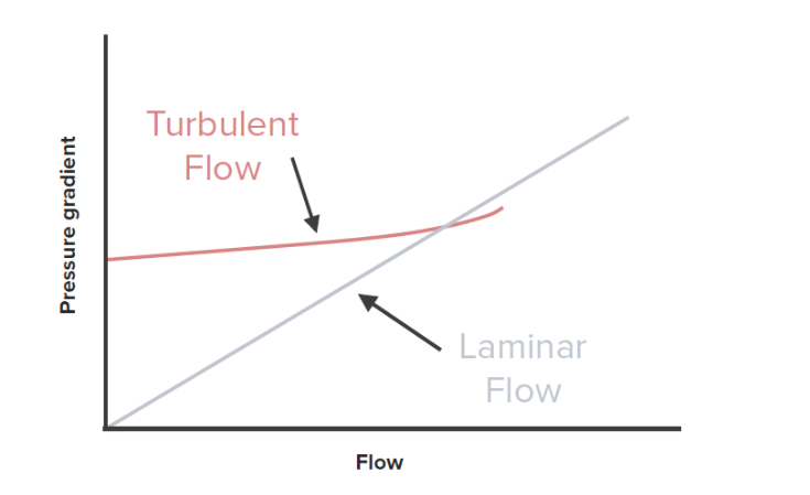
Flow versus pressure gradient: Vascular resistance is disproportionately higher in vessels with turbulent flow compared to vessels with laminar flow.
Image by Lecturio.
ResistanceResistancePhysiologically, the opposition to flow of air caused by the forces of friction. As a part of pulmonary function testing, it is the ratio of driving pressure to the rate of air flow.Ventilation: Mechanics of Breathing
ResistanceResistancePhysiologically, the opposition to flow of air caused by the forces of friction. As a part of pulmonary function testing, it is the ratio of driving pressure to the rate of air flow.Ventilation: Mechanics of Breathing: forces opposing flow:
Arises from the friction between the moving blood and vessel walls
Equation for resistanceResistancePhysiologically, the opposition to flow of air caused by the forces of friction. As a part of pulmonary function testing, it is the ratio of driving pressure to the rate of air flow.Ventilation: Mechanics of Breathing against laminar flow: R = (8 x viscosity x length) / πr4
R = resistanceResistancePhysiologically, the opposition to flow of air caused by the forces of friction. As a part of pulmonary function testing, it is the ratio of driving pressure to the rate of air flow.Ventilation: Mechanics of Breathing
Viscosity = thickness of the blood
Length = length of the vessel
r = radiusRadiusThe outer shorter of the two bones of the forearm, lying parallel to the ulna and partially revolving around it.Forearm: Anatomy of the vessel
Viscosity:
The thickness of the blood
Due primarily to:
Number of erythrocytesErythrocytesErythrocytes, or red blood cells (RBCs), are the most abundant cells in the blood. While erythrocytes in the fetus are initially produced in the yolk sac then the liver, the bone marrow eventually becomes the main site of production. Erythrocytes: Histology
AlbuminAlbuminSerum albumin from humans. It is an essential carrier of both endogenous substances, such as fatty acids and bilirubin, and of xenobiotics in the blood.Liver Function Tests levels
↓ Viscosity: due to anemiaAnemiaAnemia is a condition in which individuals have low Hb levels, which can arise from various causes. Anemia is accompanied by a reduced number of RBCs and may manifest with fatigue, shortness of breath, pallor, and weakness. Subtypes are classified by the size of RBCs, chronicity, and etiology. Anemia: Overview and Types, hypoalbuminemiaHypoalbuminemiaA condition in which albumin level in blood (serum albumin) is below the normal range. Hypoalbuminemia may be due to decreased hepatic albumin synthesis, increased albumin catabolism, altered albumin distribution, or albumin loss through the urine (albuminuria).Nephrotic Syndrome in Children, and adequate hydration
Relatively stable within individuals: The body is unable to quickly regulate flow by adjusting viscosity.
Length of the vessel:
The longer the vessel, the greater the cumulative friction encountered
Each vessel has a fairly fixed length (no ability for regulation).
RadiusRadiusThe outer shorter of the two bones of the forearm, lying parallel to the ulna and partially revolving around it.Forearm: Anatomy of the vessel:
Significant impact on resistanceResistancePhysiologically, the opposition to flow of air caused by the forces of friction. As a part of pulmonary function testing, it is the ratio of driving pressure to the rate of air flow.Ventilation: Mechanics of Breathing
Highly regulated by smooth muscle within the vessel walls
= ↑ RadiusRadiusThe outer shorter of the two bones of the forearm, lying parallel to the ulna and partially revolving around it.Forearm: Anatomy of vessel → ↓ blood in contact with the vessel wall → ↓ friction → ↓ overall resistanceResistancePhysiologically, the opposition to flow of air caused by the forces of friction. As a part of pulmonary function testing, it is the ratio of driving pressure to the rate of air flow.Ventilation: Mechanics of Breathing → ↑ blood flow through the vessel
Vasoconstriction: ↓ radiusRadiusThe outer shorter of the two bones of the forearm, lying parallel to the ulna and partially revolving around it.Forearm: Anatomy
VasodilationVasodilationThe physiological widening of blood vessels by relaxing the underlying vascular smooth muscle.Pulmonary Hypertension Drugs: ↑ radiusRadiusThe outer shorter of the two bones of the forearm, lying parallel to the ulna and partially revolving around it.Forearm: Anatomy
Pressure gradient (ΔP)
ΔP: the difference in pressure between 1 point and another
Influences the direction of blood flow (blood flows from high pressure → low pressure)
If the flow is constant (which the body tries to maintain), vessel resistanceResistancePhysiologically, the opposition to flow of air caused by the forces of friction. As a part of pulmonary function testing, it is the ratio of driving pressure to the rate of air flow.Ventilation: Mechanics of Breathing ↑ (e.g., vasoconstriction) and leads to ΔP ↑.
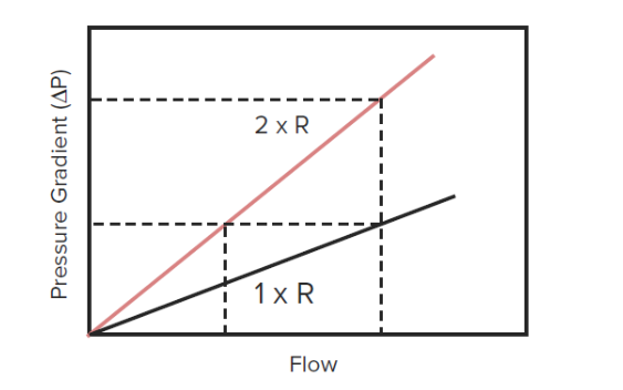
Pressure as a function of flow and resistance:
Pressure is directly related to both flow and resistance. As either flow or resistance increases, pressure increases proportionally. ΔP (pressure gradient) = R (resistance) x F (flow)
Image by Lecturio.
CapacitanceCapacitanceThe measure of a blood vessel’s ability to increase the volume of blood it holds without a large increase in blood pressure. The vascular capacitance is equal to the change in volume divided by the change in pressure.Venous Function
CapacitanceCapacitanceThe measure of a blood vessel’s ability to increase the volume of blood it holds without a large increase in blood pressure. The vascular capacitance is equal to the change in volume divided by the change in pressure.Venous Function: the amount a vessel can stretch without significantly increasing pressure:
CapacitanceCapacitanceThe measure of a blood vessel’s ability to increase the volume of blood it holds without a large increase in blood pressure. The vascular capacitance is equal to the change in volume divided by the change in pressure.Venous Function: C = ΔV / ΔP:
C: capacitanceCapacitanceThe measure of a blood vessel’s ability to increase the volume of blood it holds without a large increase in blood pressure. The vascular capacitance is equal to the change in volume divided by the change in pressure.Venous Function
ΔV: change in volume
ΔP: change in pressure
Venous capacitanceCapacitanceThe measure of a blood vessel’s ability to increase the volume of blood it holds without a large increase in blood pressure. The vascular capacitance is equal to the change in volume divided by the change in pressure.Venous Function > arterial capacitanceCapacitanceThe measure of a blood vessel’s ability to increase the volume of blood it holds without a large increase in blood pressure. The vascular capacitance is equal to the change in volume divided by the change in pressure.Venous Function
60%–80% of total blood volume is in the venous circulationCirculationThe movement of the blood as it is pumped through the cardiovascular system.ABCDE Assessment.
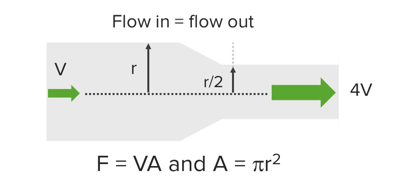
Velocity
The speed blood is traveling.
Velocity is inversely related to the radiusRadiusThe outer shorter of the two bones of the forearm, lying parallel to the ulna and partially revolving around it.Forearm: Anatomy of the blood vessel (i.e., velocity increases when diameter decreases).
Velocity is different from flow:
Velocity is a unit of distance per unit of time.
Flow is a unit of volume per unit time.
Clinical relevance: The velocity of blood moving across the valve will increase with a stenotic valve (smaller diameter), but the flow will not.
Relationship between flow and velocity:
Flow = velocity x the area of the vessel or pathway available to blood
Flow = velocity x (πr2)
The relationship between flow and velocity:
Velocity is inversely related to area. If the radius of the cylinder (r) is halved, the velocity increases 4-fold.
F: flow
V: velocity
A: area
r: radius
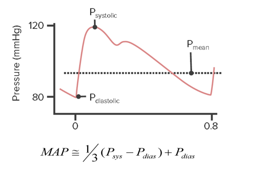
Mean arterial pressureis the average systemic arterial pressure.
MAP = (CO x SVR) + CVPCVPThe blood pressure in the central large veins of the body. It is distinguished from peripheral venous pressure which occurs in an extremity.Central Venous Catheter:
CO: cardiac outputCardiac outputThe volume of blood passing through the heart per unit of time. It is usually expressed as liters (volume) per minute so as not to be confused with stroke volume (volume per beat).Cardiac Mechanics (stroke volumeStroke volumeThe amount of blood pumped out of the heart per beat, not to be confused with cardiac output (volume/time). It is calculated as the difference between the end-diastolic volume and the end-systolic volume.Cardiac Cycle x heart rateHeart rateThe number of times the heart ventricles contract per unit of time, usually per minute.Cardiac Physiology)
SVR: systemic vascular resistanceResistancePhysiologically, the opposition to flow of air caused by the forces of friction. As a part of pulmonary function testing, it is the ratio of driving pressure to the rate of air flow.Ventilation: Mechanics of Breathing
CVPCVPThe blood pressure in the central large veins of the body. It is distinguished from peripheral venous pressure which occurs in an extremity.Central Venous Catheter: central venous pressureCentral venous pressureThe blood pressure in the central large veins of the body. It is distinguished from peripheral venous pressure which occurs in an extremity.Central Venous Catheter (close to 0; often disregarded)
Approximate using systolic blood pressure (SBPSBPAscites) and diastolic blood pressure (DBP):
Because the heart spends more time in diastoleDiastolePost-systolic relaxation of the heart, especially the heart ventricles.Cardiac Cycle than systoleSystolePeriod of contraction of the heart, especially of the heart ventricles.Cardiac Cycle, DBP contributes more to the MAP than SBPSBPAscites.
Mean arterial intravascular pressure throughout the cardiac cycle
MAP: mean arterial pressure
P: pressure Sys: systolic Dias: diastolic
Image by Lecturio.
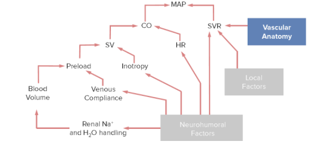
Factors affecting the MAP
Mean arterial pressure is primarily affected by the CO and SVR:
CO = heart rateHeart rateThe number of times the heart ventricles contract per unit of time, usually per minute.Cardiac Physiology x stroke volumeStroke volumeThe amount of blood pumped out of the heart per beat, not to be confused with cardiac output (volume/time). It is calculated as the difference between the end-diastolic volume and the end-systolic volume.Cardiac Cycle:
Heart rateHeart rateThe number of times the heart ventricles contract per unit of time, usually per minute.Cardiac Physiology is determined by:
The autonomic nervous systemAutonomic nervous systemThe ANS is a component of the peripheral nervous system that uses both afferent (sensory) and efferent (effector) neurons, which control the functioning of the internal organs and involuntary processes via connections with the CNS. The ANS consists of the sympathetic and parasympathetic nervous systems. Autonomic Nervous System: Anatomy (primary regulator)
Other factors:
ThyroidThyroidThe thyroid gland is one of the largest endocrine glands in the human body. The thyroid gland is a highly vascular, brownish-red gland located in the visceral compartment of the anterior region of the neck.Thyroid Gland: AnatomyhormonesHormonesHormones are messenger molecules that are synthesized in one part of the body and move through the bloodstream to exert specific regulatory effects on another part of the body. Hormones play critical roles in coordinating cellular activities throughout the body in response to the constant changes in both the internal and external environments. Hormones: Overview and Types
Circulating catecholaminesCatecholaminesA general class of ortho-dihydroxyphenylalkylamines derived from tyrosine.Adrenal Hormones
K+ levels
IschemiaIschemiaA hypoperfusion of the blood through an organ or tissue caused by a pathologic constriction or obstruction of its blood vessels, or an absence of blood circulation.Ischemic Cell Damage
Stroke volumeStroke volumeThe amount of blood pumped out of the heart per beat, not to be confused with cardiac output (volume/time). It is calculated as the difference between the end-diastolic volume and the end-systolic volume.Cardiac Cycle is determined primarily by:
Inotropy: the contractile strength of each heartbeat
AfterloadAfterloadAfterload is the resistance in the aorta that prevents blood from leaving the heart. Afterload represents the pressure the LV needs to overcome to eject blood into the aorta.Cardiac Mechanics: the pressure the left ventricle needs to overcome to eject blood into the aortaAortaThe main trunk of the systemic arteries.Mediastinum and Great Vessels: Anatomy
PreloadPreloadCardiac Mechanics: the amount the ventricles have stretched or filled with blood by the end of diastoleDiastolePost-systolic relaxation of the heart, especially the heart ventricles.Cardiac Cycle, which is affected by:
Venous complianceComplianceDistensibility measure of a chamber such as the lungs (lung compliance) or bladder. Compliance is expressed as a change in volume per unit change in pressure.Veins: Histology (the amount of blood the veinsVeinsVeins are tubular collections of cells, which transport deoxygenated blood and waste from the capillary beds back to the heart. Veins are classified into 3 types: small veins/venules, medium veins, and large veins. Each type contains 3 primary layers: tunica intima, tunica media, and tunica adventitia. Veins: Histology can hold)
Blood volume (primarily affected by renal Na+ and H2O handling)
Systemic vascular resistanceResistancePhysiologically, the opposition to flow of air caused by the forces of friction. As a part of pulmonary function testing, it is the ratio of driving pressure to the rate of air flow.Ventilation: Mechanics of Breathing is primarily affected by:
Vascular anatomy has significant effects on SVR, which directly affects MAP.
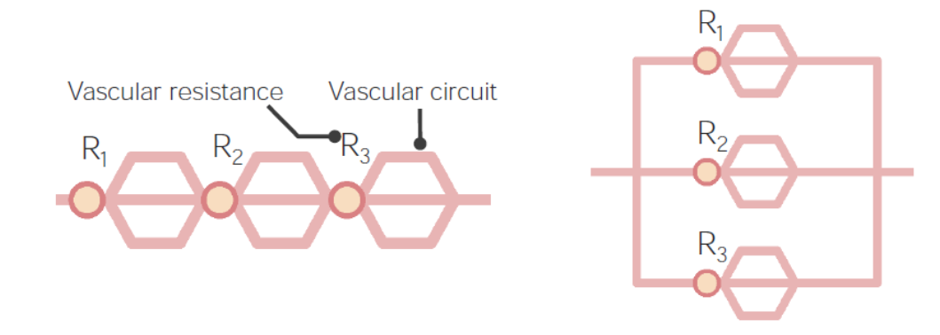
Arrangement of vessels in series or parallel
Series circuits:
Blood runs through the vessels sequentially; therefore, resistanceResistancePhysiologically, the opposition to flow of air caused by the forces of friction. As a part of pulmonary function testing, it is the ratio of driving pressure to the rate of air flow.Ventilation: Mechanics of Breathing is additive along the length of the entire vessel.
Example: a measure of the resistanceResistancePhysiologically, the opposition to flow of air caused by the forces of friction. As a part of pulmonary function testing, it is the ratio of driving pressure to the rate of air flow.Ventilation: Mechanics of Breathing along a single path from aortaAortaThe main trunk of the systemic arteries.Mediastinum and Great Vessels: Anatomy → large artery → arteriole → a single capillary
Total resistanceResistancePhysiologically, the opposition to flow of air caused by the forces of friction. As a part of pulmonary function testing, it is the ratio of driving pressure to the rate of air flow.Ventilation: Mechanics of Breathing (RT) = R1 + R2 + R3…
Parallel circuits:
Multiple paths are available to the blood when vessels divide.
The total area through which blood can flow is increased, even if the individual vessels have a smaller diameter.
ResistanceResistancePhysiologically, the opposition to flow of air caused by the forces of friction. As a part of pulmonary function testing, it is the ratio of driving pressure to the rate of air flow.Ventilation: Mechanics of Breathing decreases with each available path for blood to follow.
Example: ResistanceResistancePhysiologically, the opposition to flow of air caused by the forces of friction. As a part of pulmonary function testing, it is the ratio of driving pressure to the rate of air flow.Ventilation: Mechanics of Breathing changes as blood moves farther from the heart:
Blood flows from the aortaAortaThe main trunk of the systemic arteries.Mediastinum and Great Vessels: Anatomy (a single vessel) → all the capillariesCapillariesCapillaries are the primary structures in the circulatory system that allow the exchange of gas, nutrients, and other materials between the blood and the extracellular fluid (ECF). Capillaries are the smallest of the blood vessels. Because a capillary diameter is so small, only 1 RBC may pass through at a time.Capillaries: Histology in the systemic circulationCirculationThe movement of the blood as it is pumped through the cardiovascular system.ABCDE Assessment (millions of pathways)
The cross-sectional area of all capillariesCapillariesCapillaries are the primary structures in the circulatory system that allow the exchange of gas, nutrients, and other materials between the blood and the extracellular fluid (ECF). Capillaries are the smallest of the blood vessels. Because a capillary diameter is so small, only 1 RBC may pass through at a time.Capillaries: Histology combined: approximately 4,500–6,000 cm2
Total resistanceResistancePhysiologically, the opposition to flow of air caused by the forces of friction. As a part of pulmonary function testing, it is the ratio of driving pressure to the rate of air flow.Ventilation: Mechanics of Breathing for vessels in parallel: 1/RT = 1/R1 + 1/R2 + 1/R3…
Mathematical example:
Assume: a circuit with 3 points of resistanceResistancePhysiologically, the opposition to flow of air caused by the forces of friction. As a part of pulmonary function testing, it is the ratio of driving pressure to the rate of air flow.Ventilation: Mechanics of Breathing, all equal to 10 arbitrary units
If a 4th circuit is added, resistanceResistancePhysiologically, the opposition to flow of air caused by the forces of friction. As a part of pulmonary function testing, it is the ratio of driving pressure to the rate of air flow.Ventilation: Mechanics of Breathing increases to 40 in series and drops to 2.5 in parallel.
Left: a vascular circuit in series with 3 different points of resistance Right: a vascular circuit in parallel
Image by Lecturio.
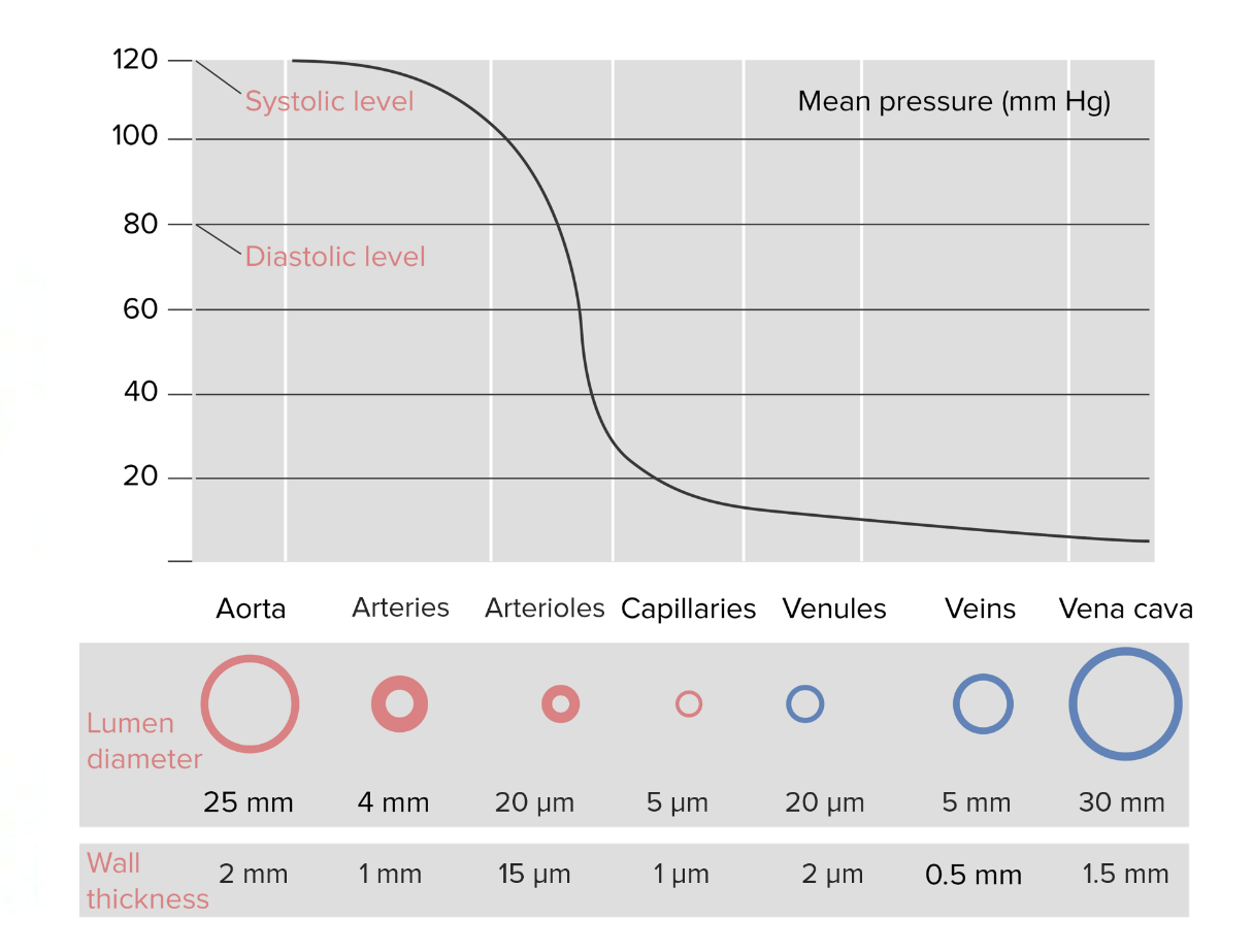
Anatomy of vessel walls
Vessels have:
Different functions at different points in the circuit. For example:
Distribution of blood to the body: aortaAortaThe main trunk of the systemic arteries.Mediastinum and Great Vessels: Anatomy, large arteriesArteriesArteries are tubular collections of cells that transport oxygenated blood and nutrients from the heart to the tissues of the body. The blood passes through the arteries in order of decreasing luminal diameter, starting in the largest artery (the aorta) and ending in the small arterioles. Arteries are classified into 3 types: large elastic arteries, medium muscular arteries, and small arteries and arterioles. Arteries: Histology
ResistanceResistancePhysiologically, the opposition to flow of air caused by the forces of friction. As a part of pulmonary function testing, it is the ratio of driving pressure to the rate of air flow.Ventilation: Mechanics of Breathing (regulates pressure and flow): small arteriesSmall arteriesArteries: Histology and arteriolesArteriolesThe smallest divisions of the arteries located between the muscular arteries and the capillaries.Arteries: Histology
Gas, nutrient, and waste exchange: capillariesCapillariesCapillaries are the primary structures in the circulatory system that allow the exchange of gas, nutrients, and other materials between the blood and the extracellular fluid (ECF). Capillaries are the smallest of the blood vessels. Because a capillary diameter is so small, only 1 RBC may pass through at a time.Capillaries: Histology
Collection: venulesVenulesThe minute vessels that collect blood from the capillary plexuses and join together to form veins.Veins: Histology
CapacitanceCapacitanceThe measure of a blood vessel’s ability to increase the volume of blood it holds without a large increase in blood pressure. The vascular capacitance is equal to the change in volume divided by the change in pressure.Venous Function (holding blood volume): venulesVenulesThe minute vessels that collect blood from the capillary plexuses and join together to form veins.Veins: Histology, veinsVeinsVeins are tubular collections of cells, which transport deoxygenated blood and waste from the capillary beds back to the heart. Veins are classified into 3 types: small veins/venules, medium veins, and large veins. Each type contains 3 primary layers: tunica intima, tunica media, and tunica adventitia. Veins: Histology, and vena cava
Different amounts of smooth muscle in the wall depending on the function
Different diameters
Distribution of pressure:
As the blood leaves the heart and moves through the body, pressure continually decreases until the blood returns to the heart → ΔP is always causing forward flow of blood through the circuit
In arteriesArteriesArteries are tubular collections of cells that transport oxygenated blood and nutrients from the heart to the tissues of the body. The blood passes through the arteries in order of decreasing luminal diameter, starting in the largest artery (the aorta) and ending in the small arterioles. Arteries are classified into 3 types: large elastic arteries, medium muscular arteries, and small arteries and arterioles. Arteries: Histology: walls are thicker/more smooth muscle:
Protect against higher pressures
Ability to control flow
In capillariesCapillariesCapillaries are the primary structures in the circulatory system that allow the exchange of gas, nutrients, and other materials between the blood and the extracellular fluid (ECF). Capillaries are the smallest of the blood vessels. Because a capillary diameter is so small, only 1 RBC may pass through at a time.Capillaries: Histology:
Capillary bedsCapillary bedsGroups of 10–100 individual capillary vessels supplied by a single metarteriole.Capillaries: Histology serve as parallel circuits.
Minimal resistanceResistancePhysiologically, the opposition to flow of air caused by the forces of friction. As a part of pulmonary function testing, it is the ratio of driving pressure to the rate of air flow.Ventilation: Mechanics of Breathing is created, allowing for lower pressures.
Thicker wall muscles are not needed to protect against high pressures.
Thin walls allow for gas exchangeGas exchangeHuman cells are primarily reliant on aerobic metabolism. The respiratory system is involved in pulmonary ventilation and external respiration, while the circulatory system is responsible for transport and internal respiration. Pulmonary ventilation (breathing) represents movement of air into and out of the lungs. External respiration, or gas exchange, is represented by the O2 and CO2 exchange between the lungs and the blood.Gas Exchange.
Pressure is higher at the arteriole end of the capillariesCapillariesCapillaries are the primary structures in the circulatory system that allow the exchange of gas, nutrients, and other materials between the blood and the extracellular fluid (ECF). Capillaries are the smallest of the blood vessels. Because a capillary diameter is so small, only 1 RBC may pass through at a time.Capillaries: Histology than at the venulesVenulesThe minute vessels that collect blood from the capillary plexuses and join together to form veins.Veins: Histology → pushes blood through capillariesCapillariesCapillaries are the primary structures in the circulatory system that allow the exchange of gas, nutrients, and other materials between the blood and the extracellular fluid (ECF). Capillaries are the smallest of the blood vessels. Because a capillary diameter is so small, only 1 RBC may pass through at a time.Capillaries: Histology
In veinsVeinsVeins are tubular collections of cells, which transport deoxygenated blood and waste from the capillary beds back to the heart. Veins are classified into 3 types: small veins/venules, medium veins, and large veins. Each type contains 3 primary layers: tunica intima, tunica media, and tunica adventitia. Veins: Histology:
Thinner walls allow veinsVeinsVeins are tubular collections of cells, which transport deoxygenated blood and waste from the capillary beds back to the heart. Veins are classified into 3 types: small veins/venules, medium veins, and large veins. Each type contains 3 primary layers: tunica intima, tunica media, and tunica adventitia. Veins: Histology to stretch (capacitanceCapacitanceThe measure of a blood vessel’s ability to increase the volume of blood it holds without a large increase in blood pressure. The vascular capacitance is equal to the change in volume divided by the change in pressure.Venous Function)
Lowest pressures
Respective intraluminal pressures:
Intraluminal pressure decreases as blood moves from the arterial to the venous system.
Endothelial cells lining blood vessels can secrete a number of factors causing vasodilationVasodilationThe physiological widening of blood vessels by relaxing the underlying vascular smooth muscle.Pulmonary Hypertension Drugs or vasoconstriction. Changing the radiusRadiusThe outer shorter of the two bones of the forearm, lying parallel to the ulna and partially revolving around it.Forearm: Anatomy of the vessel changes the SVR, which changes MAP.
Vasoconstriction
Primarily occurs by increasing intracellular calciumCalciumA basic element found in nearly all tissues. It is a member of the alkaline earth family of metals with the atomic symbol ca, atomic number 20, and atomic weight 40. Calcium is the most abundant mineral in the body and combines with phosphorus to form calcium phosphate in the bones and teeth. It is essential for the normal functioning of nerves and muscles and plays a role in blood coagulation (as factor IV) and in many enzymatic processes.Electrolytes (CaCACondylomata acuminata are a clinical manifestation of genital HPV infection. Condylomata acuminata are described as raised, pearly, flesh-colored, papular, cauliflower-like lesions seen in the anogenital region that may cause itching, pain, or bleeding.Condylomata Acuminata (Genital Warts)2+) levels, which is required for myofilament (actinActinFilamentous proteins that are the main constituent of the thin filaments of muscle fibers. The filaments (known also as filamentous or f-actin) can be dissociated into their globular subunits; each subunit is composed of a single polypeptide 375 amino acids long. This is known as globular or g-actin. In conjunction with myosins, actin is responsible for the contraction and relaxation of muscle.Skeletal Muscle Contraction and myosinMyosinA diverse superfamily of proteins that function as translocating proteins. They share the common characteristics of being able to bind actins and hydrolyze mgATP. Myosins generally consist of heavy chains which are involved in locomotion, and light chains which are involved in regulation. Within the structure of myosin heavy chain are three domains: the head, the neck and the tail. The head region of the heavy chain contains the actin binding domain and mgATPase domain which provides energy for locomotion. The neck region is involved in binding the light-chains. The tail region provides the anchoring point that maintains the position of the heavy chain. The superfamily of myosins is organized into structural classes based upon the type and arrangement of the subunits they contain.Skeletal Muscle Contraction) contraction within muscle cells.
Factors causing ↑ intracellular CaCACondylomata acuminata are a clinical manifestation of genital HPV infection. Condylomata acuminata are described as raised, pearly, flesh-colored, papular, cauliflower-like lesions seen in the anogenital region that may cause itching, pain, or bleeding.Condylomata Acuminata (Genital Warts)2+ include:
EndothelinsEndothelins21-amino-acid peptides produced by vascular endothelial cells and functioning as potent vasoconstrictors. The endothelin family consists of three members, endothelin-1; endothelin-2; and endothelin-3. All three peptides contain 21 amino acids, but vary in amino acid composition. The three peptides produce vasoconstrictor and pressor responses in various parts of the body. However, the quantitative profiles of the pharmacological activities are considerably different among the three isopeptides.Hemostasis
ThromboxanesThromboxanesPhysiologically active compounds found in many organs of the body. They are formed in vivo from the prostaglandin endoperoxides and cause platelet aggregation, contraction of arteries, and other biological effects. Thromboxanes are important mediators of the actions of polyunsaturated fatty acids transformed by cyclooxygenase.Eicosanoids
Angiotensin IIAngiotensin IIAn octapeptide that is a potent but labile vasoconstrictor. It is produced from angiotensin I after the removal of two amino acids at the c-terminal by angiotensin converting enzyme. The amino acid in position 5 varies in different species. To block vasoconstriction and hypertension effect of angiotensin II, patients are often treated with ace inhibitors or with angiotensin II type 1 receptor blockers.Renal Sodium and Water Regulation
↓ Intracellular CaCACondylomata acuminata are a clinical manifestation of genital HPV infection. Condylomata acuminata are described as raised, pearly, flesh-colored, papular, cauliflower-like lesions seen in the anogenital region that may cause itching, pain, or bleeding.Condylomata Acuminata (Genital Warts)2+ levels
↑ MyosinMyosinA diverse superfamily of proteins that function as translocating proteins. They share the common characteristics of being able to bind actins and hydrolyze mgATP. Myosins generally consist of heavy chains which are involved in locomotion, and light chains which are involved in regulation. Within the structure of myosin heavy chain are three domains: the head, the neck and the tail. The head region of the heavy chain contains the actin binding domain and mgATPase domain which provides energy for locomotion. The neck region is involved in binding the light-chains. The tail region provides the anchoring point that maintains the position of the heavy chain. The superfamily of myosins is organized into structural classes based upon the type and arrangement of the subunits they contain.Skeletal Muscle Contraction light chain (MLC) phosphatase activity → leads to ↑ dephosphorylation of contracted actinActinFilamentous proteins that are the main constituent of the thin filaments of muscle fibers. The filaments (known also as filamentous or f-actin) can be dissociated into their globular subunits; each subunit is composed of a single polypeptide 375 amino acids long. This is known as globular or g-actin. In conjunction with myosins, actin is responsible for the contraction and relaxation of muscle.Skeletal Muscle Contraction → relaxation
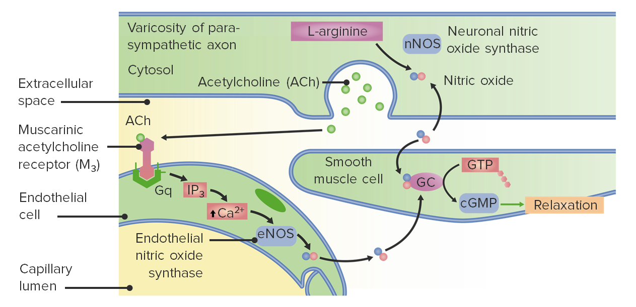
Neuronal NO synthase (nNOS) in parasympathetic neuronsNeuronsThe basic cellular units of nervous tissue. Each neuron consists of a body, an axon, and dendrites. Their purpose is to receive, conduct, and transmit impulses in the nervous system.Nervous System: Histology
cGMPcGMPGuanosine cyclic 3.Phosphodiesterase Inhibitors → ↓ intracellular CaCACondylomata acuminata are a clinical manifestation of genital HPV infection. Condylomata acuminata are described as raised, pearly, flesh-colored, papular, cauliflower-like lesions seen in the anogenital region that may cause itching, pain, or bleeding.Condylomata Acuminata (Genital Warts)2+ and ↑ MLC phosphatase activity → vasodilationVasodilationThe physiological widening of blood vessels by relaxing the underlying vascular smooth muscle.Pulmonary Hypertension Drugs
Production of prostacyclinProstacyclinA prostaglandin that is a powerful vasodilator and inhibits platelet aggregation. It is biosynthesized enzymatically from prostaglandin endoperoxides in human vascular tissue. The sodium salt has been also used to treat primary pulmonary hypertension.Eicosanoids:
Synthesized by cyclooxygenaseCyclooxygenaseNonsteroidal Antiinflammatory Drugs (NSAIDs) (COX) enzymesEnzymesEnzymes are complex protein biocatalysts that accelerate chemical reactions without being consumed by them. Due to the body’s constant metabolic needs, the absence of enzymes would make life unsustainable, as reactions would occur too slowly without these molecules. Basics of Enzymes
↑ Levels of cAMPcAMPAn adenine nucleotide containing one phosphate group which is esterified to both the 3′- and 5′-positions of the sugar moiety. It is a second messenger and a key intracellular regulator, functioning as a mediator of activity for a number of hormones, including epinephrine, glucagon, and acth.Phosphodiesterase Inhibitors
cAMPcAMPAn adenine nucleotide containing one phosphate group which is esterified to both the 3′- and 5′-positions of the sugar moiety. It is a second messenger and a key intracellular regulator, functioning as a mediator of activity for a number of hormones, including epinephrine, glucagon, and acth.Phosphodiesterase Inhibitors → ↓ intracellular CaCACondylomata acuminata are a clinical manifestation of genital HPV infection. Condylomata acuminata are described as raised, pearly, flesh-colored, papular, cauliflower-like lesions seen in the anogenital region that may cause itching, pain, or bleeding.Condylomata Acuminata (Genital Warts)2+ and ↑ MLC phosphatase activity → vasodilationVasodilationThe physiological widening of blood vessels by relaxing the underlying vascular smooth muscle.Pulmonary Hypertension Drugs
Factors stimulating the production of NO and/or prostacyclinProstacyclinA prostaglandin that is a powerful vasodilator and inhibits platelet aggregation. It is biosynthesized enzymatically from prostaglandin endoperoxides in human vascular tissue. The sodium salt has been also used to treat primary pulmonary hypertension.Eicosanoids:
AcetylcholineAcetylcholineA neurotransmitter found at neuromuscular junctions, autonomic ganglia, parasympathetic effector junctions, a subset of sympathetic effector junctions, and at many sites in the central nervous system.Receptors and Neurotransmitters of the CNS
ATP
Substance P
BradykininBradykininA nonapeptide messenger that is enzymatically produced from kallidin in the blood where it is a potent but short-lived agent of arteriolar dilation and increased capillary permeability. Bradykinin is also released from mast cells during asthma attacks, from gut walls as a gastrointestinal vasodilator, from damaged tissues as a pain signal, and may be a neurotransmitter.Hereditary Angioedema (C1 Esterase Inhibitor Deficiency)
ThrombinThrombinAn enzyme formed from prothrombin that converts fibrinogen to fibrin.Hemostasis
Histamine
Bacterial endotoxinsEndotoxinsToxins closely associated with the living cytoplasm or cell wall of certain microorganisms, which do not readily diffuse into the culture medium, but are released upon lysis of the cells.Bacteriology
Shearing forces
Chemical pathways lead to the production of nitric oxide, which ultimately causes smooth muscle relaxation and vasodilation. GTP: guanosine triphosphate Gq: gq protein GC: guanylyl cyclase cGMP: cyclic guanosine monophosphate Ca2+: calcium
Overview of Neurohumoral Factors Affecting Mean Arterial Pressure
Effects on the arterial system
Neurohumoral factors can affect both CO and SVR and include:
Stimulation from the ANSANSThe ans is a component of the peripheral nervous system that uses both afferent (sensory) and efferent (effector) neurons, which control the functioning of the internal organs and involuntary processes via connections with the CNS. The ans consists of the sympathetic and parasympathetic nervous systems.Autonomic Nervous System: Anatomy:
Sympathetic stimulation: ↑ CO and SVR → ↑ MAP
Parasympathetic stimulation: ↓ CO and SVR → ↓ MAP
Arterial baroreceptor reflexArterial Baroreceptor reflexThe baroreceptor reflex is the most important mechanism for acute BP regulation.Arterial Pressure Regulation: senses pressures and adjusts CO to maintain blood pressure homeostasisHomeostasisThe processes whereby the internal environment of an organism tends to remain balanced and stable.Cell Injury and Death
Circulating catecholaminesCatecholaminesA general class of ortho-dihydroxyphenylalkylamines derived from tyrosine.Adrenal Hormones:
Secreted by the adrenal medullaAdrenal MedullaThe inner portion of the adrenal gland. Derived from ectoderm, adrenal medulla consists mainly of chromaffin cells that produces and stores a number of neurotransmitters, mainly adrenaline (epinephrine) and norepinephrine. The activity of the adrenal medulla is regulated by the sympathetic nervous system.Adrenal Glands: Anatomy into the blood
Similar effect as sympathetic stimulation: ↑ CO and SVR → ↑ MAP
HormonesHormonesHormones are messenger molecules that are synthesized in one part of the body and move through the bloodstream to exert specific regulatory effects on another part of the body. Hormones play critical roles in coordinating cellular activities throughout the body in response to the constant changes in both the internal and external environments. Hormones: Overview and Types:
EpinephrineEpinephrineThe active sympathomimetic hormone from the adrenal medulla. It stimulates both the alpha- and beta- adrenergic systems, causes systemic vasoconstriction and gastrointestinal relaxation, stimulates the heart, and dilates bronchi and cerebral vessels.Sympathomimetic Drugs
NorepinephrineNorepinephrinePrecursor of epinephrine that is secreted by the adrenal medulla and is a widespread central and autonomic neurotransmitter. Norepinephrine is the principal transmitter of most postganglionic sympathetic fibers, and of the diffuse projection system in the brain that arises from the locus ceruleus.Receptors and Neurotransmitters of the CNS
RAASRAASA blood pressure regulating system of interacting components that include renin; angiotensinogen; angiotensin converting enzyme; angiotensin i; angiotensin ii; and angiotensinase. Renin, an enzyme produced in the kidney, acts on angiotensinogen, an alpha-2 globulin produced by the liver, forming angiotensin I. Angiotensin-converting enzyme, contained in the lung, acts on angiotensin I in the plasma converting it to angiotensin II, an extremely powerful vasoconstrictor. Angiotensin II causes contraction of the arteriolar and renal vascular smooth muscle, leading to retention of salt and water in the kidney and increased arterial blood pressure. In addition, angiotensin II stimulates the release of aldosterone from the adrenal cortex, which in turn also increases salt and water retention in the kidney. Angiotensin-converting enzyme also breaks down bradykinin, a powerful vasodilator and component of the kallikrein-kinin system.Adrenal Hormones:
The primary mechanism to control Na+ and level of body water
Activation of the RAASRAASA blood pressure regulating system of interacting components that include renin; angiotensinogen; angiotensin converting enzyme; angiotensin i; angiotensin ii; and angiotensinase. Renin, an enzyme produced in the kidney, acts on angiotensinogen, an alpha-2 globulin produced by the liver, forming angiotensin I. Angiotensin-converting enzyme, contained in the lung, acts on angiotensin I in the plasma converting it to angiotensin II, an extremely powerful vasoconstrictor. Angiotensin II causes contraction of the arteriolar and renal vascular smooth muscle, leading to retention of salt and water in the kidney and increased arterial blood pressure. In addition, angiotensin II stimulates the release of aldosterone from the adrenal cortex, which in turn also increases salt and water retention in the kidney. Angiotensin-converting enzyme also breaks down bradykinin, a powerful vasodilator and component of the kallikrein-kinin system.Adrenal Hormones → ↑ Na+ and water retention by the kidneysKidneysThe kidneys are a pair of bean-shaped organs located retroperitoneally against the posterior wall of the abdomen on either side of the spine. As part of the urinary tract, the kidneys are responsible for blood filtration and excretion of water-soluble waste in the urine.Kidneys: Anatomy → ↑ blood volume → ↑ preloadPreloadCardiac Mechanics → ↑ CO → ↑ MAP
Antidiuretic hormoneAntidiuretic hormoneAntidiuretic hormones released by the neurohypophysis of all vertebrates (structure varies with species) to regulate water balance and osmolarity. In general, vasopressin is a nonapeptide consisting of a six-amino-acid ring with a cysteine 1 to cysteine 6 disulfide bridge or an octapeptide containing a cystine. All mammals have arginine vasopressin except the pig with a lysine at position 8. Vasopressin, a vasoconstrictor, acts on the kidney collecting ducts to increase water reabsorption, increase blood volume and blood pressure.Hypernatremia (ADH):
SecretionSecretionCoagulation Studies of ADH from the posterior pituitaryPituitaryA small, unpaired gland situated in the sella turcica. It is connected to the hypothalamus by a short stalk which is called the infundibulum.Hormones: Overview and Types → ↓ water excretion → ↑ blood volume → ↑ CO → ↑ MAP
Additionally, ADH binds to receptorsReceptorsReceptors are proteins located either on the surface of or within a cell that can bind to signaling molecules known as ligands (e.g., hormones) and cause some type of response within the cell.Receptors on vascular smooth muscle → vasoconstriction
Natriuretic peptidesNatriuretic peptidesPeptides that regulate the water-electrolyte balance in the body, also known as natriuretic peptide hormones. Several have been sequenced (atrial natriuretic factor; brain natriuretic peptide; c-type natriuretic peptide).Arterial Pressure Regulation:
HormonesHormonesHormones are messenger molecules that are synthesized in one part of the body and move through the bloodstream to exert specific regulatory effects on another part of the body. Hormones play critical roles in coordinating cellular activities throughout the body in response to the constant changes in both the internal and external environments. Hormones: Overview and Types secreted by the heart inhibit the RAASRAASA blood pressure regulating system of interacting components that include renin; angiotensinogen; angiotensin converting enzyme; angiotensin i; angiotensin ii; and angiotensinase. Renin, an enzyme produced in the kidney, acts on angiotensinogen, an alpha-2 globulin produced by the liver, forming angiotensin I. Angiotensin-converting enzyme, contained in the lung, acts on angiotensin I in the plasma converting it to angiotensin II, an extremely powerful vasoconstrictor. Angiotensin II causes contraction of the arteriolar and renal vascular smooth muscle, leading to retention of salt and water in the kidney and increased arterial blood pressure. In addition, angiotensin II stimulates the release of aldosterone from the adrenal cortex, which in turn also increases salt and water retention in the kidney. Angiotensin-converting enzyme also breaks down bradykinin, a powerful vasodilator and component of the kallikrein-kinin system.Adrenal Hormones.
↓ Na+ and water retention by the kidneysKidneysThe kidneys are a pair of bean-shaped organs located retroperitoneally against the posterior wall of the abdomen on either side of the spine. As part of the urinary tract, the kidneys are responsible for blood filtration and excretion of water-soluble waste in the urine.Kidneys: Anatomy → ↓ blood volume → ↓ preloadPreloadCardiac Mechanics → ↓ CO → ↓ MAP
HormonesHormonesHormones are messenger molecules that are synthesized in one part of the body and move through the bloodstream to exert specific regulatory effects on another part of the body. Hormones play critical roles in coordinating cellular activities throughout the body in response to the constant changes in both the internal and external environments. Hormones: Overview and Types:
Atrial natriuretic peptideAtrial natriuretic peptideA potent natriuretic and vasodilatory peptide or mixture of different-sized low molecular weight peptides derived from a common precursor and secreted mainly by the heart atrium. All these peptides share a sequence of about 20 amino acids.Renal Sodium and Water Regulation: secreted by atrial myocytesMyocytesMature contractile cells, commonly known as myocytes, that form one of three kinds of muscle. The three types of muscle cells are skeletal, cardiac, and smooth. They are derived from embryonic (precursor) muscle cells called myoblasts.Muscle Tissue: Histology
BrainBrainThe part of central nervous system that is contained within the skull (cranium). Arising from the neural tube, the embryonic brain is comprised of three major parts including prosencephalon (the forebrain); mesencephalon (the midbrain); and rhombencephalon (the hindbrain). The developed brain consists of cerebrum; cerebellum; and other structures in the brain stem.Nervous System: Anatomy, Structure, and Classification natriuretic peptide: secreted by ventricular myocytesMyocytesMature contractile cells, commonly known as myocytes, that form one of three kinds of muscle. The three types of muscle cells are skeletal, cardiac, and smooth. They are derived from embryonic (precursor) muscle cells called myoblasts.Muscle Tissue: Histology
Effects on the venous system
Neurohumoral factors (primarily via the ANSANSThe ans is a component of the peripheral nervous system that uses both afferent (sensory) and efferent (effector) neurons, which control the functioning of the internal organs and involuntary processes via connections with the CNS. The ans consists of the sympathetic and parasympathetic nervous systems.Autonomic Nervous System: Anatomy) can affect venous capacitanceCapacitanceThe measure of a blood vessel’s ability to increase the volume of blood it holds without a large increase in blood pressure. The vascular capacitance is equal to the change in volume divided by the change in pressure.Venous Function, which can affect preloadPreloadCardiac Mechanics and, as a result, CO and MAP:
VenoconstrictionVenoconstrictionVenous Function → ↓ venous capacitanceCapacitanceThe measure of a blood vessel’s ability to increase the volume of blood it holds without a large increase in blood pressure. The vascular capacitance is equal to the change in volume divided by the change in pressure.Venous Function → ↑ venous return to the heart → ↑ preloadPreloadCardiac Mechanics → ↑ CO → ↑ MAP
VenodilationVenodilationVenous Function → ↑ venous capacitanceCapacitanceThe measure of a blood vessel’s ability to increase the volume of blood it holds without a large increase in blood pressure. The vascular capacitance is equal to the change in volume divided by the change in pressure.Venous Function → ↓ venous return to the heart → ↓ preloadPreloadCardiac Mechanics → ↓ CO → ↓ MAP
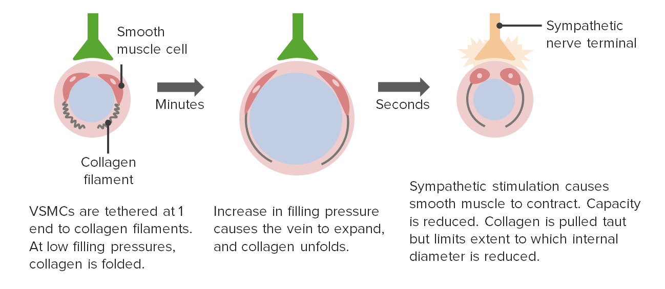
Changes in venous tone and the effect on capacitance
VSMC: vascular smooth muscle cell
The information presented below explains factors that determine blood pressure and how blood moves throughout the body. The foundational topics are critical to understanding how and why the body adjusts to different situations in order to maintain appropriate perfusion.
HypertensionHypertensionHypertension, or high blood pressure, is a common disease that manifests as elevated systemic arterial pressures. Hypertension is most often asymptomatic and is found incidentally as part of a routine physical examination or during triage for an unrelated medical encounter. Hypertension: chronically increased pressure in the arterial system. The increased pressures are due to narrower vessels (i.e., smaller radiusRadiusThe outer shorter of the two bones of the forearm, lying parallel to the ulna and partially revolving around it.Forearm: Anatomy). The pressures can cause damage to more delicate capillariesCapillariesCapillaries are the primary structures in the circulatory system that allow the exchange of gas, nutrients, and other materials between the blood and the extracellular fluid (ECF). Capillaries are the smallest of the blood vessels. Because a capillary diameter is so small, only 1 RBC may pass through at a time.Capillaries: Histology, which is especially problematic in the kidneysKidneysThe kidneys are a pair of bean-shaped organs located retroperitoneally against the posterior wall of the abdomen on either side of the spine. As part of the urinary tract, the kidneys are responsible for blood filtration and excretion of water-soluble waste in the urine.Kidneys: Anatomy and eyes. In addition, hypertensionHypertensionHypertension, or high blood pressure, is a common disease that manifests as elevated systemic arterial pressures. Hypertension is most often asymptomatic and is found incidentally as part of a routine physical examination or during triage for an unrelated medical encounter. Hypertension is a state of persistently increased afterloadAfterloadAfterload is the resistance in the aorta that prevents blood from leaving the heart. Afterload represents the pressure the LV needs to overcome to eject blood into the aorta.Cardiac Mechanics, requiring the heart to pumpPumpACES and RUSH: Resuscitation Ultrasound Protocols harder to eject the same volume of blood to maintain flow rates. Therefore, hypertensionHypertensionHypertension, or high blood pressure, is a common disease that manifests as elevated systemic arterial pressures. Hypertension is most often asymptomatic and is found incidentally as part of a routine physical examination or during triage for an unrelated medical encounter. Hypertension is a major risk factor for both heart disease and peripheral vascular disease.
Hemorrhage: excessive blood loss, which results in decreased blood volume and leads to ↓ preloadPreloadCardiac Mechanics, ↓ stroke volumeStroke volumeThe amount of blood pumped out of the heart per beat, not to be confused with cardiac output (volume/time). It is calculated as the difference between the end-diastolic volume and the end-systolic volume.Cardiac Cycle, ↓ cardiac outputCardiac outputThe volume of blood passing through the heart per unit of time. It is usually expressed as liters (volume) per minute so as not to be confused with stroke volume (volume per beat).Cardiac Mechanics, ↓ MAP, and, as a result, ↓ perfusion to vital organs. To maintain perfusion, the body attempts to increase MAP by boosting CO through increases in heart rateHeart rateThe number of times the heart ventricles contract per unit of time, usually per minute.Cardiac Physiology and contractility, and by vasoconstriction to increase SVR. Intravenous fluidsIntravenous FluidsIntravenous fluids are one of the most common interventions administered in medicine to approximate physiologic bodily fluids. Intravenous fluids are divided into 2 categories: crystalloid and colloid solutions. Intravenous fluids have a wide variety of indications, including intravascular volume expansion, electrolyte manipulation, and maintenance fluids. Intravenous Fluids and/or blood transfusionsBlood transfusionsThe introduction of whole blood or blood component directly into the bloodstream.Transfusion Products can help to restore blood volume.
References
Mohrman, D. E., & Heller, L. J. (2018). Overview of the cardiovascular system. Cardiovascular physiology, 9e (). New York, NY: McGraw-Hill Education. Retrieved from accessmedicine.mhmedical.com/content.aspx?aid=1153946098
Mohrman, D. E., & Heller, L. J. (2018). Vascular control. Cardiovascular physiology, (9e). New York, NY: McGraw-Hill Education. Retrieved from accessmedicine.mhmedical.com/content.aspx?aid=1153946722
Mohrman, D. E., & Heller, L. J. (2018). Regulation of arterial pressure. Cardiovascular physiology, 9e (). New York, NY: McGraw-Hill Education. Retrieved from accessmedicine.mhmedical.com/content.aspx?aid=1153946898
Baumann, B. M. (2016). Systemic hypertension. In J. E. Tintinalli, J. S. Stapczynski, O. J. Ma, D. M. Yealy, G. D. Meckler & D. M. Cline (Eds.), Tintinalli’s emergency medicine: A comprehensive study guide, 8e (). New York, NY: McGraw-Hill Education. Retrieved from accessmedicine.mhmedical.com/content.aspx?aid=1121496251
Klabunde R. E. (2021). Cardiovascular Physiology Concepts. Retrieved 10 June 2021, from https://www.cvphysiology.com/
Saladin, K. S., Miller, L. (2004). Anatomy and physiology. (3rd Ed. Pp. 753–760).
Create your free account or log in to continue reading!