Capillaries are the primary structures in the circulatory system that allow the exchange of gas, nutrients, and other materials between the blood and the extracellular fluidExtracellular fluidThe fluid of the body that is outside of cells. It is the external environment for the cells.Body Fluid Compartments (ECF). Capillaries are the smallest of the blood vessels. Because a capillary diameter is so small, only 1 RBC may pass through at a time. Capillaries are organized into capillary beds, which are extensive networks of branches and anastomoses. Blood flows from the metarteriolesMetarteriolesShort vessels linking arterioles and capillaries.Arteries: Histology, into the capillaries, out the thoroughfare channel, and into venulesVenulesThe minute vessels that collect blood from the capillary plexuses and join together to form veins.Veins: Histology. Continuous, fenestrated, and sinusoid (discontinuous) capillaries are the 3 primary types, and each has a slightly different structure. Capillary dysfunction can occur either as a result of or a contribution to the clinical manifestation of many clinical conditions.
Capillaries are the smallest of the blood vessels. Capillaries are the primary structures in the circulatory system that allow the exchange of gas, nutrients, and other materials between the blood and the extracellular fluidExtracellular fluidThe fluid of the body that is outside of cells. It is the external environment for the cells.Body Fluid Compartments (ECF).
Structure
Simple tubes made up of a single layer of endothelial cells
Diameter:
Approximately 5 µm in diameter at the arterial end
Approximately 9 µm in diameter at the venous end
RBCsRBCsErythrocytes, or red blood cells (RBCs), are the most abundant cells in the blood. While erythrocytes in the fetus are initially produced in the yolk sac then the liver, the bone marrow eventually becomes the main site of production.Erythrocytes: Histology are approximately 7 µm in diameter → RBCsRBCsErythrocytes, or red blood cells (RBCs), are the most abundant cells in the blood. While erythrocytes in the fetus are initially produced in the yolk sac then the liver, the bone marrow eventually becomes the main site of production.Erythrocytes: Histology are forced through capillaries 1 at a time
Endothelial cells are separated from surrounding tissue by basal lamina.
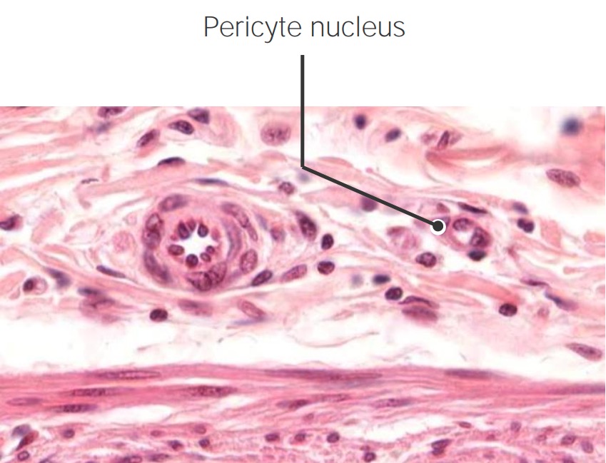
Capillaries are surrounded by pericytes (epithelial cells within the endothelial basal lamina, which play a role in neurovascular signaling).
Arranged in vast networks known as capillary beds (groups of 10–100 individual capillary vessels supplied by a single metarteriole)
Massive total surface area: estimated at > 6,300 m2
Cross-section of an arteriole (left) and a capillary (right) with a surrounding pericyte (labeled pericyte nucleus)
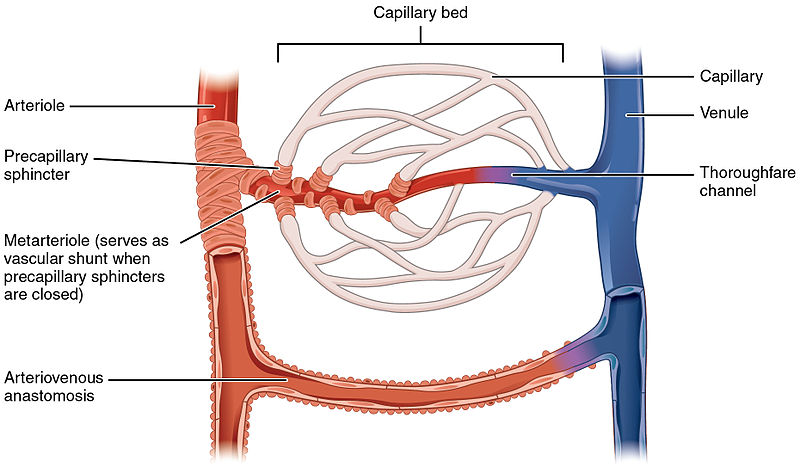
Blood enters the capillary beds through the arteriolesArteriolesThe smallest divisions of the arteries located between the muscular arteries and the capillaries.Arteries: Histology → metarteriolesMetarteriolesShort vessels linking arterioles and capillaries.Arteries: Histology → capillaries
Blood drains into the thoroughfare channel → venulesVenulesThe minute vessels that collect blood from the capillary plexuses and join together to form veins.Veins: Histology
MetarteriolesMetarteriolesShort vessels linking arterioles and capillaries.Arteries: Histology contain precapillary sphincters of smooth muscle at the entrance to each individual capillary:
Regulate the amount of blood flowBlood flowBlood flow refers to the movement of a certain volume of blood through the vasculature over a given unit of time (e.g., mL per minute).Vascular Resistance, Flow, and Mean Arterial Pressure into the capillary bed
When sphincters are closed, blood bypasses the capillaries and flows straight into the thoroughfare channel.
AV shunts are present when precapillary sphincters are closed.
Numerous in the dermisDermisA layer of vascularized connective tissue underneath the epidermis. The surface of the dermis contains innervated papillae. Embedded in or beneath the dermis are sweat glands; hair follicles; and sebaceous glands.Skin: Structure and Functions (help regulate body heatHeatInflammation)
Capillary bed demonstrating arteriole, metarteriole, and precapillary sphincters
Image: “Capillary bed” by OpenStax College. License: CC BY 3.0
Location
Capillaries are the connection between the smallest arteriesArteriesArteries are tubular collections of cells that transport oxygenated blood and nutrients from the heart to the tissues of the body. The blood passes through the arteries in order of decreasing luminal diameter, starting in the largest artery (the aorta) and ending in the small arterioles. Arteries are classified into 3 types: large elastic arteries, medium muscular arteries, and small arteries and arterioles. Arteries: Histology (arteriolesArteriolesThe smallest divisions of the arteries located between the muscular arteries and the capillaries.Arteries: Histology) and the smallest veinsVeinsVeins are tubular collections of cells, which transport deoxygenated blood and waste from the capillary beds back to the heart. Veins are classified into 3 types: small veins/venules, medium veins, and large veins. Each type contains 3 primary layers: tunica intima, tunica media, and tunica adventitia. Veins: Histology (venulesVenulesThe minute vessels that collect blood from the capillary plexuses and join together to form veins.Veins: Histology).
Found within 60–80 µm of essentially every cell in the body
Capillary beds are located in all tissues except:
CartilageCartilageCartilage is a type of connective tissue derived from embryonic mesenchyme that is responsible for structural support, resilience, and the smoothness of physical actions. Perichondrium (connective tissue membrane surrounding cartilage) compensates for the absence of vasculature in cartilage by providing nutrition and support. Cartilage: Histology
Epithelia
Eye corneaCorneaThe transparent anterior portion of the fibrous coat of the eye consisting of five layers: stratified squamous corneal epithelium; bowman membrane; corneal stroma; descemet membrane; and mesenchymal corneal endothelium. It serves as the first refracting medium of the eye.Eye: Anatomy and lensLensA transparent, biconvex structure of the eye, enclosed in a capsule and situated behind the iris and in front of the vitreous humor (vitreous body). It is slightly overlapped at its margin by the ciliary processes. Adaptation by the ciliary body is crucial for ocular accommodation.Eye: Anatomy
Tendons and ligaments have some capillaries, but much less than most other tissue.
Physiology
Functions:
Gas exchangeGas exchangeHuman cells are primarily reliant on aerobic metabolism. The respiratory system is involved in pulmonary ventilation and external respiration, while the circulatory system is responsible for transport and internal respiration. Pulmonary ventilation (breathing) represents movement of air into and out of the lungs. External respiration, or gas exchange, is represented by the O2 and CO2 exchange between the lungs and the blood.Gas Exchange: Oxygen exits RBCsRBCsErythrocytes, or red blood cells (RBCs), are the most abundant cells in the blood. While erythrocytes in the fetus are initially produced in the yolk sac then the liver, the bone marrow eventually becomes the main site of production.Erythrocytes: Histology, carbon dioxide enters RBCsRBCsErythrocytes, or red blood cells (RBCs), are the most abundant cells in the blood. While erythrocytes in the fetus are initially produced in the yolk sac then the liver, the bone marrow eventually becomes the main site of production.Erythrocytes: Histology.
Nutrient delivery
Blood picks up cellular and interstitial waste.
Mechanisms of exchange:
Transcytosis/pinocytosisPinocytosisThe engulfing of liquids by cells by a process of invagination and closure of the cell membrane to form fluid-filled vacuoles.Pharmacokinetics and Pharmacodynamics: Substances are taken into the endothelial cells in vesiclesVesiclesFemale Genitourinary Examination via endocytosisEndocytosisCellular uptake of extracellular materials within membrane-limited vacuoles or microvesicles. Endosomes play a central role in endocytosis.The Cell: Cell Membrane, transported across the cell, and released on the other side.
Direct filtration: relies on Starling forces
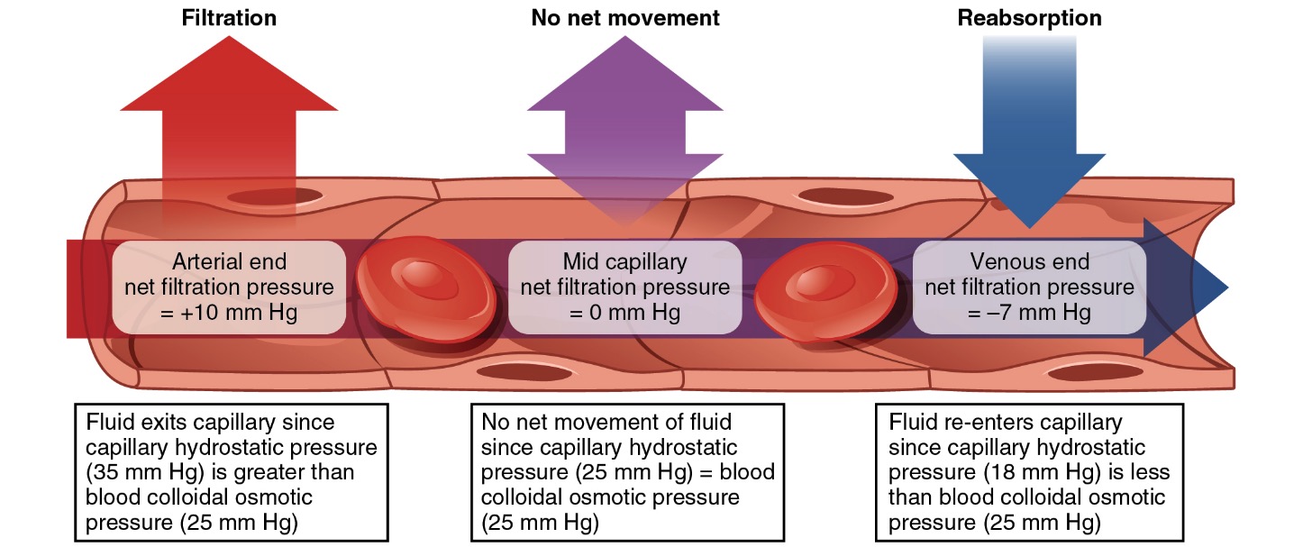
Starling forces applied to capillaries:
Relatively higher hydrostatic pressureHydrostatic pressureThe pressure due to the weight of fluid.Edema in the arteriolesArteriolesThe smallest divisions of the arteries located between the muscular arteries and the capillaries.Arteries: Histology pushes fluid, nutrients, and other cellular material into the surrounding ECF.
PlasmaPlasmaThe residual portion of blood that is left after removal of blood cells by centrifugation without prior blood coagulation.Transfusion ProductsproteinsProteinsLinear polypeptides that are synthesized on ribosomes and may be further modified, crosslinked, cleaved, or assembled into complex proteins with several subunits. The specific sequence of amino acids determines the shape the polypeptide will take, during protein folding, and the function of the protein.Energy Homeostasis generally cannot pass through the capillary walls → plasmaPlasmaThe residual portion of blood that is left after removal of blood cells by centrifugation without prior blood coagulation.Transfusion Productsoncotic pressureOncotic PressureEdema ↑ towards the venous end of the capillary
Relatively higher oncotic pressureOncotic PressureEdema in the venulesVenulesThe minute vessels that collect blood from the capillary plexuses and join together to form veins.Veins: Histology allows waste to be absorbed into the vessels
Starling forces: Starling forces within a capillary determine the flow of molecules into and out of the vessel.
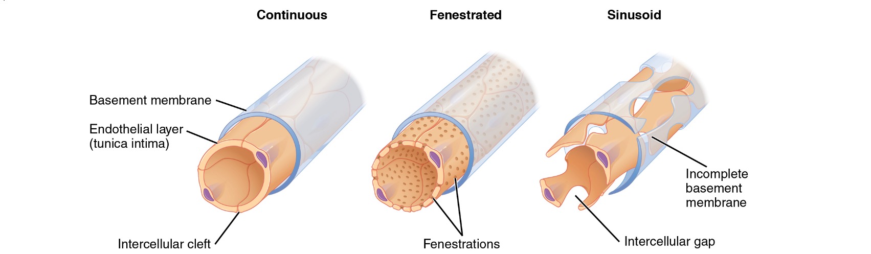
Image: “Types of capillaries” by Phil Schatz. License: CC BY 4.0
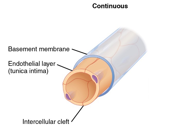
Continuous capillaries
Continuous capillaries are the most common type of capillary.
Structure:
Endothelial cells are connected via occluding tight junctionsTight junctionsCell-cell junctions that seal adjacent epithelial cells together, preventing the passage of most dissolved molecules from one side of the epithelial sheet to the other.The Cell: Cell Junctions.
Continuous basal lamina
Prevents diffusionDiffusionThe tendency of a gas or solute to pass from a point of higher pressure or concentration to a point of lower pressure or concentration and to distribute itself throughout the available space. Diffusion, especially facilitated diffusion, is a major mechanism of biological transport.Peritoneal Dialysis and Hemodialysis of fluid, protein, and other molecules
Some tissues contain small clefts (approximately 4 nm wide) → allow the passage of very small molecules (e.g., glucoseGlucoseA primary source of energy for living organisms. It is naturally occurring and is found in fruits and other parts of plants in its free state. It is used therapeutically in fluid and nutrient replacement.Lactose Intolerance)
Primary mechanism of exchange: transcytosis/pinocytosisPinocytosisThe engulfing of liquids by cells by a process of invagination and closure of the cell membrane to form fluid-filled vacuoles.Pharmacokinetics and Pharmacodynamics
Location (found in organs requiring passage of only select molecules):
LungsLungsLungs are the main organs of the respiratory system. Lungs are paired viscera located in the thoracic cavity and are composed of spongy tissue. The primary function of the lungs is to oxygenate blood and eliminate CO2. Lungs: Anatomy
All muscle: cardiac, skeletal, and smooth
Connective tissueConnective tissueConnective tissues originate from embryonic mesenchyme and are present throughout the body except inside the brain and spinal cord. The main function of connective tissues is to provide structural support to organs. Connective tissues consist of cells and an extracellular matrix.Connective Tissue: Histology
SkinSkinThe skin, also referred to as the integumentary system, is the largest organ of the body. The skin is primarily composed of the epidermis (outer layer) and dermis (deep layer). The epidermis is primarily composed of keratinocytes that undergo rapid turnover, while the dermis contains dense layers of connective tissue.Skin: Structure and Functions
Diagram of a continuous capillary
Image: “Continuous capillaries” by Phil Schatz. License: CC BY 4.0
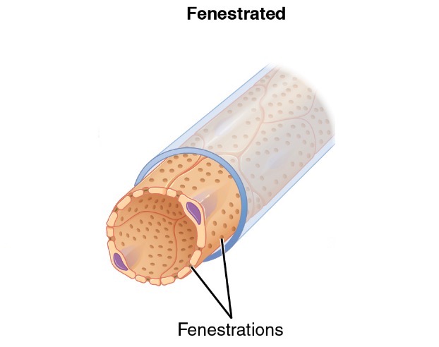
Fenestrated capillaries
Fenestrated capillaries are important in organs requiring rapid absorptionAbsorptionAbsorption involves the uptake of nutrient molecules and their transfer from the lumen of the GI tract across the enterocytes and into the interstitial space, where they can be taken up in the venous or lymphatic circulation.Digestion and Absorption and filtration, or with high metabolic activity.
Structure:
Wall contains multiple fenestrations, or “pores”, with continuous basal lamina.
Fenestrations are approximately 20–100 nm in diameter.
Allows for the rapid passage of small molecules, but keeps proteinsProteinsLinear polypeptides that are synthesized on ribosomes and may be further modified, crosslinked, cleaved, or assembled into complex proteins with several subunits. The specific sequence of amino acids determines the shape the polypeptide will take, during protein folding, and the function of the protein.Energy Homeostasis and larger particles within the blood vessel
Primary mechanism of exchange: filtration
Location:
KidneysKidneysThe kidneys are a pair of bean-shaped organs located retroperitoneally against the posterior wall of the abdomen on either side of the spine. As part of the urinary tract, the kidneys are responsible for blood filtration and excretion of water-soluble waste in the urine.Kidneys: Anatomy
Endocrine organs (e.g., pancreasPancreasThe pancreas lies mostly posterior to the stomach and extends across the posterior abdominal wall from the duodenum on the right to the spleen on the left. This organ has both exocrine and endocrine tissue. Pancreas: Anatomy)
Intestinal tract
Diagram of a fenestrated capillary
Image: “Fenestrated capillaries” by Phil Schatz. License: CC BY 4.0
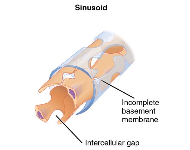
Sinusoid (discontinuous) capillaries
Sinusoid, or discontinuous, capillaries allow larger proteinsProteinsLinear polypeptides that are synthesized on ribosomes and may be further modified, crosslinked, cleaved, or assembled into complex proteins with several subunits. The specific sequence of amino acids determines the shape the polypeptide will take, during protein folding, and the function of the protein.Energy Homeostasis and full cells to pass through larger gaps.
Structure:
Large gaps (up to 0.5 µm) in the cytoplasm of the endotheliumEndotheliumA layer of epithelium that lines the heart, blood vessels (vascular endothelium), lymph vessels (lymphatic endothelium), and the serous cavities of the body.Arteries: Histology
Basal lamina has large gaps or may be completely absent.
May appear as larger, blood-filled spaces between other tissues
Mechanism of exchange: direct filtration/diffusionDiffusionThe tendency of a gas or solute to pass from a point of higher pressure or concentration to a point of lower pressure or concentration and to distribute itself throughout the available space. Diffusion, especially facilitated diffusion, is a major mechanism of biological transport.Peritoneal Dialysis and Hemodialysis
Locations:
LiverLiverThe liver is the largest gland in the human body. The liver is found in the superior right quadrant of the abdomen and weighs approximately 1.5 kilograms. Its main functions are detoxification, metabolism, nutrient storage (e.g., iron and vitamins), synthesis of coagulation factors, formation of bile, filtration, and storage of blood. Liver: Anatomy
SpleenSpleenThe spleen is the largest lymphoid organ in the body, located in the LUQ of the abdomen, superior to the left kidney and posterior to the stomach at the level of the 9th-11th ribs just below the diaphragm. The spleen is highly vascular and acts as an important blood filter, cleansing the blood of pathogens and damaged erythrocytes. Spleen: Anatomy
Bone marrowBone marrowThe soft tissue filling the cavities of bones. Bone marrow exists in two types, yellow and red. Yellow marrow is found in the large cavities of large bones and consists mostly of fat cells and a few primitive blood cells. Red marrow is a hematopoietic tissue and is the site of production of erythrocytes and granular leukocytes. Bone marrow is made up of a framework of connective tissue containing branching fibers with the frame being filled with marrow cells.Bone Marrow: Composition and Hematopoiesis
Specialized functions:
Allows larger structures to enter circulationCirculationThe movement of the blood as it is pumped through the cardiovascular system.ABCDE Assessment, for example:
ProteinsProteinsLinear polypeptides that are synthesized on ribosomes and may be further modified, crosslinked, cleaved, or assembled into complex proteins with several subunits. The specific sequence of amino acids determines the shape the polypeptide will take, during protein folding, and the function of the protein.Energy Homeostasis synthesized in the liverLiverThe liver is the largest gland in the human body. The liver is found in the superior right quadrant of the abdomen and weighs approximately 1.5 kilograms. Its main functions are detoxification, metabolism, nutrient storage (e.g., iron and vitamins), synthesis of coagulation factors, formation of bile, filtration, and storage of blood. Liver: Anatomy (e.g., albuminAlbuminSerum albumin from humans. It is an essential carrier of both endogenous substances, such as fatty acids and bilirubin, and of xenobiotics in the blood.Liver Function Tests and clotting factors)
Blood synthesized in the bone marrowBone marrowThe soft tissue filling the cavities of bones. Bone marrow exists in two types, yellow and red. Yellow marrow is found in the large cavities of large bones and consists mostly of fat cells and a few primitive blood cells. Red marrow is a hematopoietic tissue and is the site of production of erythrocytes and granular leukocytes. Bone marrow is made up of a framework of connective tissue containing branching fibers with the frame being filled with marrow cells.Bone Marrow: Composition and Hematopoiesis
Allows for “aggressive” communicationCommunicationThe exchange or transmission of ideas, attitudes, or beliefs between individuals or groups.Decision-making Capacity and Legal Competence between the perivascular cells and the blood itself
Diagram of a sinusoid capillary
Image: “Sinusoid capillaries” by Phil Schatz. License: CC BY 4.0
Thrombotic microangiopathies (TMAs) are a group of conditions characterized by abnormalities in the walls of arteriolesArteriolesThe smallest divisions of the arteries located between the muscular arteries and the capillaries.Arteries: Histology and capillaries, which lead to microvascular thrombosisThrombosisFormation and development of a thrombus or blood clot in the blood vessel.Epidemic Typhus. The most common primary TMAs are thrombotic thrombocytopenic purpuraThrombotic thrombocytopenic purpuraThrombotic thrombocytopenic purpura (TTP) is a life-threatening condition due to either a congenital or an acquired deficiency of ADAMTS-13, a metalloproteinase that cleaves multimers of von Willebrand factor (VWF). The large multimers then aggregate excessive platelets resulting in microvascular thrombosis and an increase in consumption of platelets. Thrombotic Thrombocytopenic Purpura (TTPTTPThrombotic thrombocytopenic purpura (TTP) is a life-threatening condition due to either a congenital or an acquired deficiency of adamts-13, a metalloproteinase that cleaves multimers of von Willebrand factor (vWF). The large multimers then aggregate excessive platelets resulting in microvascular thrombosis and an increase in consumption of platelets.Thrombotic Thrombocytopenic Purpura) and hemolytic uremic syndromeHemolytic uremic syndromeA syndrome that is associated with microvascular diseases of the kidney, such as renal cortical necrosis. It is characterized by hemolytic anemia; thrombocytopenia; and acute renal failure.Hypocoagulable Conditions (HUSHUSHemolytic uremic syndrome (HUS) is a clinical phenomenon most commonly seen in children that consists of a classic triad of microangiopathic hemolytic anemia, thrombocytopenia, and acute kidney injury. Hemolytic uremic syndrome is a major cause of acute kidney injury in children and is most commonly associated with a prodrome of diarrheal illness caused by shiga-like toxin-producing bacteria.Hemolytic Uremic Syndrome). Drugs can also induce a TMA.
Thrombotic thrombocytopenic purpuraThrombotic thrombocytopenic purpuraThrombotic thrombocytopenic purpura (TTP) is a life-threatening condition due to either a congenital or an acquired deficiency of ADAMTS-13, a metalloproteinase that cleaves multimers of von Willebrand factor (VWF). The large multimers then aggregate excessive platelets resulting in microvascular thrombosis and an increase in consumption of platelets. Thrombotic Thrombocytopenic Purpura: a life-threatening condition due to either a congenital or an acquired deficiency of ADAMTS-13, a metalloproteinase cleaving multimer of von Willebrand’s Factor (vWF). Without metalloproteinase, the large multimers aggregate excessive plateletsPlateletsPlatelets are small cell fragments involved in hemostasis. Thrombopoiesis takes place primarily in the bone marrow through a series of cell differentiation and is influenced by several cytokines. Platelets are formed after fragmentation of the megakaryocyte cytoplasm. Platelets: Histology, resulting in microvascular thrombosisThrombosisFormation and development of a thrombus or blood clot in the blood vessel.Epidemic Typhus and an increase in consumption of plateletsPlateletsPlatelets are small cell fragments involved in hemostasis. Thrombopoiesis takes place primarily in the bone marrow through a series of cell differentiation and is influenced by several cytokines. Platelets are formed after fragmentation of the megakaryocyte cytoplasm. Platelets: Histology. The classic clinical presentation includes thrombocytopeniaThrombocytopeniaThrombocytopenia occurs when the platelet count is < 150,000 per microliter. The normal range for platelets is usually 150,000-450,000/µL of whole blood. Thrombocytopenia can be a result of decreased production, increased destruction, or splenic sequestration of platelets. Patients are often asymptomatic until platelet counts are < 50,000/µL. Thrombocytopenia, hemolytic anemiaHemolytic AnemiaHemolytic anemia (HA) is the term given to a large group of anemias that are caused by the premature destruction/hemolysis of circulating red blood cells (RBCs). Hemolysis can occur within (intravascular hemolysis) or outside the blood vessels (extravascular hemolysis). Hemolytic Anemia, kidney disease, neurological symptoms, and feverFeverFever is defined as a measured body temperature of at least 38°C (100.4°F). Fever is caused by circulating endogenous and/or exogenous pyrogens that increase levels of prostaglandin E2 in the hypothalamus. Fever is commonly associated with chills, rigors, sweating, and flushing of the skin. Fever.
Hemolytic uremic syndromeHemolytic uremic syndromeA syndrome that is associated with microvascular diseases of the kidney, such as renal cortical necrosis. It is characterized by hemolytic anemia; thrombocytopenia; and acute renal failure.Hypocoagulable Conditions: a clinical phenomenon most commonly seen in children consisting of the classic triad of microangiopathic hemolytic anemiaMicroangiopathic Hemolytic AnemiaHemolytic Uremic Syndrome, thrombocytopeniaThrombocytopeniaThrombocytopenia occurs when the platelet count is < 150,000 per microliter. The normal range for platelets is usually 150,000-450,000/µL of whole blood. Thrombocytopenia can be a result of decreased production, increased destruction, or splenic sequestration of platelets. Patients are often asymptomatic until platelet counts are < 50,000/µL. Thrombocytopenia, and acute kidney injuryAcute Kidney InjuryAcute kidney injury refers to sudden and often reversible loss of renal function, which develops over days or weeks. Azotemia refers to elevated levels of nitrogen-containing substances in the blood that accompany AKI, which include BUN and creatinine. Acute Kidney Injury. Hemolytic uremic syndromeHemolytic uremic syndromeA syndrome that is associated with microvascular diseases of the kidney, such as renal cortical necrosis. It is characterized by hemolytic anemia; thrombocytopenia; and acute renal failure.Hypocoagulable Conditions is most commonly associated with a prodromeProdromeSymptoms that appear 24–48 hours prior to migraine onset.Migraine Headache of diarrheal illness caused by Shiga-like, toxin-producing bacteriaBacteriaBacteria are prokaryotic single-celled microorganisms that are metabolically active and divide by binary fission. Some of these organisms play a significant role in the pathogenesis of diseases. Bacteriology.
Increased hydrostatic pressureHydrostatic pressureThe pressure due to the weight of fluid.Edema within the capillaries
Any condition preventing blood flowBlood flowBlood flow refers to the movement of a certain volume of blood through the vasculature over a given unit of time (e.g., mL per minute).Vascular Resistance, Flow, and Mean Arterial Pressure from moving forward can lead to an increase in hydrostatic pressureHydrostatic pressureThe pressure due to the weight of fluid.Edema within the vessels, which ultimately may get transmitted to the capillaries. Increasing the hydrostatic pressureHydrostatic pressureThe pressure due to the weight of fluid.Edema within the capillaries affects the exchange of substances through the capillaries, pushing more fluid and substrates into the ECF. The types of conditions may include:
Heart failureHeart FailureA heterogeneous condition in which the heart is unable to pump out sufficient blood to meet the metabolic need of the body. Heart failure can be caused by structural defects, functional abnormalities (ventricular dysfunction), or a sudden overload beyond its capacity. Chronic heart failure is more common than acute heart failure which results from sudden insult to cardiac function, such as myocardial infarction.Total Anomalous Pulmonary Venous Return (TAPVR): the inability of the heart to supply the body with the cardiac outputCardiac outputThe volume of blood passing through the heart per unit of time. It is usually expressed as liters (volume) per minute so as not to be confused with stroke volume (volume per beat).Cardiac Mechanics required to meet the body’s metabolic needs. PatientsPatientsIndividuals participating in the health care system for the purpose of receiving therapeutic, diagnostic, or preventive procedures.Clinician–Patient Relationship typically present with dyspneaDyspneaDyspnea is the subjective sensation of breathing discomfort. Dyspnea is a normal manifestation of heavy physical or psychological exertion, but also may be caused by underlying conditions (both pulmonary and extrapulmonary). Dyspnea on exertion and/or at rest, orthopneaOrthopneaPulmonary Edema, and peripheral edemaPeripheral edemaPeripheral edema is the swelling of the lower extremities, namely, legs, feet, and ankles.Edema. Because blood is not effectively pumped out of the heart, pressure builds up in venous circulationCirculationThe movement of the blood as it is pumped through the cardiovascular system.ABCDE Assessment and ultimately backs up into the capillaries. Diagnosis can be made with an echocardiogramEchocardiogramTransposition of the Great Arteries.
CirrhosisCirrhosisCirrhosis is a late stage of hepatic parenchymal necrosis and scarring (fibrosis) most commonly due to hepatitis C infection and alcoholic liver disease. Patients may present with jaundice, ascites, and hepatosplenomegaly. Cirrhosis can also cause complications such as hepatic encephalopathy, portal hypertension, portal vein thrombosis, and hepatorenal syndrome. Cirrhosis: late stage of hepatic necrosisNecrosisThe death of cells in an organ or tissue due to disease, injury or failure of the blood supply.Ischemic Cell Damage and scarringScarringInflammation. Chronic cellular damage causes extensive distortionDistortionDefense Mechanisms of the normal hepatic architecture, which can lead to impairment of normal blood flowBlood flowBlood flow refers to the movement of a certain volume of blood through the vasculature over a given unit of time (e.g., mL per minute).Vascular Resistance, Flow, and Mean Arterial Pressure through the liverLiverThe liver is the largest gland in the human body. The liver is found in the superior right quadrant of the abdomen and weighs approximately 1.5 kilograms. Its main functions are detoxification, metabolism, nutrient storage (e.g., iron and vitamins), synthesis of coagulation factors, formation of bile, filtration, and storage of blood. Liver: Anatomy. The most common causes of cirrhosisCirrhosisCirrhosis is a late stage of hepatic parenchymal necrosis and scarring (fibrosis) most commonly due to hepatitis C infection and alcoholic liver disease. Patients may present with jaundice, ascites, and hepatosplenomegaly. Cirrhosis can also cause complications such as hepatic encephalopathy, portal hypertension, portal vein thrombosis, and hepatorenal syndrome. Cirrhosis are chronic, excessive alcohol use, viral hepatitis, and nonalcoholic steatohepatitisNonalcoholic SteatohepatitisMetabolic Dysfunction-associated Steatotic Liver Disease (MASLD) (NASH). Decompensation occurs late in the disease with manifestations including jaundiceJaundiceJaundice is the abnormal yellowing of the skin and/or sclera caused by the accumulation of bilirubin. Hyperbilirubinemia is caused by either an increase in bilirubin production or a decrease in the hepatic uptake, conjugation, or excretion of bilirubin. Jaundice, ascitesAscitesAscites is the pathologic accumulation of fluid within the peritoneal cavity that occurs due to an osmotic and/or hydrostatic pressure imbalance secondary to portal hypertension (cirrhosis, heart failure) or non-portal hypertension (hypoalbuminemia, malignancy, infection).Ascites, portal hypertensionPortal hypertensionPortal hypertension is increased pressure in the portal venous system. This increased pressure can lead to splanchnic vasodilation, collateral blood flow through portosystemic anastomoses, and increased hydrostatic pressure. There are a number of etiologies, including cirrhosis, right-sided congestive heart failure, schistosomiasis, portal vein thrombosis, hepatitis, and Budd-Chiari syndrome. Portal Hypertension, and liver failureLiver failureSevere inability of the liver to perform its normal metabolic functions, as evidenced by severe jaundice and abnormal serum levels of ammonia; bilirubin; alkaline phosphatase; aspartate aminotransferase; lactate dehydrogenases; and albumin/globulin ratio.Autoimmune Hepatitis.
Lower-extremity deep vein thrombosisThrombosisFormation and development of a thrombus or blood clot in the blood vessel.Epidemic Typhus (DVTDVTDeep vein thrombosis (DVT) usually occurs in the deep veins of the lower extremities. The affected veins include the femoral, popliteal, iliofemoral, and pelvic veins. Proximal DVT is more likely to cause a pulmonary embolism (PE) and is generally considered more serious. Deep Vein Thrombosis): occlusion of a deep vein by a thrombosisThrombosisFormation and development of a thrombus or blood clot in the blood vessel.Epidemic Typhus, most commonly occurring in the calvesCalvesErythema Nodosum. The affected veinsVeinsVeins are tubular collections of cells, which transport deoxygenated blood and waste from the capillary beds back to the heart. Veins are classified into 3 types: small veins/venules, medium veins, and large veins. Each type contains 3 primary layers: tunica intima, tunica media, and tunica adventitia. Veins: Histology may include the femoral, popliteal, iliofemoral, or pelvic veinsVeinsVeins are tubular collections of cells, which transport deoxygenated blood and waste from the capillary beds back to the heart. Veins are classified into 3 types: small veins/venules, medium veins, and large veins. Each type contains 3 primary layers: tunica intima, tunica media, and tunica adventitia. Veins: Histology. Hydrostatic pressureHydrostatic pressureThe pressure due to the weight of fluid.Edema increases distal to the DVTDVTDeep vein thrombosis (DVT) usually occurs in the deep veins of the lower extremities. The affected veins include the femoral, popliteal, iliofemoral, and pelvic veins. Proximal DVT is more likely to cause a pulmonary embolism (PE) and is generally considered more serious. Deep Vein Thrombosis, which leads to edemaEdemaEdema is a condition in which excess serous fluid accumulates in the body cavity or interstitial space of connective tissues. Edema is a symptom observed in several medical conditions. It can be categorized into 2 types, namely, peripheral (in the extremities) and internal (in an organ or body cavity). Edema and painPainAn unpleasant sensation induced by noxious stimuli which are detected by nerve endings of nociceptive neurons.Pain: Types and Pathways seen on presentation. Ultrasound can visualize the thrombus and anticoagulationAnticoagulationPulmonary Hypertension Drugs is the primary mode of treatment.
Decreased capillary oncotic pressureOncotic PressureEdema (usually due to a loss of albuminAlbuminSerum albumin from humans. It is an essential carrier of both endogenous substances, such as fatty acids and bilirubin, and of xenobiotics in the blood.Liver Function Tests) is the failure to retain fluid within the capillaries, leading to increased capillary leakage. HypoalbuminemiaHypoalbuminemiaA condition in which albumin level in blood (serum albumin) is below the normal range. Hypoalbuminemia may be due to decreased hepatic albumin synthesis, increased albumin catabolism, altered albumin distribution, or albumin loss through the urine (albuminuria).Nephrotic Syndrome in Children may result from:
Nephrotic syndromeNephrotic syndromeNephrotic syndrome is characterized by severe proteinuria, hypoalbuminemia, and peripheral edema. In contrast, the nephritic syndromes present with hematuria, variable loss of renal function, and hypertension, although there is sometimes overlap of > 1 glomerular disease in the same individual. Nephrotic Syndrome: a broad category of glomerular diseases characterized by severe proteinuriaProteinuriaThe presence of proteins in the urine, an indicator of kidney diseases.Nephrotic Syndrome in Children, hypoalbuminemiaHypoalbuminemiaA condition in which albumin level in blood (serum albumin) is below the normal range. Hypoalbuminemia may be due to decreased hepatic albumin synthesis, increased albumin catabolism, altered albumin distribution, or albumin loss through the urine (albuminuria).Nephrotic Syndrome in Children, edemaEdemaEdema is a condition in which excess serous fluid accumulates in the body cavity or interstitial space of connective tissues. Edema is a symptom observed in several medical conditions. It can be categorized into 2 types, namely, peripheral (in the extremities) and internal (in an organ or body cavity). Edema, and hyperlipidemia. In most cases, a kidney biopsyBiopsyRemoval and pathologic examination of specimens from the living body.Ewing Sarcoma is necessary for diagnosis. Management varies with etiology and usually involves glucocorticoidsGlucocorticoidsGlucocorticoids are a class within the corticosteroid family. Glucocorticoids are chemically and functionally similar to endogenous cortisol. There are a wide array of indications, which primarily benefit from the antiinflammatory and immunosuppressive effects of this class of drugs.Glucocorticoids.
CirrhosisCirrhosisCirrhosis is a late stage of hepatic parenchymal necrosis and scarring (fibrosis) most commonly due to hepatitis C infection and alcoholic liver disease. Patients may present with jaundice, ascites, and hepatosplenomegaly. Cirrhosis can also cause complications such as hepatic encephalopathy, portal hypertension, portal vein thrombosis, and hepatorenal syndrome. Cirrhosis (above): Severe liverLiverThe liver is the largest gland in the human body. The liver is found in the superior right quadrant of the abdomen and weighs approximately 1.5 kilograms. Its main functions are detoxification, metabolism, nutrient storage (e.g., iron and vitamins), synthesis of coagulation factors, formation of bile, filtration, and storage of blood. Liver: Anatomy disease may lead to a decrease in albuminAlbuminSerum albumin from humans. It is an essential carrier of both endogenous substances, such as fatty acids and bilirubin, and of xenobiotics in the blood.Liver Function TestssynthesisSynthesisPolymerase Chain Reaction (PCR).
Increased capillary permeability
Some conditions lead to increases in capillary permeability independent of changes in hydrostatic or oncotic pressureOncotic PressureEdema. The conditions are often due to the release of inflammatory cytokinesCytokinesNon-antibody proteins secreted by inflammatory leukocytes and some non-leukocytic cells, that act as intercellular mediators. They differ from classical hormones in that they are produced by a number of tissue or cell types rather than by specialized glands. They generally act locally in a paracrine or autocrine rather than endocrine manner.Adaptive Immune Response. Some conditions include:
SepsisSepsisSystemic inflammatory response syndrome with a proven or suspected infectious etiology. When sepsis is associated with organ dysfunction distant from the site of infection, it is called severe sepsis. When sepsis is accompanied by hypotension despite adequate fluid infusion, it is called septic shock.Sepsis and Septic Shock: a clinical syndrome resulting from a dysregulated, systemic, host response to infection. Systemic release of inflammatory molecules leads to activation of endothelial cells and an increase in capillary permeability. SepsisSepsisSystemic inflammatory response syndrome with a proven or suspected infectious etiology. When sepsis is associated with organ dysfunction distant from the site of infection, it is called severe sepsis. When sepsis is accompanied by hypotension despite adequate fluid infusion, it is called septic shock.Sepsis and Septic Shock also results in a significant decrease in the number of functioning capillaries (likely due to compressionCompressionBlunt Chest Trauma by surrounding tissue edemaEdemaEdema is a condition in which excess serous fluid accumulates in the body cavity or interstitial space of connective tissues. Edema is a symptom observed in several medical conditions. It can be categorized into 2 types, namely, peripheral (in the extremities) and internal (in an organ or body cavity). Edema) and plugging of the capillaries by blood cells, which lose their deformability.
AngioedemaAngioedemaAngioedema is a localized, self-limited (but potentially life-threatening), nonpitting, asymmetrical edema occurring in the deep layers of the skin and mucosal tissue. The common underlying pathophysiology involves inflammatory mediators triggering significant vasodilation and increased capillary permeability. Angioedema: a localized, self-limited, potentially life-threatening, nonpitting, asymmetrical edemaEdemaEdema is a condition in which excess serous fluid accumulates in the body cavity or interstitial space of connective tissues. Edema is a symptom observed in several medical conditions. It can be categorized into 2 types, namely, peripheral (in the extremities) and internal (in an organ or body cavity). Edema occurring in the deep layers of the skinSkinThe skin, also referred to as the integumentary system, is the largest organ of the body. The skin is primarily composed of the epidermis (outer layer) and dermis (deep layer). The epidermis is primarily composed of keratinocytes that undergo rapid turnover, while the dermis contains dense layers of connective tissue.Skin: Structure and Functions and mucosal tissue. The common underlying pathophysiology involves inflammatory mediators, which triggerTriggerThe type of signal that initiates the inspiratory phase by the ventilatorInvasive Mechanical Ventilation significant vasodilationVasodilationThe physiological widening of blood vessels by relaxing the underlying vascular smooth muscle.Pulmonary Hypertension Drugs and increased capillary permeability. AngioedemaAngioedemaAngioedema is a localized, self-limited (but potentially life-threatening), nonpitting, asymmetrical edema occurring in the deep layers of the skin and mucosal tissue. The common underlying pathophysiology involves inflammatory mediators triggering significant vasodilation and increased capillary permeability. Angioedema presentation includes swellingSwellingInflammation around the eyes, lipsLipsThe lips are the soft and movable most external parts of the oral cavity. The blood supply of the lips originates from the external carotid artery, and the innervation is through cranial nerves.Lips and Tongue: Anatomy, tongueTongueThe tongue, on the other hand, is a complex muscular structure that permits tasting and facilitates the process of mastication and communication. The blood supply of the tongue originates from the external carotid artery, and the innervation is through cranial nerves.Lips and Tongue: Anatomy, mouth, bowel wall, extremities, or genitalia. The airwayAirwayABCDE Assessment may be compromised.
IdiopathicIdiopathicDermatomyositis systemic capillary leak syndromeCapillary leak syndromeA condition characterized by recurring episodes of fluid leaking from capillaries into extravascular compartments causing hematocrit to rise precipitously. If not treated, generalized vascular leak can lead to generalized edema; shock; cardiovascular collapse; and multiple organ failure.Hematopoietic Growth Factors: a rare disorder characterized by episodes of severe hypotensionHypotensionHypotension is defined as low blood pressure, specifically < 90/60 mm Hg, and is most commonly a physiologic response. Hypotension may be mild, serious, or life threatening, depending on the cause. Hypotension, hypoalbuminemiaHypoalbuminemiaA condition in which albumin level in blood (serum albumin) is below the normal range. Hypoalbuminemia may be due to decreased hepatic albumin synthesis, increased albumin catabolism, altered albumin distribution, or albumin loss through the urine (albuminuria).Nephrotic Syndrome in Children, and hemoconcentrationHemoconcentrationNeonatal Polycythemia. The etiology is unknown. Clinical presentation results from systemic, capillary leakage of fluid with protein and larger molecule retention within the vessels.
Other clinical conditions associated with abnormal capillaries
DiabetesDiabetesDiabetes mellitus (DM) is a metabolic disease characterized by hyperglycemia and dysfunction of the regulation of glucose metabolism by insulin. Type 1 DM is diagnosed mostly in children and young adults as the result of autoimmune destruction of β cells in the pancreas and the resulting lack of insulin. Type 2 DM has a significant association with obesity and is characterized by insulin resistance.Diabetes Mellitus mellitus: Chronic hyperglycemiaHyperglycemiaAbnormally high blood glucose level.Diabetes Mellitus can cause diabetic microangiopathy, a thickening of the capillary basal lamina reducing the metabolic exchange between blood and tissues. The microangiopathy may ultimately lead to tissue ischemiaIschemiaA hypoperfusion of the blood through an organ or tissue caused by a pathologic constriction or obstruction of its blood vessels, or an absence of blood circulation.Ischemic Cell Damage (especially in the kidneysKidneysThe kidneys are a pair of bean-shaped organs located retroperitoneally against the posterior wall of the abdomen on either side of the spine. As part of the urinary tract, the kidneys are responsible for blood filtration and excretion of water-soluble waste in the urine.Kidneys: Anatomy, eyes, and limbs) and result in renal failureRenal failureConditions in which the kidneys perform below the normal level in the ability to remove wastes, concentrate urine, and maintain electrolyte balance; blood pressure; and calcium metabolism. Renal insufficiency can be classified by the degree of kidney damage (as measured by the level of proteinuria) and reduction in glomerular filtration rate.Crush Syndrome, blindnessBlindnessThe inability to see or the loss or absence of perception of visual stimuli. This condition may be the result of eye diseases; optic nerve diseases; optic chiasm diseases; or brain diseases affecting the visual pathways or occipital lobe.Retinopathy of Prematurity, and/or limb amputations, respectively.
TelangiectasiaTelangiectasiaPermanent dilation of preexisting blood vessels creating small focal red lesions, most commonly in the skin or mucous membranes. It is characterized by the prominence of skin blood vessels, such as vascular spiders.Chronic Venous Insufficiency: Small, dilated blood vessels (usually arteriolesArteriolesThe smallest divisions of the arteries located between the muscular arteries and the capillaries.Arteries: Histology, venulesVenulesThe minute vessels that collect blood from the capillary plexuses and join together to form veins.Veins: Histology, or capillaries) appear as thin, red lines on the skinSkinThe skin, also referred to as the integumentary system, is the largest organ of the body. The skin is primarily composed of the epidermis (outer layer) and dermis (deep layer). The epidermis is primarily composed of keratinocytes that undergo rapid turnover, while the dermis contains dense layers of connective tissue.Skin: Structure and Functions or mucous membranes.
References
Aird, W. C. (2023). Endothelial cell heterogeneity and capillary bed diversity. Journal of Thrombosis and Haemostasis, 21(9), 2402-2414. https://doi.org/10.1111/jth.15948
Augustin, H. G., & Koh, G. Y. (2024). Organotypic vasculature: From descriptive heterogeneity to functional specialization. Science, 373(6545), eabc9005. https://doi.org/10.1126/science.abc9005
Bélanger, M., & Magistretti, P. J. (2023). The role of astroglia in neuroprotection. Neuroscience Research, 175, 29-34. https://doi.org/10.1016/j.neures.2021.12.014
Choi, Y. K., & Kim, K. W. (2023). Blood-neural barrier: its diversity and coordinated cell-to-cell communication. BMB Reports, 41(5), 345-352.
De Bock, K., Georgiadou, M., & Carmeliet, P. (2022). Role of endothelial cell metabolism in vessel sprouting. Cell Metabolism, 37(1), 121-145. https://doi.org/10.1016/j.cmet.2020.08.008
Hall, J. E., & Hall, M. E. (2021). Guyton and Hall Textbook of Medical Physiology (14th ed.). Elsevier.
Kilianski, J., & Peeples, M. E. (2024). Microvascular structure and function: Basis for tissue perfusion and exchange. Physiological Reviews, 104(2), 753-810. https://doi.org/10.1152/physrev.00032.2023
Moore, K. L., Dalley, A. F., & Agur, A. M. R. (2022). Clinically Oriented Anatomy (9th ed.). Wolters Kluwer.
Ross, M. H., & Pawlina, W. (2024). Histology: A Text and Atlas with Correlated Cell and Molecular Biology (9th ed.). Wolters Kluwer.
Russo, E., Teijeira, A., & Detmar, M. (2023). Lymphatic vessels and their functional interactions with other tissues. Annual Review of Cell and Developmental Biology, 38, 51-80. https://doi.org/10.1146/annurev-cellbio-120219-025844
Valdez-Jasso, D. (2024). Structure-function relationships in the micro-vasculature under pathological conditions. American Journal of Physiology – Heart and Circulatory Physiology, 326(2), H309-H320. https://doi.org/10.1152/ajpheart.00553.2023
Yang, Y., & Rosenberg, G. A. (2023). Blood-brain barrier breakdown in acute and chronic cerebrovascular disease. Stroke, 52(6), 1989-2001. https://doi.org/10.1161/STROKEAHA.120.032138
Create your free account or log in to continue reading!