Achieve Mastery of Medical Concepts

Study for medical school and boards with Lecturio

Thrombotic Thrombocytopenic Purpura

Thrombotic thrombocytopenic purpura (TTP) is a life-threatening condition due to either a congenital Congenital Chorioretinitis or an acquired deficiency of ADAMTS-13, a metalloproteinase that cleaves multimers of von Willebrand factor von Willebrand factor A high-molecular-weight plasma protein, produced by endothelial cells and megakaryocytes, that is part of the factor VIII/von Willebrand factor complex. The von Willebrand factor has receptors for collagen, platelets, and ristocetin activity as well as the immunologically distinct antigenic determinants. It functions in adhesion of platelets to collagen and hemostatic plug formation. The prolonged bleeding time in von Willebrand diseases is due to the deficiency of this factor. Hemostasis (VWF). The large multimers then aggregate excessive platelets Platelets Platelets are small cell fragments involved in hemostasis. Thrombopoiesis takes place primarily in the bone marrow through a series of cell differentiation and is influenced by several cytokines. Platelets are formed after fragmentation of the megakaryocyte cytoplasm. Platelets: Histology resulting in microvascular thrombosis Thrombosis Formation and development of a thrombus or blood clot in the blood vessel. Epidemic Typhus and an increase in consumption of platelets Platelets Platelets are small cell fragments involved in hemostasis. Thrombopoiesis takes place primarily in the bone marrow through a series of cell differentiation and is influenced by several cytokines. Platelets are formed after fragmentation of the megakaryocyte cytoplasm. Platelets: Histology. Clinical presentation can consist of thrombocytopenia Thrombocytopenia Thrombocytopenia occurs when the platelet count is < 150,000 per microliter. The normal range for platelets is usually 150,000-450,000/µL of whole blood. Thrombocytopenia can be a result of decreased production, increased destruction, or splenic sequestration of platelets. Patients are often asymptomatic until platelet counts are < 50,000/µL. Thrombocytopenia, hemolytic anemia Hemolytic Anemia Hemolytic anemia (HA) is the term given to a large group of anemias that are caused by the premature destruction/hemolysis of circulating red blood cells (RBCs). Hemolysis can occur within (intravascular hemolysis) or outside the blood vessels (extravascular hemolysis). Hemolytic Anemia, hematuria Hematuria Presence of blood in the urine. Renal Cell Carcinoma, gastrointestinal symptoms, neurological symptoms, and renal involvement. Diagnosis is established based on a combination of clinical symptoms and laboratory tests. Thrombotic thrombocytopenic purpura is a medical emergency and almost always fatal if appropriate treatment is not initiated promptly. Emergency management includes plasma Plasma The residual portion of blood that is left after removal of blood cells by centrifugation without prior blood coagulation. Transfusion Products exchange and immunosuppressive therapies.

Last updated: Oct 10, 2022

Editorial responsibility: Stanley Oiseth, Lindsay Jones, Evelin Maza

Overview

Definition

Thrombotic thrombocytopenic purpura (TTP) is a thrombotic microangiopathy caused by deficiency of ADAMTS-13, a von Willebrand factor von Willebrand factor A high-molecular-weight plasma protein, produced by endothelial cells and megakaryocytes, that is part of the factor VIII/von Willebrand factor complex. The von Willebrand factor has receptors for collagen, platelets, and ristocetin activity as well as the immunologically distinct antigenic determinants. It functions in adhesion of platelets to collagen and hemostatic plug formation. The prolonged bleeding time in von Willebrand diseases is due to the deficiency of this factor. Hemostasis (VWF) cleaving protease Protease Enzyme of the human immunodeficiency virus that is required for post-translational cleavage of gag and gag-pol precursor polyproteins into functional products needed for viral assembly. HIV protease is an aspartic protease encoded by the amino terminus of the pol gene. HIV Infection and AIDS.

Epidemiology

  • Annual incidence Incidence The number of new cases of a given disease during a given period in a specified population. It also is used for the rate at which new events occur in a defined population. It is differentiated from prevalence, which refers to all cases in the population at a given time. Measures of Disease Frequency: 3 cases per million in the United States (rare)
  • Woman:man ratio is 2:1.
  • More common in African Americans
  • Affects adults predominantly:
    • Average age: 41 years old
    • Age range: 9–78 years old

Etiology

  • Deficiency of active metalloproteinase ADAMTS-13 (< 10% activity)
  • Congenital Congenital Chorioretinitis/hereditary (rare): 
    • Also known as Upshaw-Schulman syndrome
    • Autosomal recessive Autosomal recessive Autosomal inheritance, both dominant and recessive, refers to the transmission of genes from the 22 autosomal chromosomes. Autosomal recessive diseases are only expressed when 2 copies of the recessive allele are inherited. Autosomal Recessive and Autosomal Dominant Inheritance condition (> 200 pathogenic mutations)
    • TTP in children is more likely hereditary.
    • May 1st manifest in young women during pregnancy Pregnancy The status during which female mammals carry their developing young (embryos or fetuses) in utero before birth, beginning from fertilization to birth. Pregnancy: Diagnosis, Physiology, and Care 
  • Acquired: 
    • Development of autoantibody (inhibitor) to ADAMTS-13
    • 30x more common than hereditary TTP
    • Predisposing conditions:
      • Cancer 
      • Pregnancy Pregnancy The status during which female mammals carry their developing young (embryos or fetuses) in utero before birth, beginning from fertilization to birth. Pregnancy: Diagnosis, Physiology, and Care
      • Bone marrow transplant Bone marrow transplant Transfer of hematopoietic stem cells from bone marrow or blood between individuals within the same species (homologous transplantation) or transfer within the same individual (autologous transplantation). Hematopoietic stem cell transplantation has been used as an alternative to bone marrow transplantation in the treatment of a variety of neoplasms. Organ Transplantation
      • HIV HIV Anti-HIV Drugs
      • Drugs ( acyclovir Acyclovir A guanosine analog that acts as an antimetabolite. Viruses are especially susceptible. Used especially against herpes. Herpes Zoster (Shingles), antiplatelet agents Antiplatelet agents Antiplatelet agents are medications that inhibit platelet aggregation, a critical step in the formation of the initial platelet plug. Abnormal, or inappropriate, platelet aggregation is a key step in the pathophysiology of arterial ischemic events. The primary categories of antiplatelet agents include aspirin, ADP inhibitors, phosphodiesterase/adenosine uptake inhibitors, and glycoprotein IIb/IIIa inhibitors. Antiplatelet Drugs ( clopidogrel Clopidogrel A ticlopidine analog and platelet purinergic p2y receptor antagonist that inhibits adenosine diphosphate-mediated platelet aggregation. It is used to prevent thromboembolism in patients with arterial occlusive diseases; myocardial infarction; stroke; or atrial fibrillation. Antiplatelet Drugs), quinine Quinine An alkaloid derived from the bark of the cinchona tree. It is used as an antimalarial drug, and is the active ingredient in extracts of the cinchona that have been used for that purpose since before 1633. Quinine is also a mild antipyretic and analgesic and has been used in common cold preparations for that purpose. It was used commonly and as a bitter and flavoring agent, and is still useful for the treatment of babesiosis. Quinine is also useful in some muscular disorders, especially nocturnal leg cramps and myotonia congenita, because of its direct effects on muscle membrane and sodium channels. The mechanisms of its antimalarial effects are not well understood. Antimalarial Drugs, oral contraceptives, chemotherapeutics)

Pathophysiology

  • ADAMTS-13 is a protease Protease Enzyme of the human immunodeficiency virus that is required for post-translational cleavage of gag and gag-pol precursor polyproteins into functional products needed for viral assembly. HIV protease is an aspartic protease encoded by the amino terminus of the pol gene. HIV Infection and AIDS that cleaves large multimers of VWF.
  • Accumulation of large multimers results in activation of platelet aggregation Platelet aggregation The attachment of platelets to one another. This clumping together can be induced by a number of agents (e.g., thrombin; collagen) and is part of the mechanism leading to the formation of a thrombus. Hemostasis.
  • Activation and recruitment Recruitment Skeletal Muscle Contraction of excessive amounts of platelets Platelets Platelets are small cell fragments involved in hemostasis. Thrombopoiesis takes place primarily in the bone marrow through a series of cell differentiation and is influenced by several cytokines. Platelets are formed after fragmentation of the megakaryocyte cytoplasm. Platelets: Histology will cause thrombocytopenia Thrombocytopenia Thrombocytopenia occurs when the platelet count is < 150,000 per microliter. The normal range for platelets is usually 150,000-450,000/µL of whole blood. Thrombocytopenia can be a result of decreased production, increased destruction, or splenic sequestration of platelets. Patients are often asymptomatic until platelet counts are < 50,000/µL. Thrombocytopenia.
  • Excess circulating multimers of VWF promote adhesion Adhesion The process whereby platelets adhere to something other than platelets, e.g., collagen; basement membrane; microfibrils; or other ‘foreign’ surfaces. Coagulation Studies of platelets Platelets Platelets are small cell fragments involved in hemostasis. Thrombopoiesis takes place primarily in the bone marrow through a series of cell differentiation and is influenced by several cytokines. Platelets are formed after fragmentation of the megakaryocyte cytoplasm. Platelets: Histology to endothelium Endothelium A layer of epithelium that lines the heart, blood vessels (vascular endothelium), lymph vessels (lymphatic endothelium), and the serous cavities of the body. Arteries: Histology.
  • Aggregation Aggregation The attachment of platelets to one another. This clumping together can be induced by a number of agents (e.g., thrombin; collagen) and is part of the mechanism leading to the formation of a thrombus. Coagulation Studies of platelets Platelets Platelets are small cell fragments involved in hemostasis. Thrombopoiesis takes place primarily in the bone marrow through a series of cell differentiation and is influenced by several cytokines. Platelets are formed after fragmentation of the megakaryocyte cytoplasm. Platelets: Histology leads to thrombosis Thrombosis Formation and development of a thrombus or blood clot in the blood vessel. Epidemic Typhus and narrowing of the vessel.
  • Narrowing of vessels → fragmentation Fragmentation Chronic Apophyseal Injury of RBCs RBCs Erythrocytes, or red blood cells (RBCs), are the most abundant cells in the blood. While erythrocytes in the fetus are initially produced in the yolk sac then the liver, the bone marrow eventually becomes the main site of production. Erythrocytes: Histology ( schistocytes Schistocytes Hemolytic Uremic Syndrome)
  • Fragmented RBCs RBCs Erythrocytes, or red blood cells (RBCs), are the most abundant cells in the blood. While erythrocytes in the fetus are initially produced in the yolk sac then the liver, the bone marrow eventually becomes the main site of production. Erythrocytes: Histology are then cleared by the spleen Spleen The spleen is the largest lymphoid organ in the body, located in the LUQ of the abdomen, superior to the left kidney and posterior to the stomach at the level of the 9th-11th ribs just below the diaphragm. The spleen is highly vascular and acts as an important blood filter, cleansing the blood of pathogens and damaged erythrocytes. Spleen: Anatomy (hemolysis).
Thrombotic thrombocytopenic purpura

ADAMTS-13 in the diagnosis and management of thrombotic microangiopathies:
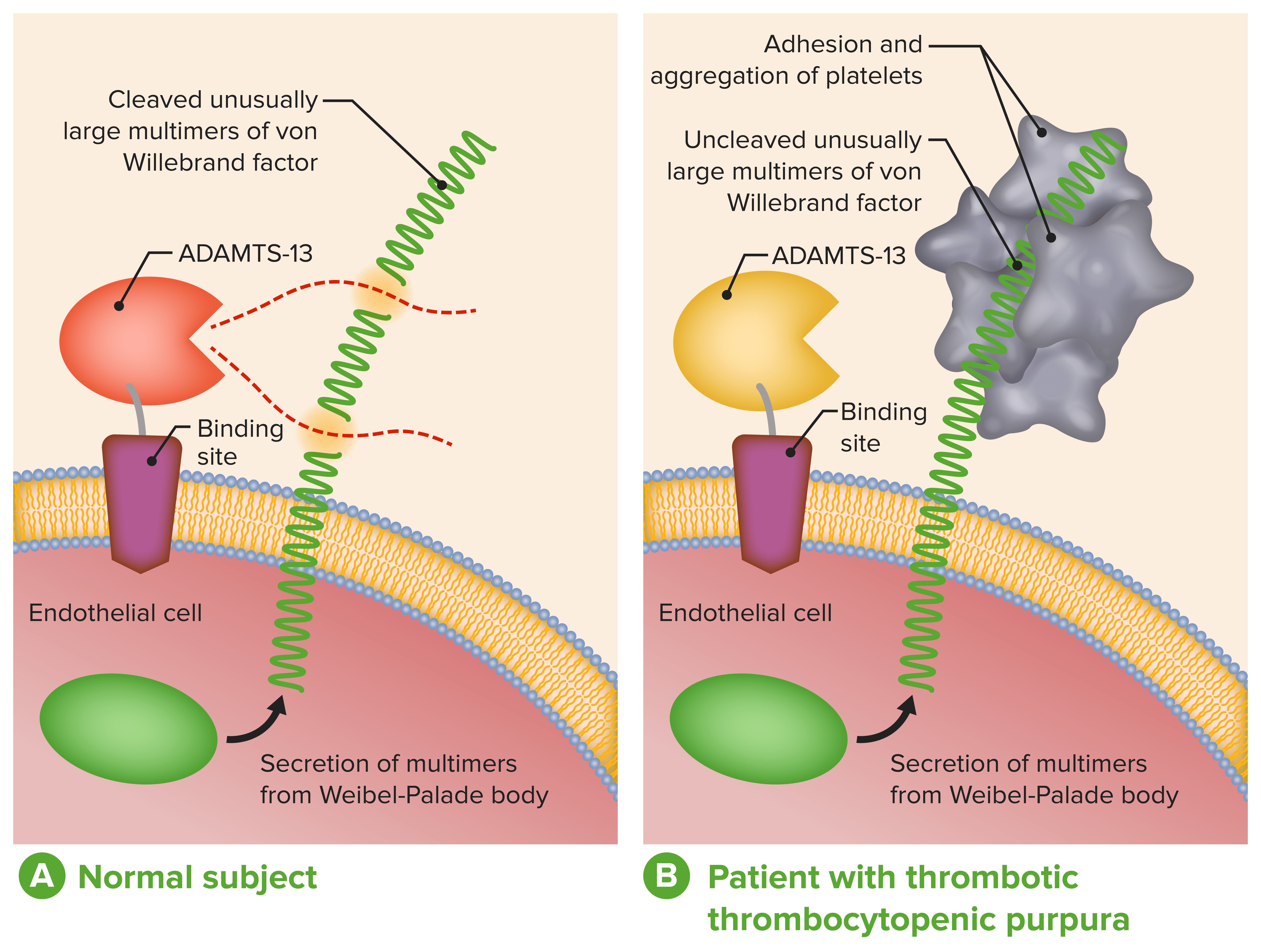
A: Normal physiological function of ADAMTS-13
B: Absent or severely reduced activity of ADAMTS-13 in patients with TTP prevents timely cleavage of unusually large multimers of VWF during secretion by endothelial cells.

Image by Lecturio.

Clinical Presentation

Acquired TTP

Classic TTP pentad:

  • Only present in < 5% of patients Patients Individuals participating in the health care system for the purpose of receiving therapeutic, diagnostic, or preventive procedures. Clinician–Patient Relationship
  • A complete pentad usually indicates late, untreated disease with high mortality Mortality All deaths reported in a given population. Measures of Health Status.
  • Findings include:
    • Microangiopathic hemolytic anemia Microangiopathic Hemolytic Anemia Hemolytic Uremic Syndrome
    • Thrombocytopenia Thrombocytopenia Thrombocytopenia occurs when the platelet count is < 150,000 per microliter. The normal range for platelets is usually 150,000-450,000/µL of whole blood. Thrombocytopenia can be a result of decreased production, increased destruction, or splenic sequestration of platelets. Patients are often asymptomatic until platelet counts are < 50,000/µL. Thrombocytopenia
    • Fever Fever Fever is defined as a measured body temperature of at least 38°C (100.4°F). Fever is caused by circulating endogenous and/or exogenous pyrogens that increase levels of prostaglandin E2 in the hypothalamus. Fever is commonly associated with chills, rigors, sweating, and flushing of the skin. Fever
    • Acute renal failure Renal failure Conditions in which the kidneys perform below the normal level in the ability to remove wastes, concentrate urine, and maintain electrolyte balance; blood pressure; and calcium metabolism. Renal insufficiency can be classified by the degree of kidney damage (as measured by the level of proteinuria) and reduction in glomerular filtration rate. Crush Syndrome
    • Severe neurologic symptoms

Organ-system manifestations:

  • Bleeding: 
  • Gastrointestinal symptoms:
    • Abdominal pain Abdominal Pain Acute Abdomen
    • Diarrhea Diarrhea Diarrhea is defined as ≥ 3 watery or loose stools in a 24-hour period. There are a multitude of etiologies, which can be classified based on the underlying mechanism of disease. The duration of symptoms (acute or chronic) and characteristics of the stools (e.g., watery, bloody, steatorrheic, mucoid) can help guide further diagnostic evaluation. Diarrhea
    • Nausea Nausea An unpleasant sensation in the stomach usually accompanied by the urge to vomit. Common causes are early pregnancy, sea and motion sickness, emotional stress, intense pain, food poisoning, and various enteroviruses. Antiemetics
    • Vomiting Vomiting The forcible expulsion of the contents of the stomach through the mouth. Hypokalemia
  • Minor neurologic findings:
  • Major neurologic findings:
    • Coma Coma Coma is defined as a deep state of unarousable unresponsiveness, characterized by a score of 3 points on the GCS. A comatose state can be caused by a multitude of conditions, making the precise epidemiology and prognosis of coma difficult to determine. Coma
    • Stroke
    • Seizures Seizures A seizure is abnormal electrical activity of the neurons in the cerebral cortex that can manifest in numerous ways depending on the region of the brain affected. Seizures consist of a sudden imbalance that occurs between the excitatory and inhibitory signals in cortical neurons, creating a net excitation. The 2 major classes of seizures are focal and generalized. Seizures
    • Transient focal abnormalities (speech impairment, weakness/numbness)
  • Renal involvement:
    • Renal insufficiency, edema Edema Edema is a condition in which excess serous fluid accumulates in the body cavity or interstitial space of connective tissues. Edema is a symptom observed in several medical conditions. It can be categorized into 2 types, namely, peripheral (in the extremities) and internal (in an organ or body cavity). Edema
    • Acute anuric renal failure Renal failure Conditions in which the kidneys perform below the normal level in the ability to remove wastes, concentrate urine, and maintain electrolyte balance; blood pressure; and calcium metabolism. Renal insufficiency can be classified by the degree of kidney damage (as measured by the level of proteinuria) and reduction in glomerular filtration rate. Crush Syndrome is rare.
  • Cardiac symptoms:
    • Arrhythmia
    • Heart failure Heart Failure A heterogeneous condition in which the heart is unable to pump out sufficient blood to meet the metabolic need of the body. Heart failure can be caused by structural defects, functional abnormalities (ventricular dysfunction), or a sudden overload beyond its capacity. Chronic heart failure is more common than acute heart failure which results from sudden insult to cardiac function, such as myocardial infarction. Total Anomalous Pulmonary Venous Return (TAPVR)
    • MI MI MI is ischemia and death of an area of myocardial tissue due to insufficient blood flow and oxygenation, usually from thrombus formation on a ruptured atherosclerotic plaque in the epicardial arteries. Clinical presentation is most commonly with chest pain, but women and patients with diabetes may have atypical symptoms. Myocardial Infarction
    • Sudden cardiac death Sudden cardiac death Cardiac arrest is the sudden, complete cessation of cardiac output with hemodynamic collapse. Patients present as pulseless, unresponsive, and apneic. Rhythms associated with cardiac arrest are ventricular fibrillation/tachycardia, asystole, or pulseless electrical activity. Cardiac Arrest
  • Constitutional:
    • Fever Fever Fever is defined as a measured body temperature of at least 38°C (100.4°F). Fever is caused by circulating endogenous and/or exogenous pyrogens that increase levels of prostaglandin E2 in the hypothalamus. Fever is commonly associated with chills, rigors, sweating, and flushing of the skin. Fever (rare)
    • Weakness
    • Not all patients Patients Individuals participating in the health care system for the purpose of receiving therapeutic, diagnostic, or preventive procedures. Clinician–Patient Relationship are critically ill (some present with vague complaints).
Typical petechial rash due to ttp

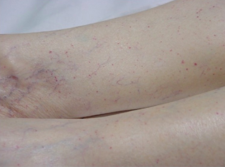
Typical petechial rash on a patient’s lower extremities

Image: “Typical petechial rash” by Department of Emergency Medicine, University of Florida, Gainesville, FL 32610, USA. License: CC BY 3.0

Congenital Congenital Chorioretinitis TTP

Unlike acquired TTP, congenital Congenital Chorioretinitis TTP does not present with acute episodes.

Symptoms are usually mild, vague, and continuous:

  • Lethargy Lethargy A general state of sluggishness, listless, or uninterested, with being tired, and having difficulty concentrating and doing simple tasks. It may be related to depression or drug addiction. Hyponatremia
  • Headache Headache The symptom of pain in the cranial region. It may be an isolated benign occurrence or manifestation of a wide variety of headache disorders. Brain Abscess
  • Loss of concentration
  • Abdominal discomfort

Severe and life-threatening exacerbations are most likely during 2 life periods:

  • Neonatal:
    • Most critical are the 1st few days of life.
    • Severe hemolysis
    • Hyperbilirubinemia Hyperbilirubinemia A condition characterized by an abnormal increase of bilirubin in the blood, which may result in jaundice. Bilirubin, a breakdown product of heme, is normally excreted in the bile or further catabolized before excretion in the urine. Jaundice
    • May subsequently present with intermittent thrombocytopenia Thrombocytopenia Thrombocytopenia occurs when the platelet count is < 150,000 per microliter. The normal range for platelets is usually 150,000-450,000/µL of whole blood. Thrombocytopenia can be a result of decreased production, increased destruction, or splenic sequestration of platelets. Patients are often asymptomatic until platelet counts are < 50,000/µL. Thrombocytopenia 
    • May remain asymptomatic until adulthood
  • Pregnancy Pregnancy The status during which female mammals carry their developing young (embryos or fetuses) in utero before birth, beginning from fertilization to birth. Pregnancy: Diagnosis, Physiology, and Care:
    • Severe preeclampsia Preeclampsia A complication of pregnancy, characterized by a complex of symptoms including maternal hypertension and proteinuria with or without pathological edema. Symptoms may range between mild and severe. Pre-eclampsia usually occurs after the 20th week of gestation, but may develop before this time in the presence of trophoblastic disease. Hypertensive Pregnancy Disorders before 20-weeks gestation
    • Thrombocytopenia Thrombocytopenia Thrombocytopenia occurs when the platelet count is < 150,000 per microliter. The normal range for platelets is usually 150,000-450,000/µL of whole blood. Thrombocytopenia can be a result of decreased production, increased destruction, or splenic sequestration of platelets. Patients are often asymptomatic until platelet counts are < 50,000/µL. Thrombocytopenia
    • Hemolysis
    • Transient neurologic symptoms
    • Acute kidney injury Acute Kidney Injury Acute kidney injury refers to sudden and often reversible loss of renal function, which develops over days or weeks. Azotemia refers to elevated levels of nitrogen-containing substances in the blood that accompany AKI, which include BUN and creatinine. Acute Kidney Injury
    • Fetal death

Diagnosis

History

  • Easy bleeding/bruising
  • Abdominal pain Abdominal Pain Acute Abdomen/discomfort
  • Fatigue Fatigue The state of weariness following a period of exertion, mental or physical, characterized by a decreased capacity for work and reduced efficiency to respond to stimuli. Fibromyalgia, headaches, lethargy Lethargy A general state of sluggishness, listless, or uninterested, with being tired, and having difficulty concentrating and doing simple tasks. It may be related to depression or drug addiction. Hyponatremia
  • Transient neurologic impairments
  • Disorders of immune system Immune system The body’s defense mechanism against foreign organisms or substances and deviant native cells. It includes the humoral immune response and the cell-mediated response and consists of a complex of interrelated cellular, molecular, and genetic components. Primary Lymphatic Organs/malignancies
  • Family history Family History Adult Health Maintenance of bleeding disorders Bleeding disorders Hypocoagulable conditions, also known as bleeding disorders or bleeding diathesis, are a diverse group of diseases that result in abnormal hemostasis. Physiologic hemostasis is dependent on the integrity of endothelial cells, subendothelial matrix, platelets, and coagulation factors. The hypocoagulable states result from abnormalities in one or more of these contributors, resulting in ineffective thrombosis and bleeding. Hypocoagulable Conditions

Physical exam

  • Can be nonrevealing
  • Skin Skin The skin, also referred to as the integumentary system, is the largest organ of the body. The skin is primarily composed of the epidermis (outer layer) and dermis (deep layer). The epidermis is primarily composed of keratinocytes that undergo rapid turnover, while the dermis contains dense layers of connective tissue. Skin: Structure and Functions: petechiae Petechiae Primary Skin Lesions, purpura, bruising
  • Generalized edema Edema Edema is a condition in which excess serous fluid accumulates in the body cavity or interstitial space of connective tissues. Edema is a symptom observed in several medical conditions. It can be categorized into 2 types, namely, peripheral (in the extremities) and internal (in an organ or body cavity). Edema (renal insufficiency)
  • Focal neurologic abnormalities
  • Abnormal mental status

Laboratory studies

  • CBC:
    • Thrombocytopenia Thrombocytopenia Thrombocytopenia occurs when the platelet count is < 150,000 per microliter. The normal range for platelets is usually 150,000-450,000/µL of whole blood. Thrombocytopenia can be a result of decreased production, increased destruction, or splenic sequestration of platelets. Patients are often asymptomatic until platelet counts are < 50,000/µL. Thrombocytopenia ( platelets Platelets Platelets are small cell fragments involved in hemostasis. Thrombopoiesis takes place primarily in the bone marrow through a series of cell differentiation and is influenced by several cytokines. Platelets are formed after fragmentation of the megakaryocyte cytoplasm. Platelets: Histology < 50 X 109/L)
    • ↓ Hemoglobin (< 10 g/dL; hemolysis)
  • Coagulation studies Coagulation studies Coagulation studies are a group of hematologic laboratory studies that reflect the function of blood vessels, platelets, and coagulation factors, which all interact with one another to achieve hemostasis. Coagulation studies are usually ordered to evaluate patients with bleeding or hypercoagulation disorders. Coagulation Studies:
    • Increased bleeding time Bleeding time Duration of blood flow after skin puncture. This test is used as a measure of capillary and platelet function. Hemostasis 
    • Normal PT, aPTT, INR, and fibrinogen Fibrinogen Plasma glycoprotein clotted by thrombin, composed of a dimer of three non-identical pairs of polypeptide chains (alpha, beta, gamma) held together by disulfide bonds. Fibrinogen clotting is a sol-gel change involving complex molecular arrangements: whereas fibrinogen is cleaved by thrombin to form polypeptides a and b, the proteolytic action of other enzymes yields different fibrinogen degradation products. Hemostasis
  • Renal function:
    • ↑ Creatinine 
    • ↑ BUN
  • Liver function tests Liver function tests Liver function tests, also known as hepatic function panels, are one of the most commonly performed screening blood tests. Such tests are also used to detect, evaluate, and monitor acute and chronic liver diseases. Liver Function Tests
  • ADAMTS-13 activity assays:
    • < 10% activity
    • > 60% activity usually rules out TTP diagnosis.
    • Inhibitor levels can be measured.
    • If no inhibitors detected, genetic testing Genetic Testing Detection of a mutation; genotype; karyotype; or specific alleles associated with genetic traits, heritable diseases, or predisposition to a disease, or that may lead to the disease in descendants. It includes prenatal genetic testing. Myotonic Dystrophies for hereditary TTP
  • Blood smear Blood smear Myeloperoxidase Deficiency:
    •  Fragmented RBCs RBCs Erythrocytes, or red blood cells (RBCs), are the most abundant cells in the blood. While erythrocytes in the fetus are initially produced in the yolk sac then the liver, the bone marrow eventually becomes the main site of production. Erythrocytes: Histology ( schistocytes Schistocytes Hemolytic Uremic Syndrome)
    •   Thrombocytopenia Thrombocytopenia Thrombocytopenia occurs when the platelet count is < 150,000 per microliter. The normal range for platelets is usually 150,000-450,000/µL of whole blood. Thrombocytopenia can be a result of decreased production, increased destruction, or splenic sequestration of platelets. Patients are often asymptomatic until platelet counts are < 50,000/µL. Thrombocytopenia
Peripheral blood film showing schistocytes

Thrombotic thrombocytopenic purpura (TTP):
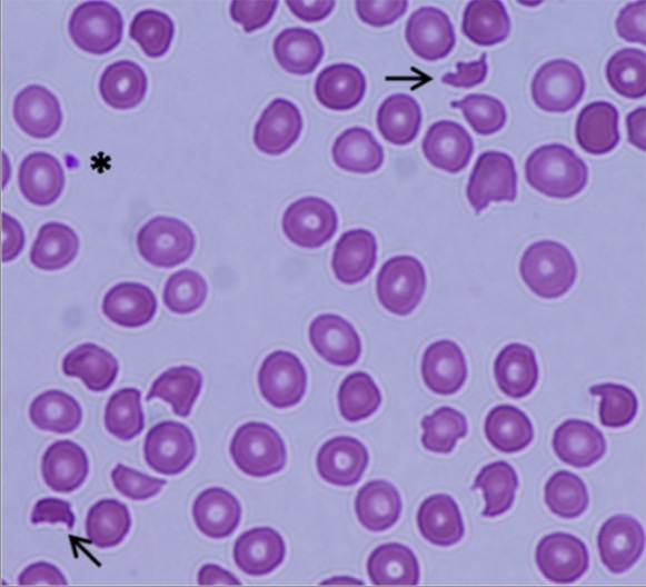
Peripheral blood smear showing schistocytes (arrows) and a marked decrease in platelet numbers, indicating microangiopathic hemolytic anemia

Image: “Peripheral blood film showing schistocytes” by Department of Medicine, Division of Hematology, Division of Hematopathology, Queen Elizabeth II Health Sciences Centre, Capital District Health, Authority and Dalhousie University, Halifax, Nova Scotia Canada. License: CC BY 4.0

Imaging

  • CT or MRI of the brain Brain The part of central nervous system that is contained within the skull (cranium). Arising from the neural tube, the embryonic brain is comprised of three major parts including prosencephalon (the forebrain); mesencephalon (the midbrain); and rhombencephalon (the hindbrain). The developed brain consists of cerebrum; cerebellum; and other structures in the brain stem. Nervous System: Anatomy, Structure, and Classification
  • Indicated for neurologic symptoms
  • Rule out ischemic/ hemorrhagic stroke Hemorrhagic stroke Stroke due to rupture of a weakened blood vessel in the brain (e.g., cerebral hemispheres; cerebellum; subarachnoid space). Subarachnoid Hemorrhage.
Emergent head ct demonstrating the larger of two frontoparietal hemorrhages

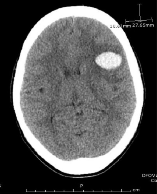
Head CT in a patient demonstrating the larger of 2 frontoparietal hemorrhages

Image: “Emergent head CT demonstrating the larger of two frontoparietal hemorrhages” by Divisions of Infectious Disease, The George Washington University, School of Medicine and Health Sciences, Washington, DC, USA. License: CC BY 3.0

PLASMIC score

The PLASMIC score predicts the probability Probability Probability is a mathematical tool used to study randomness and provide predictions about the likelihood of something happening. There are several basic rules of probability that can be used to help determine the probability of multiple events happening together, separately, or sequentially. Basics of Probability of TTP in the presence of schistocytes Schistocytes Hemolytic Uremic Syndrome on blood smear Blood smear Myeloperoxidase Deficiency.

PLASMIC criteria (1 point for each):

  • Platelet count < 30,000/μL
  • Hemolysis (defined by reticulocyte Reticulocyte Immature erythrocytes. In humans, these are erythroid cells that have just undergone extrusion of their cell nucleus. They still contain some organelles that gradually decrease in number as the cells mature. Ribosomes are last to disappear. Certain staining techniques cause components of the ribosomes to precipitate into characteristic ‘reticulum’ (not the same as the endoplasmic reticulum), hence the name reticulocytes. Erythrocytes: Histology count > 2.5%, undetectable haptoglobin, or indirect bilirubin Indirect Bilirubin Liver Function Tests > 2 mg/dL)
  • No active cancer
  • No solid organ or stem cell transplant
  • MCV < 90 fL
  • INR < 1.5
  • Creatinine < 2.0 mg/dL

Score:

  • 0–4 points: low probability Probability Probability is a mathematical tool used to study randomness and provide predictions about the likelihood of something happening. There are several basic rules of probability that can be used to help determine the probability of multiple events happening together, separately, or sequentially. Basics of Probability of TTP
  • 5 points: intermediate probability Probability Probability is a mathematical tool used to study randomness and provide predictions about the likelihood of something happening. There are several basic rules of probability that can be used to help determine the probability of multiple events happening together, separately, or sequentially. Basics of Probability of TTP
  • 6–7 points: high probability Probability Probability is a mathematical tool used to study randomness and provide predictions about the likelihood of something happening. There are several basic rules of probability that can be used to help determine the probability of multiple events happening together, separately, or sequentially. Basics of Probability of TTP

Management

  • TTP episode/exacerbation should be treated emergently.
  • Almost always fatal if not promptly treated
  • With appropriate treatment, survival is > 90%.
  • The mainstay of treatment is plasma Plasma The residual portion of blood that is left after removal of blood cells by centrifugation without prior blood coagulation. Transfusion Products exchange (PEX):
    • Replenishes functional ADAMTS-13
    • Removes offending antibodies Antibodies Immunoglobulins (Igs), also known as antibodies, are glycoprotein molecules produced by plasma cells that act in immune responses by recognizing and binding particular antigens. The various Ig classes are IgG (the most abundant), IgM, IgE, IgD, and IgA, which differ in their biologic features, structure, target specificity, and distribution. Immunoglobulins: Types and Functions
    • Performed daily until recovery
  • Plasma Plasma The residual portion of blood that is left after removal of blood cells by centrifugation without prior blood coagulation. Transfusion Products infusion (temporizing measure if no plasma Plasma The residual portion of blood that is left after removal of blood cells by centrifugation without prior blood coagulation. Transfusion Products exchange available)
  • Additional therapies:
    • Steroids Steroids A group of polycyclic compounds closely related biochemically to terpenes. They include cholesterol, numerous hormones, precursors of certain vitamins, bile acids, alcohols (sterols), and certain natural drugs and poisons. Steroids have a common nucleus, a fused, reduced 17-carbon atom ring system, cyclopentanoperhydrophenanthrene. Most steroids also have two methyl groups and an aliphatic side-chain attached to the nucleus. Benign Liver Tumors
    • Rituximab Rituximab A murine-derived monoclonal antibody and antineoplastic agent that binds specifically to the cd20 antigen and is used in the treatment of leukemia; lymphoma and rheumatoid arthritis. Immunosuppressants
    • Caplacizumab (anti-VWF antibody)
    • Platelet transfusion for thrombocytopenia Thrombocytopenia Thrombocytopenia occurs when the platelet count is < 150,000 per microliter. The normal range for platelets is usually 150,000-450,000/µL of whole blood. Thrombocytopenia can be a result of decreased production, increased destruction, or splenic sequestration of platelets. Patients are often asymptomatic until platelet counts are < 50,000/µL. Thrombocytopenia

Differential Diagnosis

  • Hemolytic uremic syndrome Hemolytic uremic syndrome A syndrome that is associated with microvascular diseases of the kidney, such as renal cortical necrosis. It is characterized by hemolytic anemia; thrombocytopenia; and acute renal failure. Hypocoagulable Conditions: a disease mainly in children or the elderly. Shiga toxin Shiga toxin A class of toxins that inhibit protein synthesis by blocking the interaction of ribosomal RNA; with peptide elongation factors. They include shiga toxin which is produced by Shigella dysenteriae and a variety of shiga-like toxins that are produced by pathologic strains of Escherichia coli such as Escherichia coli o157. Diarrheagenic E. coli results in severe thrombocytopenia Severe Thrombocytopenia Thrombocytopenia, hemolytic anemia Hemolytic Anemia Hemolytic anemia (HA) is the term given to a large group of anemias that are caused by the premature destruction/hemolysis of circulating red blood cells (RBCs). Hemolysis can occur within (intravascular hemolysis) or outside the blood vessels (extravascular hemolysis). Hemolytic Anemia, and resultant kidney injury. Patients Patients Individuals participating in the health care system for the purpose of receiving therapeutic, diagnostic, or preventive procedures. Clinician–Patient Relationship usually have a gastrointestinal prodrome Prodrome Symptoms that appear 24–48 hours prior to migraine onset. Migraine Headache before the development of thrombocytopenia Thrombocytopenia Thrombocytopenia occurs when the platelet count is < 150,000 per microliter. The normal range for platelets is usually 150,000-450,000/µL of whole blood. Thrombocytopenia can be a result of decreased production, increased destruction, or splenic sequestration of platelets. Patients are often asymptomatic until platelet counts are < 50,000/µL. Thrombocytopenia and anemia Anemia Anemia is a condition in which individuals have low Hb levels, which can arise from various causes. Anemia is accompanied by a reduced number of RBCs and may manifest with fatigue, shortness of breath, pallor, and weakness. Subtypes are classified by the size of RBCs, chronicity, and etiology. Anemia: Overview and Types. Hemolytic uremic syndrome Hemolytic uremic syndrome A syndrome that is associated with microvascular diseases of the kidney, such as renal cortical necrosis. It is characterized by hemolytic anemia; thrombocytopenia; and acute renal failure. Hypocoagulable Conditions has symptoms very similar to TTP; the difference is gastrointestinal prodrome Prodrome Symptoms that appear 24–48 hours prior to migraine onset. Migraine Headache and management (mainly supportive).
  • Immune-mediated thrombocytopenic purpura (previously known as idiopathic Idiopathic Dermatomyositis thrombocytopenic purpura): a condition developing secondary to immune-mediated destruction of platelets Platelets Platelets are small cell fragments involved in hemostasis. Thrombopoiesis takes place primarily in the bone marrow through a series of cell differentiation and is influenced by several cytokines. Platelets are formed after fragmentation of the megakaryocyte cytoplasm. Platelets: Histology resulting in thrombocytopenia Thrombocytopenia Thrombocytopenia occurs when the platelet count is < 150,000 per microliter. The normal range for platelets is usually 150,000-450,000/µL of whole blood. Thrombocytopenia can be a result of decreased production, increased destruction, or splenic sequestration of platelets. Patients are often asymptomatic until platelet counts are < 50,000/µL. Thrombocytopenia. IgG IgG The major immunoglobulin isotype class in normal human serum. There are several isotype subclasses of igg, for example, igg1, igg2a, and igg2b. Hypersensitivity Pneumonitis antibodies Antibodies Immunoglobulins (Igs), also known as antibodies, are glycoprotein molecules produced by plasma cells that act in immune responses by recognizing and binding particular antigens. The various Ig classes are IgG (the most abundant), IgM, IgE, IgD, and IgA, which differ in their biologic features, structure, target specificity, and distribution. Immunoglobulins: Types and Functions tag the platelets Platelets Platelets are small cell fragments involved in hemostasis. Thrombopoiesis takes place primarily in the bone marrow through a series of cell differentiation and is influenced by several cytokines. Platelets are formed after fragmentation of the megakaryocyte cytoplasm. Platelets: Histology, which are then cleared by the spleen Spleen The spleen is the largest lymphoid organ in the body, located in the LUQ of the abdomen, superior to the left kidney and posterior to the stomach at the level of the 9th-11th ribs just below the diaphragm. The spleen is highly vascular and acts as an important blood filter, cleansing the blood of pathogens and damaged erythrocytes. Spleen: Anatomy. Immune-mediated thrombocytopenic purpura is a diagnosis of exclusion and is managed with steroids Steroids A group of polycyclic compounds closely related biochemically to terpenes. They include cholesterol, numerous hormones, precursors of certain vitamins, bile acids, alcohols (sterols), and certain natural drugs and poisons. Steroids have a common nucleus, a fused, reduced 17-carbon atom ring system, cyclopentanoperhydrophenanthrene. Most steroids also have two methyl groups and an aliphatic side-chain attached to the nucleus. Benign Liver Tumors and IV immune globulin.
  • Von Willebrand’s disease ( VWD vWD Von Willebrand disease (vWD) is a bleeding disorder characterized by a qualitative or quantitative deficiency of von Willebrand factor (vWF). Von Willebrand factor is a multimeric protein involved in the plate adhesion phase of hemostasis by forming a bridge between platelets and damaged portions of the vessel wall. Von Willebrand Disease): The most common inherited bleeding disorder, VWD vWD Von Willebrand disease (vWD) is a bleeding disorder characterized by a qualitative or quantitative deficiency of von Willebrand factor (vWF). Von Willebrand factor is a multimeric protein involved in the plate adhesion phase of hemostasis by forming a bridge between platelets and damaged portions of the vessel wall. Von Willebrand Disease is caused by an abnormality of VWF. Bruising and mucocutaneous bleeding are common manifestations. Most individuals with VWD vWD Von Willebrand disease (vWD) is a bleeding disorder characterized by a qualitative or quantitative deficiency of von Willebrand factor (vWF). Von Willebrand factor is a multimeric protein involved in the plate adhesion phase of hemostasis by forming a bridge between platelets and damaged portions of the vessel wall. Von Willebrand Disease have a normal CBC, and many have normal coagulation studies Coagulation studies Coagulation studies are a group of hematologic laboratory studies that reflect the function of blood vessels, platelets, and coagulation factors, which all interact with one another to achieve hemostasis. Coagulation studies are usually ordered to evaluate patients with bleeding or hypercoagulation disorders. Coagulation Studies. However, some individuals have a prolonged aPTT. Treatment is usually with desmopressin Desmopressin Hemophilia
  • Disseminated intravascular coagulation Disseminated intravascular coagulation Disseminated intravascular coagulation (DIC) is a condition characterized by systemic bodywide activation of the coagulation cascade. This cascade results in both widespread microvascular thrombi contributing to multiple organ dysfunction and consumption of clotting factors and platelets, leading to hemorrhage. Disseminated Intravascular Coagulation ( DIC DIC Disseminated intravascular coagulation (DIC) is a condition characterized by systemic bodywide activation of the coagulation cascade. This cascade results in both widespread microvascular thrombi contributing to multiple organ dysfunction and consumption of clotting factors and platelets, leading to hemorrhage. Disseminated Intravascular Coagulation): A complication of many diseases, DIC DIC Disseminated intravascular coagulation (DIC) is a condition characterized by systemic bodywide activation of the coagulation cascade. This cascade results in both widespread microvascular thrombi contributing to multiple organ dysfunction and consumption of clotting factors and platelets, leading to hemorrhage. Disseminated Intravascular Coagulation is an imbalance between procoagulants and anticoagulants Anticoagulants Anticoagulants are drugs that retard or interrupt the coagulation cascade. The primary classes of available anticoagulants include heparins, vitamin K-dependent antagonists (e.g., warfarin), direct thrombin inhibitors, and factor Xa inhibitors. Anticoagulants. There is an excessive consumption of platelets Platelets Platelets are small cell fragments involved in hemostasis. Thrombopoiesis takes place primarily in the bone marrow through a series of cell differentiation and is influenced by several cytokines. Platelets are formed after fragmentation of the megakaryocyte cytoplasm. Platelets: Histology, coagulation factors Coagulation factors Endogenous substances, usually proteins, that are involved in the blood coagulation process. Hemostasis, and fibrinogen Fibrinogen Plasma glycoprotein clotted by thrombin, composed of a dimer of three non-identical pairs of polypeptide chains (alpha, beta, gamma) held together by disulfide bonds. Fibrinogen clotting is a sol-gel change involving complex molecular arrangements: whereas fibrinogen is cleaved by thrombin to form polypeptides a and b, the proteolytic action of other enzymes yields different fibrinogen degradation products. Hemostasis due to thrombus formation. Presentation includes signs and symptoms of thrombocytopenia Thrombocytopenia Thrombocytopenia occurs when the platelet count is < 150,000 per microliter. The normal range for platelets is usually 150,000-450,000/µL of whole blood. Thrombocytopenia can be a result of decreased production, increased destruction, or splenic sequestration of platelets. Patients are often asymptomatic until platelet counts are < 50,000/µL. Thrombocytopenia. The disease has high morbidity Morbidity The proportion of patients with a particular disease during a given year per given unit of population. Measures of Health Status and mortality Mortality All deaths reported in a given population. Measures of Health Status and requires emergent care, which involves blood product Product A molecule created by the enzymatic reaction. Basics of Enzymes transfusion and treatment of the underlying process. 
  • Henoch-Schönlein purpura Henoch-Schönlein Purpura Henoch-Schönlein purpura (HSP), also known as immunoglobulin A vasculitis, is an autoimmune small-vessel vasculitis that typically presents as a tetrad of abdominal pain, arthralgia, hematuria, and purpuric rash. Henoch-Schönlein Purpura: an autoimmune small-vessel vasculitis Small-Vessel Vasculitis Henoch-Schönlein Purpura typically presenting as a tetrad of abdominal pain Abdominal Pain Acute Abdomen, hematuria Hematuria Presence of blood in the urine. Renal Cell Carcinoma, arthritis Arthritis Acute or chronic inflammation of joints. Osteoarthritis, and purpuric rash Rash Rocky Mountain Spotted Fever. Diagnosis is commonly established clinically and management is mostly supportive with a focus on symptoms.
  • Infections Infections Invasion of the host organism by microorganisms or their toxins or by parasites that can cause pathological conditions or diseases. Chronic Granulomatous Disease and drugs: many infections Infections Invasion of the host organism by microorganisms or their toxins or by parasites that can cause pathological conditions or diseases. Chronic Granulomatous Disease and medications can cause thrombocytopenia Thrombocytopenia Thrombocytopenia occurs when the platelet count is < 150,000 per microliter. The normal range for platelets is usually 150,000-450,000/µL of whole blood. Thrombocytopenia can be a result of decreased production, increased destruction, or splenic sequestration of platelets. Patients are often asymptomatic until platelet counts are < 50,000/µL. Thrombocytopenia by several nonimmune mechanisms. Examples include bone marrow Bone marrow The soft tissue filling the cavities of bones. Bone marrow exists in two types, yellow and red. Yellow marrow is found in the large cavities of large bones and consists mostly of fat cells and a few primitive blood cells. Red marrow is a hematopoietic tissue and is the site of production of erythrocytes and granular leukocytes. Bone marrow is made up of a framework of connective tissue containing branching fibers with the frame being filled with marrow cells. Bone Marrow: Composition and Hematopoiesis suppression Suppression Defense Mechanisms, hypersplenism Hypersplenism Condition characterized by splenomegaly, some reduction in the number of circulating blood cells in the presence of a normal or hyperactive bone marrow, and the potential for reversal by splenectomy. Splenomegaly, and platelet consumption.

References

  1. George, J., and Cuker, A. (2021), Acquired TTP: Initial treatment. In Leung, L. (Ed.), UpToDate. Retrieved March 06, 2021, from https://www.uptodate.com/contents/acquired-ttp-initial-treatment
  2. George, J., and Cuker, A. (2021), Acquired TTP: Clinical manifestations and diagnosis. In Leung, L. (Ed.), UpToDate. Retrieved March 06, 2021, from https://www.uptodate.com/contents/acquired-ttp-clinical-manifestations-and-diagnosis
  3. George, J., and Nester, C. (2021), Approach to the patient with suspected TTP, or other thrombotic microangiopathy (TMA). In Leung, L. (Ed.), UpToDate. Retrieved March 06, 2021, from https://www.uptodate.com/contents/approach-to-the-patient-with-suspected-ttp-hus-or-other-thrombotic-microangiopathy-tma
  4. Vesely SK, George JN, Lämmle B, et al. (2003). ADAMTS13 activity in thrombotic thrombocytopenic purpura-hemolytic uremic syndrome: relation to presenting features and clinical outcomes in a prospective cohort of 142 patients. Blood 2003; 102:60.
  5. Page EE, Kremer Hovinga JA, Terrell DR, et al. (2017). Thrombotic thrombocytopenic purpura: diagnostic criteria, clinical features, and long-term outcomes from 1995 through 2015. Blood Adv 2017; 1:590.
  6. Reese JA, Muthurajah DS, Kremer Hovinga JA, et al. (2013). Children and adults with thrombotic thrombocytopenic purpura associated with severe, acquired Adamts13 deficiency: comparison of incidence, demographic and clinical features. Pediatr Blood Cancer.

Create your free account or log in to continue reading!

Sign up now and get free access to Lecturio with concept pages, medical videos, and questions for your medical education.

User Reviews

Details