Advertisement

Advertisement

Advertisement

Advertisement

Acute Kidney Injury

Acute kidney injury refers to sudden and often reversible loss of renal function, which develops over days or weeks. Azotemia refers to elevated levels of nitrogen-containing substances in the blood that accompany AKI, which include BUN and creatinine. Uremia refers specifically to the constellation of symptoms that occur with severe renal dysfunction. The etiologies of AKI are classified as prerenal, intrinsic renal, or post-renal, and there is a varied clinical presentation depending on the severity of kidney dysfunction. Acute kidney injury is diagnosed initially by a change in serum creatinine levels, and then the etiology is determined by clinical history, lab testing, imaging, and possibly kidney biopsy Biopsy Removal and pathologic examination of specimens from the living body. Ewing Sarcoma. The management of AKI depends on the etiology; however, attention Attention Focusing on certain aspects of current experience to the exclusion of others. It is the act of heeding or taking notice or concentrating. Psychiatric Assessment to the individual's volume status Volume Status ACES and RUSH: Resuscitation Ultrasound Protocols and serum electrolytes Electrolytes Electrolytes are mineral salts that dissolve in water and dissociate into charged particles called ions, which can be either be positively (cations) or negatively (anions) charged. Electrolytes are distributed in the extracellular and intracellular compartments in different concentrations. Electrolytes are essential for various basic life-sustaining functions. Electrolytes is always important. If management is unsuccessful and AKI progresses to CKD CKD Chronic kidney disease (CKD) is kidney impairment that lasts for ≥ 3 months, implying that it is irreversible. Hypertension and diabetes are the most common causes; however, there are a multitude of other etiologies. In the early to moderate stages, CKD is usually asymptomatic and is primarily diagnosed by laboratory abnormalities. Chronic Kidney Disease, then renal replacement therapy with dialysis Dialysis Renal replacement therapy refers to dialysis and/or kidney transplantation. Dialysis is a procedure by which toxins and excess water are removed from the circulation. Hemodialysis and peritoneal dialysis (PD) are the two types of dialysis, and their primary difference is the location of the filtration process (external to the body in hemodialysis versus inside the body for PD). Peritoneal Dialysis and Hemodialysis or kidney transplantation Kidney Transplantation The transference of a kidney from one human or animal to another. Organ Transplantation is necessary.

Last updated: May 17, 2024

Editorial responsibility: Stanley Oiseth, Lindsay Jones, Evelin Maza

Advertisement

Advertisement

Advertisement

Advertisement

Advertisement

Advertisement

Overview

Definition

Acute kidney injury refers to an abrupt decrease in kidney function, manifested by an increase in serum creatinine level, with or without reduced urine output.

Classification

Previously there had been more than 30 definitions of “acute renal failure Renal failure Conditions in which the kidneys perform below the normal level in the ability to remove wastes, concentrate urine, and maintain electrolyte balance; blood pressure; and calcium metabolism. Renal insufficiency can be classified by the degree of kidney damage (as measured by the level of proteinuria) and reduction in glomerular filtration rate. Crush Syndrome” in the literature, and these have been updated to the term AKI.

  • The RIFLE criteria:
    • Risk
    • Injury
    • Failure
    • Loss of kidney function
    • End-stage kidney disease
  • The Acute Kidney Injury Network (AKIN) criteria
  • Kidney Disease: Improving Global Outcomes (KDIGO) staging Staging Methods which attempt to express in replicable terms the extent of the neoplasm in the patient. Grading, Staging, and Metastasis of AKI:
    • Supersedes the other staging Staging Methods which attempt to express in replicable terms the extent of the neoplasm in the patient. Grading, Staging, and Metastasis systems; currently preferred
    • Classifies individuals according to criteria that result in the highest degree of injury, as do the other systems
    • Stage 1 Stage 1 Trypanosoma brucei/African trypanosomiasis:
      • ≥ 0.3 mg/dL increase in serum creatinine within 48 hours, or
      • Increase in serum creatinine > 1.5–1.9 times baseline in the past 7 days, or
      • Urine volume/urine output < 0.5 mL/kg/hr for 6–12 hours
    • Stage 2:
      • Serum creatinine 2.0‒2.9 times baseline, or
      • Urine volume/urine output < 0.5 mL/kg/hr for ≥ 12 hours
    • Stage 3:
      • Serum creatinine 3 times baseline, or
      • Serum creatinine ≥ 4 mg/dL, or
      • Initiation of renal replacement therapy, or
      • Decrease in estimated GFR GFR The volume of water filtered out of plasma through glomerular capillary walls into Bowman’s capsules per unit of time. It is considered to be equivalent to inulin clearance. Kidney Function Tests < 35 mL/min/1.73 m2 in individuals < 18 years of age, or
      • Urine volume/urine output < 0.3 mL/kg/hr for ≥ 24 hours, or
      • Anuria for ≥ 12 hours

Epidemiology

  • 7% of individuals who are hospitalized develop AKI.
  • 20% of acutely ill individuals develop AKI.
  • Mortality Mortality All deaths reported in a given population. Measures of Health Status is low in uncomplicated AKI.
  • Mortality Mortality All deaths reported in a given population. Measures of Health Status is 50%‒70% in AKI associated with sepsis Sepsis Systemic inflammatory response syndrome with a proven or suspected infectious etiology. When sepsis is associated with organ dysfunction distant from the site of infection, it is called severe sepsis. When sepsis is accompanied by hypotension despite adequate fluid infusion, it is called septic shock. Sepsis and Septic Shock and multiple organ failure.
  • No gender Gender Gender Dysphoria predilection
  • More common in extremes of age

Etiology and Pathogenesis

The causes of AKI are commonly organized into prerenal, intrinsic renal, or postrenal etiologies. With prerenal and postrenal causes, the kidneys Kidneys The kidneys are a pair of bean-shaped organs located retroperitoneally against the posterior wall of the abdomen on either side of the spine. As part of the urinary tract, the kidneys are responsible for blood filtration and excretion of water-soluble waste in the urine. Kidneys: Anatomy have normal anatomy and the injury is due to the effects of the inciting factor.

Normal physiology

  • Normally, GFR GFR The volume of water filtered out of plasma through glomerular capillary walls into Bowman’s capsules per unit of time. It is considered to be equivalent to inulin clearance. Kidney Function Tests is maintained by compensatory mechanisms:
  • With decreased renal perfusion, compensatory mechanisms will:
    • Activate antidiuretic hormone Antidiuretic hormone Antidiuretic hormones released by the neurohypophysis of all vertebrates (structure varies with species) to regulate water balance and osmolarity. In general, vasopressin is a nonapeptide consisting of a six-amino-acid ring with a cysteine 1 to cysteine 6 disulfide bridge or an octapeptide containing a cystine. All mammals have arginine vasopressin except the pig with a lysine at position 8. Vasopressin, a vasoconstrictor, acts on the kidney collecting ducts to increase water reabsorption, increase blood volume and blood pressure. Hypernatremia (ADH) → increased water reabsorption in the distal tubule and collecting duct Collecting duct Straight tubes commencing in the radiate part of the kidney cortex where they receive the curved ends of the distal convoluted tubules. In the medulla the collecting tubules of each pyramid converge to join a central tube (duct of bellini) which opens on the summit of the papilla. Renal Cell Carcinoma → concentrated urine
    • Increase filtration fraction Filtration fraction Fraction of the RPF that actually moves across the glomerular membrane Glomerular Filtration → increased oncotic pressure Oncotic Pressure Edema in the glomerulus

Etiology and pathophysiology

  • Prerenal causes of AKI result in renal hypoperfusion due to:
    • True volume depletion Volume depletion Volume status is a balance between water and solutes, the majority of which is Na. Volume depletion refers to a loss of both water and Na, whereas dehydration refers only to a loss of water. Volume depletion can be caused by GI losses, renal losses, bleeding, poor oral Na intake, or third spacing of fluids. Volume Depletion and Dehydration: loss of sodium Sodium A member of the alkali group of metals. It has the atomic symbol na, atomic number 11, and atomic weight 23. Hyponatremia from the extracellular fluid Extracellular fluid The fluid of the body that is outside of cells. It is the external environment for the cells. Body Fluid Compartments (ECF) due to:
      • Acute blood loss/hemorrhage
      • GI losses with vomiting Vomiting The forcible expulsion of the contents of the stomach through the mouth. Hypokalemia or diarrhea Diarrhea Diarrhea is defined as ≥ 3 watery or loose stools in a 24-hour period. There are a multitude of etiologies, which can be classified based on the underlying mechanism of disease. The duration of symptoms (acute or chronic) and characteristics of the stools (e.g., watery, bloody, steatorrheic, mucoid) can help guide further diagnostic evaluation. Diarrhea
      • Diuretics Diuretics Agents that promote the excretion of urine through their effects on kidney function. Heart Failure and Angina Medication
    • Perceived decreased blood volume by the kidney/glomerular afferent Afferent Neurons which conduct nerve impulses to the central nervous system. Nervous System: Histology arterioles Arterioles The smallest divisions of the arteries located between the muscular arteries and the capillaries. Arteries: Histology (although total ECF may be increased) due to:
      • Hypotension Hypotension Hypotension is defined as low blood pressure, specifically < 90/60 mm Hg, and is most commonly a physiologic response. Hypotension may be mild, serious, or life threatening, depending on the cause. Hypotension
      • Sepsis Sepsis Systemic inflammatory response syndrome with a proven or suspected infectious etiology. When sepsis is associated with organ dysfunction distant from the site of infection, it is called severe sepsis. When sepsis is accompanied by hypotension despite adequate fluid infusion, it is called septic shock. Sepsis and Septic Shock
      • Heart failure Heart Failure A heterogeneous condition in which the heart is unable to pump out sufficient blood to meet the metabolic need of the body. Heart failure can be caused by structural defects, functional abnormalities (ventricular dysfunction), or a sudden overload beyond its capacity. Chronic heart failure is more common than acute heart failure which results from sudden insult to cardiac function, such as myocardial infarction. Total Anomalous Pulmonary Venous Return (TAPVR)
      • Cirrhosis Cirrhosis Cirrhosis is a late stage of hepatic parenchymal necrosis and scarring (fibrosis) most commonly due to hepatitis C infection and alcoholic liver disease. Patients may present with jaundice, ascites, and hepatosplenomegaly. Cirrhosis can also cause complications such as hepatic encephalopathy, portal hypertension, portal vein thrombosis, and hepatorenal syndrome. Cirrhosis
      • Severe burns Burns A burn is a type of injury to the skin and deeper tissues caused by exposure to heat, electricity, chemicals, friction, or radiation. Burns are classified according to their depth as superficial (1st-degree), partial-thickness (2nd-degree), full-thickness (3rd-degree), and 4th-degree burns. Burns/3rd spacing of fluids
    • Effects of medications:
    • Often rapidly reversible
  • Intrarenal causes of AKI: direct damage to the glomeruli and/or tubulointerstitial structures
    • Acute tubular necrosis Necrosis The death of cells in an organ or tissue due to disease, injury or failure of the blood supply. Ischemic Cell Damage (ATN): most common cause of intrarenal AKI:
      • Decreased renal perfusion due to patch Patch Nonpalpable lesion > 1 cm in diameter Generalized and Localized Rashes necrosis Necrosis The death of cells in an organ or tissue due to disease, injury or failure of the blood supply. Ischemic Cell Damage of the proximal tubule Proximal tubule The renal tubule portion that extends from the bowman capsule in the kidney cortex into the kidney medulla. The proximal tubule consists of a convoluted proximal segment in the cortex, and a distal straight segment descending into the medulla where it forms the u-shaped loop of henle. Tubular System and thick ascending limb Thick ascending limb Renal Sodium and Water Regulation of the loop of Henle Loop of Henle The U-shaped portion of the renal tubule in the kidney medulla, consisting of a descending limb and an ascending limb. It is situated between the proximal kidney tubule and the distal kidney tubule. Tubular System
      • Endothelial AND epithelial cell injury Cell injury The cell undergoes a variety of changes in response to injury, which may or may not lead to cell death. Injurious stimuli trigger the process of cellular adaptation, whereby cells respond to withstand the harmful changes in their environment. Overwhelmed adaptive mechanisms lead to cell injury. Mild stimuli produce reversible injury. If the stimulus is severe or persistent, injury becomes irreversible. Cell Injury and Death
      • Tubular obstruction due to casts
      • Microvascular ischemic changes in blood flow Blood flow Blood flow refers to the movement of a certain volume of blood through the vasculature over a given unit of time (e.g., mL per minute). Vascular Resistance, Flow, and Mean Arterial Pressure and reperfusion injuries
      • Immunological factors
      • Risk factors include volume depletion Volume depletion Volume status is a balance between water and solutes, the majority of which is Na. Volume depletion refers to a loss of both water and Na, whereas dehydration refers only to a loss of water. Volume depletion can be caused by GI losses, renal losses, bleeding, poor oral Na intake, or third spacing of fluids. Volume Depletion and Dehydration, underlying kidney disease, NSAIDs NSAIDS Primary vs Secondary Headaches, and diabetes Diabetes Diabetes mellitus (DM) is a metabolic disease characterized by hyperglycemia and dysfunction of the regulation of glucose metabolism by insulin. Type 1 DM is diagnosed mostly in children and young adults as the result of autoimmune destruction of β cells in the pancreas and the resulting lack of insulin. Type 2 DM has a significant association with obesity and is characterized by insulin resistance. Diabetes Mellitus mellitus.
    • Acute interstitial nephritis:
      • Inflammatory cell infiltration into the kidney interstitium
      • Caused by medications, infections Infections Invasion of the host organism by microorganisms or their toxins or by parasites that can cause pathological conditions or diseases. Chronic Granulomatous Disease, or autoimmune diseases Autoimmune diseases Disorders that are characterized by the production of antibodies that react with host tissues or immune effector cells that are autoreactive to endogenous peptides. Selective IgA Deficiency
    • Acute tubular obstruction:
    • Vascular disease:
      • Atherosclerosis Atherosclerosis Atherosclerosis is a common form of arterial disease in which lipid deposition forms a plaque in the blood vessel walls. Atherosclerosis is an incurable disease, for which there are clearly defined risk factors that often can be reduced through a change in lifestyle and behavior of the patient. Atherosclerosis
      • Vasculitis Vasculitis Inflammation of any one of the blood vessels, including the arteries; veins; and rest of the vasculature system in the body. Systemic Lupus Erythematosus
    • Glomerular disease:
      • Glomerulonephritis (presents with hematuria Hematuria Presence of blood in the urine. Renal Cell Carcinoma)
      • Nephrotic syndrome Nephrotic syndrome Nephrotic syndrome is characterized by severe proteinuria, hypoalbuminemia, and peripheral edema. In contrast, the nephritic syndromes present with hematuria, variable loss of renal function, and hypertension, although there is sometimes overlap of > 1 glomerular disease in the same individual. Nephrotic Syndrome (presents with proteinuria Proteinuria The presence of proteins in the urine, an indicator of kidney diseases. Nephrotic Syndrome in Children)
  • Postrenal causes of AKI: obstruction
    • May occur at any point in its course from the renal pelvis Renal pelvis Kidneys: Anatomy to the urethra Urethra A tube that transports urine from the urinary bladder to the outside of the body in both the sexes. It also has a reproductive function in the male by providing a passage for sperm. Urinary Tract: Anatomy
    • Requires bilateral obstruction of the urinary tract Urinary tract The urinary tract is located in the abdomen and pelvis and consists of the kidneys, ureters, urinary bladder, and urethra. The structures permit the excretion of urine from the body. Urine flows from the kidneys through the ureters to the urinary bladder and out through the urethra. Urinary Tract: Anatomy if the individual has 2 functioning kidneys Kidneys The kidneys are a pair of bean-shaped organs located retroperitoneally against the posterior wall of the abdomen on either side of the spine. As part of the urinary tract, the kidneys are responsible for blood filtration and excretion of water-soluble waste in the urine. Kidneys: Anatomy
    • If only 1 functioning kidney, obstruction will cause an increase in serum creatinine
    • Mechanism:
      • Obstruction induces apoptosis Apoptosis A regulated cell death mechanism characterized by distinctive morphologic changes in the nucleus and cytoplasm, including the endonucleolytic cleavage of genomic DNA, at regularly spaced, internucleosomal sites, I.e., DNA fragmentation. It is genetically-programmed and serves as a balance to mitosis in regulating the size of animal tissues and in mediating pathologic processes associated with tumor growth. Ischemic Cell Damage of the renal tubular cells.
      • Apoptosis Apoptosis A regulated cell death mechanism characterized by distinctive morphologic changes in the nucleus and cytoplasm, including the endonucleolytic cleavage of genomic DNA, at regularly spaced, internucleosomal sites, I.e., DNA fragmentation. It is genetically-programmed and serves as a balance to mitosis in regulating the size of animal tissues and in mediating pathologic processes associated with tumor growth. Ischemic Cell Damage is mediated by caspases Caspases A family of intracellular cysteine endopeptidases that play a role in regulating inflammation and apoptosis. They specifically cleave peptides at a cysteine amino acid that follows an aspartic acid residue. Caspases are activated by proteolytic cleavage of a precursor form to yield large and small subunits that form the enzyme. Since the cleavage site within precursors matches the specificity of caspases, sequential activation of precursors by activated caspases can occur. Cell Injury and Death (a family of protease Protease Enzyme of the human immunodeficiency virus that is required for post-translational cleavage of gag and gag-pol precursor polyproteins into functional products needed for viral assembly. HIV protease is an aspartic protease encoded by the amino terminus of the pol gene. HIV Infection and AIDS enzymes Enzymes Enzymes are complex protein biocatalysts that accelerate chemical reactions without being consumed by them. Due to the body’s constant metabolic needs, the absence of enzymes would make life unsustainable, as reactions would occur too slowly without these molecules. Basics of Enzymes that play essential roles in programmed cell death Cell death Injurious stimuli trigger the process of cellular adaptation, whereby cells respond to withstand the harmful changes in their environment. Overwhelmed adaptive mechanisms lead to cell injury. Mild stimuli produce reversible injury. If the stimulus is severe or persistent, injury becomes irreversible. Apoptosis is programmed cell death, a mechanism with both physiologic and pathologic effects. Cell Injury and Death), which are generated from cytokine release and inflammatory responses.
      • Prolonged obstructive processes induce tubulointerstitial fibrosis Fibrosis Any pathological condition where fibrous connective tissue invades any organ, usually as a consequence of inflammation or other injury. Bronchiolitis Obliterans and inflammation Inflammation Inflammation is a complex set of responses to infection and injury involving leukocytes as the principal cellular mediators in the body’s defense against pathogenic organisms. Inflammation is also seen as a response to tissue injury in the process of wound healing. The 5 cardinal signs of inflammation are pain, heat, redness, swelling, and loss of function. Inflammation.
    • Causes of obstruction:
      • Bladder outlet obstruction Bladder Outlet Obstruction Benign Prostatic Hyperplasia
      • Benign Benign Fibroadenoma prostatic hyperplasia Hyperplasia An increase in the number of cells in a tissue or organ without tumor formation. It differs from hypertrophy, which is an increase in bulk without an increase in the number of cells. Cellular Adaptation ( BPH BPH Benign prostatic hyperplasia (bph) is a condition indicating an increase in the number of stromal and epithelial cells within the prostate gland (transition zone). Benign prostatic hyperplasia is common in men > 50 years of age and may greatly affect their quality of life. Benign Prostatic Hyperplasia)
      • Neurogenic bladder Bladder A musculomembranous sac along the urinary tract. Urine flows from the kidneys into the bladder via the ureters, and is held there until urination. Pyelonephritis and Perinephric Abscess
      • Gynecologic tumors ( ovarian cancer Ovarian cancer Ovarian cancer is a malignant tumor arising from the ovarian tissue and is classified according to the type of tissue from which it originates. The 3 major types of ovarian cancer are epithelial ovarian carcinomas (EOCs), ovarian germ cell tumors (OGCTs), and sex cord-stromal tumors (SCSTs). Ovarian Cancer, uterine fibroids Uterine Fibroids Gynecological Imaging, cervical cancer Cervical cancer Cervical cancer, or invasive cervical carcinoma (ICC), is the 3rd most common cancer in women in the world, with > 50% of the cases being fatal. In the United States, ICC is the 13th most common cancer and the cause of < 3% of all cancer deaths due to the slow progression of precursor lesions and, more importantly, effective cancer screening. Cervical Cancer)
      • Retroperitoneal Retroperitoneal Peritoneum: Anatomy fibrosis Fibrosis Any pathological condition where fibrous connective tissue invades any organ, usually as a consequence of inflammation or other injury. Bronchiolitis Obliterans (rare)
    • Often rapidly reversible
  • Pathophysiologic effects of AKI on electrolytes Electrolytes Electrolytes are mineral salts that dissolve in water and dissociate into charged particles called ions, which can be either be positively (cations) or negatively (anions) charged. Electrolytes are distributed in the extracellular and intracellular compartments in different concentrations. Electrolytes are essential for various basic life-sustaining functions. Electrolytes and acid-base balance:
    • Hyperkalemia Hyperkalemia Hyperkalemia is defined as a serum potassium (K+) concentration >5.2 mEq/L. Homeostatic mechanisms maintain the serum K+ concentration between 3.5 and 5.2 mEq/L, despite marked variation in dietary intake. Hyperkalemia can be due to a variety of causes, which include transcellular shifts, tissue breakdown, inadequate renal excretion, and drugs. Hyperkalemia:
      • Due to decreased renal excretion of potassium Potassium An element in the alkali group of metals with an atomic symbol k, atomic number 19, and atomic weight 39. 10. It is the chief cation in the intracellular fluid of muscle and other cells. Potassium ion is a strong electrolyte that plays a significant role in the regulation of fluid volume and maintenance of the water-electrolyte balance. Hyperkalemia
      • Increases the probability Probability Probability is a mathematical tool used to study randomness and provide predictions about the likelihood of something happening. There are several basic rules of probability that can be used to help determine the probability of multiple events happening together, separately, or sequentially. Basics of Probability of cardiac arrhythmias
    • Hyperphosphatemia Hyperphosphatemia A condition of abnormally high level of phosphates in the blood, usually significantly above the normal range of 0. 84-1. 58 mmol per liter of serum. Hypocalcemia: due to decreased renal excretion of phosphate Phosphate Inorganic salts of phosphoric acid. Electrolytes
    • Metabolic acidosis Acidosis A pathologic condition of acid accumulation or depletion of base in the body. The two main types are respiratory acidosis and metabolic acidosis, due to metabolic acid build up. Respiratory Acidosis:
      • Inability of the kidneys Kidneys The kidneys are a pair of bean-shaped organs located retroperitoneally against the posterior wall of the abdomen on either side of the spine. As part of the urinary tract, the kidneys are responsible for blood filtration and excretion of water-soluble waste in the urine. Kidneys: Anatomy to excrete acids Acids Chemical compounds which yield hydrogen ions or protons when dissolved in water, whose hydrogen can be replaced by metals or basic radicals, or which react with bases to form salts and water (neutralization). An extension of the term includes substances dissolved in media other than water. Acid-Base Balance
      • Exacerbates hyperkalemia Hyperkalemia Hyperkalemia is defined as a serum potassium (K+) concentration >5.2 mEq/L. Homeostatic mechanisms maintain the serum K+ concentration between 3.5 and 5.2 mEq/L, despite marked variation in dietary intake. Hyperkalemia can be due to a variety of causes, which include transcellular shifts, tissue breakdown, inadequate renal excretion, and drugs. Hyperkalemia
      • Increases the probability Probability Probability is a mathematical tool used to study randomness and provide predictions about the likelihood of something happening. There are several basic rules of probability that can be used to help determine the probability of multiple events happening together, separately, or sequentially. Basics of Probability of cardiac arrhythmias

Clinical Presentation

Acute kidney injury can present with a wide range of symptoms, from being asymptomatic with only laboratory abnormalities to being comatose Comatose A profound state of unconsciousness associated with depressed cerebral activity from which the individual cannot be aroused. Coma generally occurs when there is dysfunction or injury involving both cerebral hemispheres or the brain stem reticular formation. Hyponatremia due to severe uremia.
Clinical presentation with hypovolemia Hypovolemia Sepsis in Children

  • Symptoms:
    • Dizziness Dizziness An imprecise term which may refer to a sense of spatial disorientation, motion of the environment, or lightheadedness. Lateral Medullary Syndrome (Wallenberg Syndrome)
    • Fatigue Fatigue The state of weariness following a period of exertion, mental or physical, characterized by a decreased capacity for work and reduced efficiency to respond to stimuli. Fibromyalgia
    • Confusion
    • Decreased urine output ( oliguria Oliguria Decreased urine output that is below the normal range. Oliguria can be defined as urine output of less than or equal to 0. 5 or 1 ml/kg/hr depending on the age. Renal Potassium Regulation)
  • Physical exam findings:
    • Hypotension Hypotension Hypotension is defined as low blood pressure, specifically < 90/60 mm Hg, and is most commonly a physiologic response. Hypotension may be mild, serious, or life threatening, depending on the cause. Hypotension
    • Tachycardia Tachycardia Abnormally rapid heartbeat, usually with a heart rate above 100 beats per minute for adults. Tachycardia accompanied by disturbance in the cardiac depolarization (cardiac arrhythmia) is called tachyarrhythmia. Sepsis in Children
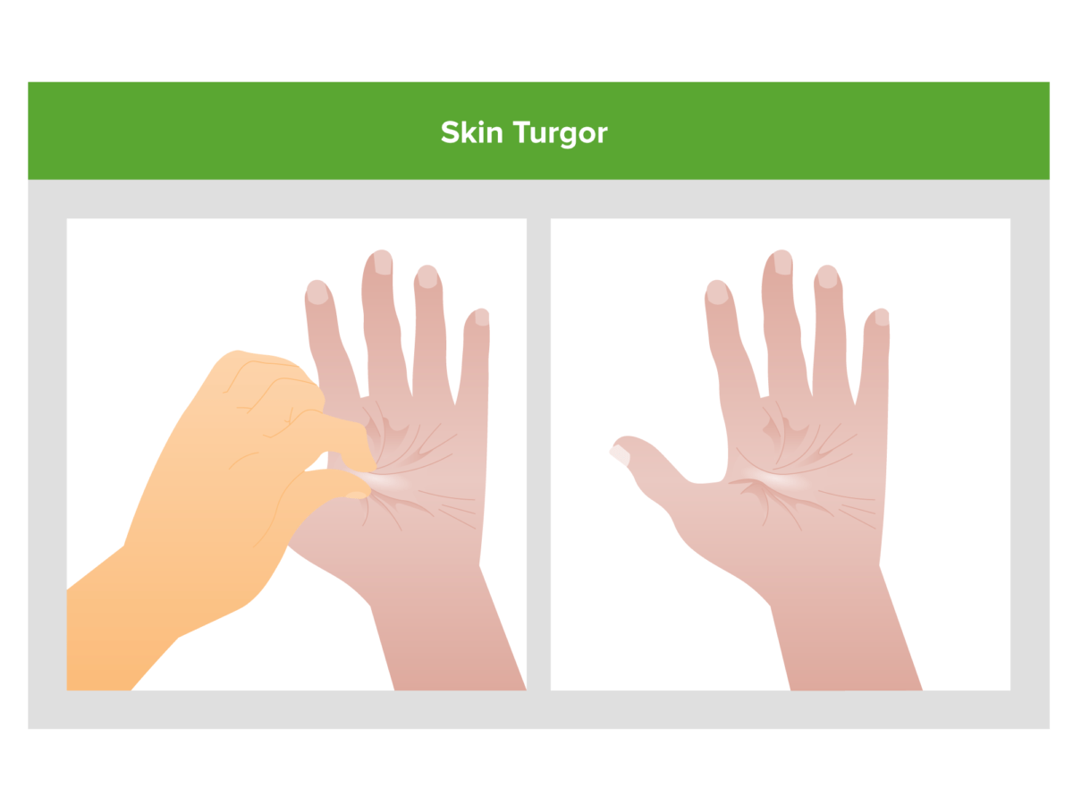
    • Poor skin turgor Skin turgor Malnutrition in children in resource-limited countries
    • Dry mucous membranes
Skin turgor

Poor skin turgor (skin remains tented after being pinched) is a potential clue that volume depletion may be the cause of an individual’s acute kidney injury.

Image by Lecturio.
Clinical presentation with hypervolemia Hypervolemia Renal Sodium and Water Regulation/fluid overload

  • Symptoms:
    • Dyspnea Dyspnea Dyspnea is the subjective sensation of breathing discomfort. Dyspnea is a normal manifestation of heavy physical or psychological exertion, but also may be caused by underlying conditions (both pulmonary and extrapulmonary). Dyspnea
    • Abdominal distension
    • Swelling Swelling Inflammation in the extremities
  • Physical exam findings:
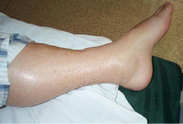
    • Edema Edema Edema is a condition in which excess serous fluid accumulates in the body cavity or interstitial space of connective tissues. Edema is a symptom observed in several medical conditions. It can be categorized into 2 types, namely, peripheral (in the extremities) and internal (in an organ or body cavity). Edema
    • Crackles
    • Ascites Ascites Ascites is the pathologic accumulation of fluid within the peritoneal cavity that occurs due to an osmotic and/or hydrostatic pressure imbalance secondary to portal hypertension (cirrhosis, heart failure) or non-portal hypertension (hypoalbuminemia, malignancy, infection). Ascites
    • Pleural effusion Pleural Effusion Pleural effusion refers to the accumulation of fluid between the layers of the parietal and visceral pleura. Common causes of this condition include infection, malignancy, autoimmune disorders, or volume overload. Clinical manifestations include chest pain, cough, and dyspnea. Pleural Effusion
    • Elevated jugular venous pressure Jugular Venous Pressure Portal Hypertension (JVP)
    • S3 S3 Heart Sounds heart sound
Leg edema

Lower extremity edema: may be seen in individuals presenting with hypervolemia as a result of acute kidney injury

Image: “Leg Edema 01” by Wang Kai-feng, Pan Hong-ming, Lou Hai-zhou, Shen Li-rong, Zhu Xi-yan. License: CC BY 2.0
Clinical presentation with hyperkalemia Hyperkalemia Hyperkalemia is defined as a serum potassium (K+) concentration >5.2 mEq/L. Homeostatic mechanisms maintain the serum K+ concentration between 3.5 and 5.2 mEq/L, despite marked variation in dietary intake. Hyperkalemia can be due to a variety of causes, which include transcellular shifts, tissue breakdown, inadequate renal excretion, and drugs. Hyperkalemia

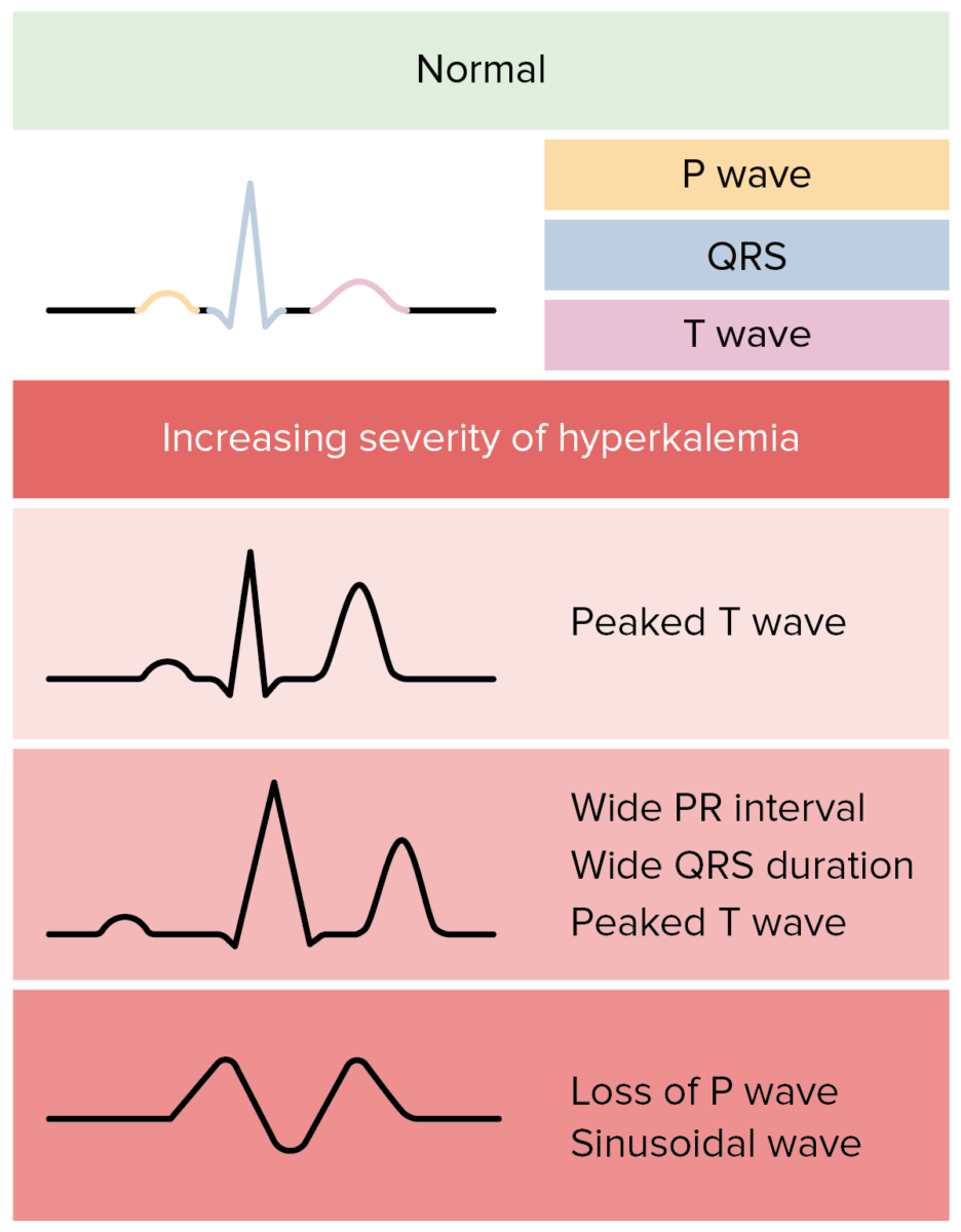
Hyperkalemia ekg

Possible ECG changes due to hyperkalemia in individuals with acute kidney injury:
In reality, ECG changes in hyperkalemia are more variable and less predictable.
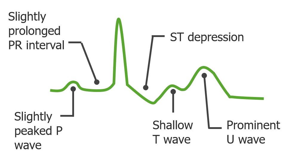
Image by Lecturio.

Clinical presentation with hypokalemia Hypokalemia Hypokalemia is defined as plasma potassium (K+) concentration < 3.5 mEq/L. Homeostatic mechanisms maintain plasma concentration between 3.5-5.2 mEq/L despite marked variation in dietary intake. Hypokalemia can be due to renal losses, GI losses, transcellular shifts, or poor dietary intake. Hypokalemia

  • Usually found in the same instances as hypovolemia Hypovolemia Sepsis in Children
  • Symptoms:
    • Muscle cramps Cramps Ion Channel Myopathy
    • Ileus Ileus A condition caused by the lack of intestinal peristalsis or intestinal motility without any mechanical obstruction. This interference of the flow of intestinal contents often leads to intestinal obstruction. Ileus may be classified into postoperative, inflammatory, metabolic, neurogenic, and drug-induced. Small Bowel Obstruction
  • Physical exam findings:
    • Usually normal
    • May have muscle weakness and rhabdomyolysis Rhabdomyolysis Rhabdomyolysis is characterized by muscle necrosis and the release of toxic intracellular contents, especially myoglobin, into the circulation. Rhabdomyolysis, if severe
  • ECG ECG An electrocardiogram (ECG) is a graphic representation of the electrical activity of the heart plotted against time. Adhesive electrodes are affixed to the skin surface allowing measurement of cardiac impulses from many angles. The ECG provides 3-dimensional information about the conduction system of the heart, the myocardium, and other cardiac structures. Electrocardiogram (ECG) changes:
    • ST depression
    • T wave T wave Electrocardiogram (ECG) flattening
    • U waves
    • QT interval QT interval Electrocardiogram (ECG) prolongation
    • May not show any overt ECG ECG An electrocardiogram (ECG) is a graphic representation of the electrical activity of the heart plotted against time. Adhesive electrodes are affixed to the skin surface allowing measurement of cardiac impulses from many angles. The ECG provides 3-dimensional information about the conduction system of the heart, the myocardium, and other cardiac structures. Electrocardiogram (ECG) manifestations
Ecg changes seen in hypokalemia

Possible ECG changes in individuals with acute kidney injury presenting with hypokalemia

Image by Lecturio. License: CC BY-NC-SA 4.0

Clinical presentation with uremia

  • Symptoms:
    • GI:
      • Anorexia Anorexia The lack or loss of appetite accompanied by an aversion to food and the inability to eat. It is the defining characteristic of the disorder anorexia nervosa. Anorexia Nervosa
      • Nausea Nausea An unpleasant sensation in the stomach usually accompanied by the urge to vomit. Common causes are early pregnancy, sea and motion sickness, emotional stress, intense pain, food poisoning, and various enteroviruses. Antiemetics/ vomiting Vomiting The forcible expulsion of the contents of the stomach through the mouth. Hypokalemia
    • Dysgeusia Dysgeusia A condition characterized by alterations of the sense of taste which may range from mild to severe, including gross distortions of taste quality. Nitroimidazoles (abnormal taste)
    • Neurologic:
      • Confusion
      • Drowsiness
      • Encephalopathy Encephalopathy Hyper-IgM Syndrome
      • Seizure
      • Coma Coma Coma is defined as a deep state of unarousable unresponsiveness, characterized by a score of 3 points on the GCS. A comatose state can be caused by a multitude of conditions, making the precise epidemiology and prognosis of coma difficult to determine. Coma
    • Bleeding due to qualitative platelet dysfunction from uremic toxins
  • Physical exam findings:
    • Asterixis Asterixis Hepatic Encephalopathy: tremor Tremor Cyclical movement of a body part that can represent either a physiologic process or a manifestation of disease. Intention or action tremor, a common manifestation of cerebellar diseases, is aggravated by movement. In contrast, resting tremor is maximal when there is no attempt at voluntary movement, and occurs as a relatively frequent manifestation of parkinson disease. Myotonic Dystrophies of the hand Hand The hand constitutes the distal part of the upper limb and provides the fine, precise movements needed in activities of daily living. It consists of 5 metacarpal bones and 14 phalanges, as well as numerous muscles innervated by the median and ulnar nerves. Hand: Anatomy when the wrist is extended
    • Myoclonus Myoclonus Involuntary shock-like contractions, irregular in rhythm and amplitude, followed by relaxation, of a muscle or a group of muscles. This condition may be a feature of some central nervous system diseases; (e.g., epilepsy-myoclonic). Nocturnal myoclonus is the principal feature of the nocturnal myoclonus syndrome. Neurological Examination: quick involuntary muscle jerks
    • Pericarditis Pericarditis Pericarditis is an inflammation of the pericardium, often with fluid accumulation. It can be caused by infection (often viral), myocardial infarction, drugs, malignancies, metabolic disorders, autoimmune disorders, or trauma. Acute, subacute, and chronic forms exist. Pericarditis/pericardial rub: audible grating sound best heard between the apex and sternum Sternum A long, narrow, and flat bone commonly known as breastbone occurring in the midsection of the anterior thoracic segment or chest region, which stabilizes the rib cage and serves as the point of origin for several muscles that move the arms, head, and neck. Chest Wall: Anatomy
    • Pericardial effusion Pericardial effusion Fluid accumulation within the pericardium. Serous effusions are associated with pericardial diseases. Hemopericardium is associated with trauma. Lipid-containing effusion (chylopericardium) results from leakage of thoracic duct. Severe cases can lead to cardiac tamponade. Pericardial Effusion and Cardiac Tamponade
Asterixis

Asterixis: a potential examination finding in an individual with uremia from acute kidney injury

Image by Lecturio.
Clinical presentations with specific etiologies of AKI

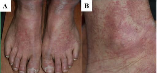
  • Classic triad of acute interstitial nephritis (all 3 are only seen in 10% of affected individuals):
  • Other causes of AKI presenting with a rash Rash Rocky Mountain Spotted Fever:
    • Petechial rash Rash Rocky Mountain Spotted Fever, palpable purpura‒glomerulonephritis/ vasculitis Vasculitis Inflammation of any one of the blood vessels, including the arteries; veins; and rest of the vasculature system in the body. Systemic Lupus Erythematosus
    • Livedo reticularis Livedo reticularis A condition characterized by a reticular or fishnet pattern on the skin of lower extremities and other parts of the body. This red and blue pattern is due to deoxygenated blood in unstable dermal blood vessels. The condition is intensified by cold exposure and relieved by rewarming. Chronic Kidney Disease cholesterol Cholesterol The principal sterol of all higher animals, distributed in body tissues, especially the brain and spinal cord, and in animal fats and oils. Cholesterol Metabolism emboli
  • Anuria: < 50‒100 mL urine volume/urine output per day:
    • Shock Shock Shock is a life-threatening condition associated with impaired circulation that results in tissue hypoxia. The different types of shock are based on the underlying cause: distributive (↑ cardiac output (CO), ↓ systemic vascular resistance (SVR)), cardiogenic (↓ CO, ↑ SVR), hypovolemic (↓ CO, ↑ SVR), obstructive (↓ CO), and mixed. Types of Shock
    • Bilateral urinary tract Urinary tract The urinary tract is located in the abdomen and pelvis and consists of the kidneys, ureters, urinary bladder, and urethra. The structures permit the excretion of urine from the body. Urine flows from the kidneys through the ureters to the urinary bladder and out through the urethra. Urinary Tract: Anatomy obstruction
    • Rapidly progressive glomerulonephritis Rapidly Progressive Glomerulonephritis Rapidly progressive glomerulonephritis (RPGN) is a syndrome of severe glomerular disease with progressive loss of kidney function within weeks to months. Histologically, crescents (the proliferation of epithelial cells and the infiltration of monocytes/macrophages in the Bowman space) are found in the glomeruli and arise from immunologic injury. Rapidly Progressive Glomerulonephritis
    • Hemolytic uremic syndrome Hemolytic uremic syndrome A syndrome that is associated with microvascular diseases of the kidney, such as renal cortical necrosis. It is characterized by hemolytic anemia; thrombocytopenia; and acute renal failure. Hypocoagulable Conditions
    • Bilateral renal artery Renal artery A branch of the abdominal aorta which supplies the kidneys, adrenal glands and ureters. Glomerular Filtration occlusion
  • Hematuria Hematuria Presence of blood in the urine. Renal Cell Carcinoma:
    • Microscopic: seen with glomerulonephritis
    • Gross: seen with IgA IgA Represents 15-20% of the human serum immunoglobulins, mostly as the 4-chain polymer in humans or dimer in other mammals. Secretory iga is the main immunoglobulin in secretions. Immunoglobulins: Types and Functions nephropathy
  • Foamy urine: very high levels of proteinuria Proteinuria The presence of proteins in the urine, an indicator of kidney diseases. Nephrotic Syndrome in Children nephrotic syndrome Nephrotic syndrome Nephrotic syndrome is characterized by severe proteinuria, hypoalbuminemia, and peripheral edema. In contrast, the nephritic syndromes present with hematuria, variable loss of renal function, and hypertension, although there is sometimes overlap of > 1 glomerular disease in the same individual. Nephrotic Syndrome
  • Urinary frequency/hesitancy → urinary tract Urinary tract The urinary tract is located in the abdomen and pelvis and consists of the kidneys, ureters, urinary bladder, and urethra. The structures permit the excretion of urine from the body. Urine flows from the kidneys through the ureters to the urinary bladder and out through the urethra. Urinary Tract: Anatomy obstruction with BPH BPH Benign prostatic hyperplasia (bph) is a condition indicating an increase in the number of stromal and epithelial cells within the prostate gland (transition zone). Benign prostatic hyperplasia is common in men > 50 years of age and may greatly affect their quality of life. Benign Prostatic Hyperplasia
Variable-sized macules and patches of erythematous on an individual with acute interstitial nephritis

Variable-sized macules and patches of erythematous on an individual with acute interstitial nephritis

Image: “F1: Gross and microscopic exam of the skin.” by Ha Yeon Kim, Sung Sun Kim, Soo Hyeon Bae, Eun Hui Bae, Seong Kwon Ma & Soo Wan Kim. License: CC BY 4.0, cropped by Lecturio.

Acute tubular necrosis Necrosis The death of cells in an organ or tissue due to disease, injury or failure of the blood supply. Ischemic Cell Damage phases

Acute tubular necrosis Necrosis The death of cells in an organ or tissue due to disease, injury or failure of the blood supply. Ischemic Cell Damage follows a characteristic clinical course involving 3 phases.
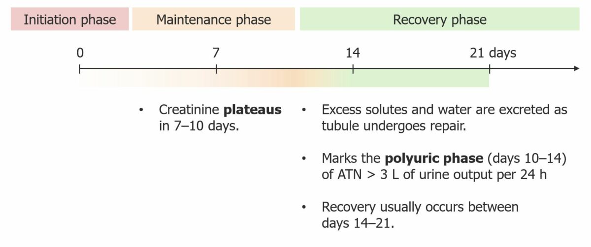
Clinical course of atn

Clinical course of acute tubular necrosis (ATN)

Image by Lecturio. License CC BY-NC-SA 4.0

Diagnosis

The diagnosis of AKI includes identifying inciting factors and determining if the etiology is prerenal, intrarenal, or postrenal, in order to plan appropriate treatment.

History and physical examination

  • Volume depletion Volume depletion Volume status is a balance between water and solutes, the majority of which is Na. Volume depletion refers to a loss of both water and Na, whereas dehydration refers only to a loss of water. Volume depletion can be caused by GI losses, renal losses, bleeding, poor oral Na intake, or third spacing of fluids. Volume Depletion and Dehydration/ dehydration Dehydration The condition that results from excessive loss of water from a living organism. Volume Depletion and Dehydration:
    • Diarrhea Diarrhea Diarrhea is defined as ≥ 3 watery or loose stools in a 24-hour period. There are a multitude of etiologies, which can be classified based on the underlying mechanism of disease. The duration of symptoms (acute or chronic) and characteristics of the stools (e.g., watery, bloody, steatorrheic, mucoid) can help guide further diagnostic evaluation. Diarrhea
    • Vomiting Vomiting The forcible expulsion of the contents of the stomach through the mouth. Hypokalemia
    • Excessive sweating
  • Common medications associated with AKI:
  • Look for common IV nephrotoxic drugs used in the hospital as a cause of AKI:
    • Aminoglycosides Aminoglycosides Aminoglycosides are a class of antibiotics including gentamicin, tobramycin, amikacin, neomycin, plazomicin, and streptomycin. The class binds the 30S ribosomal subunit to inhibit bacterial protein synthesis. Unlike other medications with a similar mechanism of action, aminoglycosides are bactericidal. Aminoglycosides
    • Amphotericin B Amphotericin B Macrolide antifungal antibiotic produced by streptomyces nodosus obtained from soil of the orinoco river region of venezuela. Polyenes
    • Platinum-based chemotherapies
    • Vancomycin Vancomycin Antibacterial obtained from streptomyces orientalis. It is a glycopeptide related to ristocetin that inhibits bacterial cell wall assembly and is toxic to kidneys and the inner ear. Glycopeptides
  • Common comorbidities Comorbidities The presence of co-existing or additional diseases with reference to an initial diagnosis or with reference to the index condition that is the subject of study. Comorbidity may affect the ability of affected individuals to function and also their survival; it may be used as a prognostic indicator for length of hospital stay, cost factors, and outcome or survival. St. Louis Encephalitis Virus:
    • Sepsis Sepsis Systemic inflammatory response syndrome with a proven or suspected infectious etiology. When sepsis is associated with organ dysfunction distant from the site of infection, it is called severe sepsis. When sepsis is accompanied by hypotension despite adequate fluid infusion, it is called septic shock. Sepsis and Septic Shock
    • Heart failure Heart Failure A heterogeneous condition in which the heart is unable to pump out sufficient blood to meet the metabolic need of the body. Heart failure can be caused by structural defects, functional abnormalities (ventricular dysfunction), or a sudden overload beyond its capacity. Chronic heart failure is more common than acute heart failure which results from sudden insult to cardiac function, such as myocardial infarction. Total Anomalous Pulmonary Venous Return (TAPVR) (HF)
    • Cirrhosis Cirrhosis Cirrhosis is a late stage of hepatic parenchymal necrosis and scarring (fibrosis) most commonly due to hepatitis C infection and alcoholic liver disease. Patients may present with jaundice, ascites, and hepatosplenomegaly. Cirrhosis can also cause complications such as hepatic encephalopathy, portal hypertension, portal vein thrombosis, and hepatorenal syndrome. Cirrhosis

Regular Regular Insulin laboratory testing

  • Serum BUN and creatinine
  • BUN/creatinine ratio:
    • Normal ratio is approximately 15.
    • Prerenal > 20
    • Intrinsic renal < 15
  • Electrolyte abnormalities/ hyperkalemia Hyperkalemia Hyperkalemia is defined as a serum potassium (K+) concentration >5.2 mEq/L. Homeostatic mechanisms maintain the serum K+ concentration between 3.5 and 5.2 mEq/L, despite marked variation in dietary intake. Hyperkalemia can be due to a variety of causes, which include transcellular shifts, tissue breakdown, inadequate renal excretion, and drugs. Hyperkalemia
  • CMP/ bicarbonate Bicarbonate Inorganic salts that contain the -HCO3 radical. They are an important factor in determining the ph of the blood and the concentration of bicarbonate ions is regulated by the kidney. Levels in the blood are an index of the alkali reserve or buffering capacity. Electrolytes for metabolic acidosis Acidosis A pathologic condition of acid accumulation or depletion of base in the body. The two main types are respiratory acidosis and metabolic acidosis, due to metabolic acid build up. Respiratory Acidosis
  • BNP BNP A peptide that is secreted by the brain and the heart atria, stored mainly in cardiac ventricular myocardium. It can cause natriuresis; diuresis; vasodilation; and inhibits secretion of renin and aldosterone. It improves heart function. It contains 32 amino acids. Renal Sodium and Water Regulation for heart failure Heart Failure A heterogeneous condition in which the heart is unable to pump out sufficient blood to meet the metabolic need of the body. Heart failure can be caused by structural defects, functional abnormalities (ventricular dysfunction), or a sudden overload beyond its capacity. Chronic heart failure is more common than acute heart failure which results from sudden insult to cardiac function, such as myocardial infarction. Total Anomalous Pulmonary Venous Return (TAPVR)/volume overload
  • Urinalysis Urinalysis Examination of urine by chemical, physical, or microscopic means. Routine urinalysis usually includes performing chemical screening tests, determining specific gravity, observing any unusual color or odor, screening for bacteriuria, and examining the sediment microscopically. Urinary Tract Infections (UTIs) in Children:
    • Proteinuria Proteinuria The presence of proteins in the urine, an indicator of kidney diseases. Nephrotic Syndrome in Children: seen with nephrotic syndrome Nephrotic syndrome Nephrotic syndrome is characterized by severe proteinuria, hypoalbuminemia, and peripheral edema. In contrast, the nephritic syndromes present with hematuria, variable loss of renal function, and hypertension, although there is sometimes overlap of > 1 glomerular disease in the same individual. Nephrotic Syndrome
    • Hematuria Hematuria Presence of blood in the urine. Renal Cell Carcinoma: seen with nephritic syndromes
    • RBC casts are a finding of glomerulonephritis.
    • WBC casts are a finding of acute interstitial nephritis.
    • Renal tubular cell casts and muddy brown casts are findings of ATN.
  • Urine sodium Sodium A member of the alkali group of metals. It has the atomic symbol na, atomic number 11, and atomic weight 23. Hyponatremia (UNa+):
    • UNa+ < 10 suggests a prerenal cause.
    • UNa+ > 20 suggests an intrinsic renal cause.
  • Fractional excretion of sodium Sodium A member of the alkali group of metals. It has the atomic symbol na, atomic number 11, and atomic weight 23. Hyponatremia (FENa) is calculated by simultaneously obtaining urine and plasma Plasma The residual portion of blood that is left after removal of blood cells by centrifugation without prior blood coagulation. Transfusion Products specimens of Na+ and creatinine:
    • FENa < 1% suggests a prerenal cause of AKI.
    • FENa > 1% suggests an intrinsic renal cause.
$$FENa \%=\frac{Urine\ Na\times serum\ creatinine}{Serum\ Na \times urine\ creatinine}\times 100$$

Other specific testing

Consider other specific testing for less common etiologies:
  • Serum CK for rhabdomyolysis Rhabdomyolysis Rhabdomyolysis is characterized by muscle necrosis and the release of toxic intracellular contents, especially myoglobin, into the circulation. Rhabdomyolysis
  • Serum free light chains Light chains Polypeptide chains, consisting of 211 to 217 amino acid residues and having a molecular weight of approximately 22 kda. There are two major types of light chains, kappa and lambda. Two ig light chains and two ig heavy chains (immunoglobulin heavy chains) make one immunoglobulin molecule. Immunoglobulins: Types and Functions, serum/urine protein electrophoresis Electrophoresis An electrochemical process in which macromolecules or colloidal particles with a net electric charge migrate in a solution under the influence of an electric current. Blotting Techniques for multiple myeloma
  • Serologies for glomerulonephritis:
    • HIV HIV Anti-HIV Drugs, hepatitis B Hepatitis B Hepatitis B virus (HBV) is a partially double-stranded DNA virus, which belongs to the Orthohepadnavirus genus and the Hepadnaviridae family. Most individuals with acute HBV infection are asymptomatic or have mild, self-limiting symptoms. Chronic infection can be asymptomatic or create hepatic inflammation, leading to liver cirrhosis and hepatocellular carcinoma (HCC). Hepatitis B Virus, and hepatitis C Hepatitis C Hepatitis C is an infection of the liver caused by the hepatitis C virus (HCV). The infection can be transmitted through infectious blood or body fluids and may be transmitted during childbirth or through IV drug use or sexual intercourse. Hepatitis C virus can cause both acute and chronic hepatitis, ranging from a mild to a serious, lifelong illness including liver cirrhosis and hepatocellular carcinoma (HCC). Hepatitis C Virus serologies
    • Serum complement (C3, C4) levels
    • PLA2R antibody for membranous nephropathy
    • Anticytoplasmic neutrophil antibodies Antibodies Immunoglobulins (Igs), also known as antibodies, are glycoprotein molecules produced by plasma cells that act in immune responses by recognizing and binding particular antigens. The various Ig classes are IgG (the most abundant), IgM, IgE, IgD, and IgA, which differ in their biologic features, structure, target specificity, and distribution. Immunoglobulins: Types and Functions ( ANCA ANCA Group of systemic vasculitis with a strong association with anca. The disorders are characterized by necrotizing inflammation of small and medium size vessels, with little or no immune-complex deposits in vessel walls. Rapidly Progressive Glomerulonephritis) for vasculitis Vasculitis Inflammation of any one of the blood vessels, including the arteries; veins; and rest of the vasculature system in the body. Systemic Lupus Erythematosus
    • Antiglomerular basement membrane Basement membrane A darkly stained mat-like extracellular matrix (ecm) that separates cell layers, such as epithelium from endothelium or a layer of connective tissue. The ecm layer that supports an overlying epithelium or endothelium is called basal lamina. Basement membrane (bm) can be formed by the fusion of either two adjacent basal laminae or a basal lamina with an adjacent reticular lamina of connective tissue. Bm, composed mainly of type IV collagen; glycoprotein laminin; and proteoglycan, provides barriers as well as channels between interacting cell layers. Thin Basement Membrane Nephropathy (TBMN) for Goodpasture disease
    • ANA and anti-dsDNA for lupus

Imaging

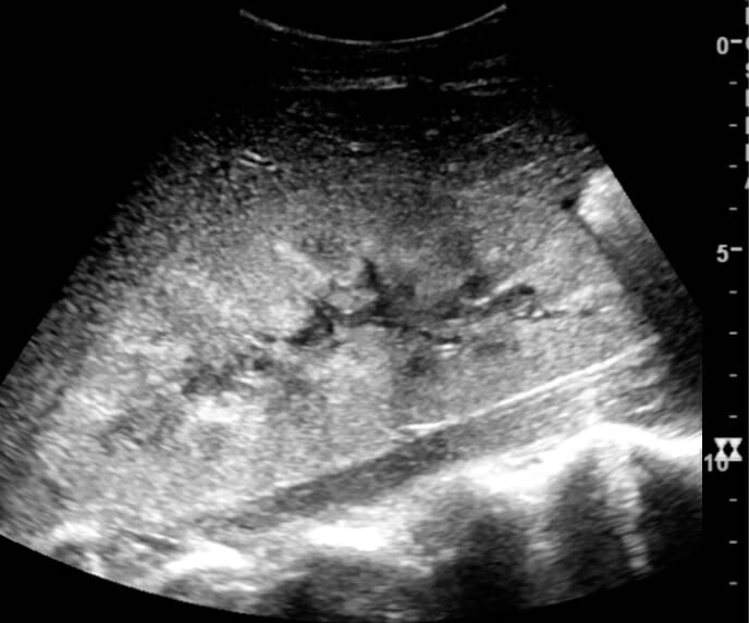
  • Consider renal artery Renal artery A branch of the abdominal aorta which supplies the kidneys, adrenal glands and ureters. Glomerular Filtration Doppler Doppler Ultrasonography applying the doppler effect, with frequency-shifted ultrasound reflections produced by moving targets (usually red blood cells) in the bloodstream along the ultrasound axis in direct proportion to the velocity of movement of the targets, to determine both direction and velocity of blood flow. Ultrasound (Sonography) ultrasound if high suspicion for renal artery Renal artery A branch of the abdominal aorta which supplies the kidneys, adrenal glands and ureters. Glomerular Filtration stenosis Stenosis Hypoplastic Left Heart Syndrome (HLHS)
  • Ultrasound for kidney stones Kidney stones Nephrolithiasis is the formation of a stone, or calculus, anywhere along the urinary tract caused by precipitations of solutes in the urine. The most common type of kidney stone is the calcium oxalate stone, but other types include calcium phosphate, struvite (ammonium magnesium phosphate), uric acid, and cystine stones. Nephrolithiasis or obstruction:
    • BPH BPH Benign prostatic hyperplasia (bph) is a condition indicating an increase in the number of stromal and epithelial cells within the prostate gland (transition zone). Benign prostatic hyperplasia is common in men > 50 years of age and may greatly affect their quality of life. Benign Prostatic Hyperplasia in men
    • Gynecologic tumors in women

Parameters to differentiate prerenal from renal AKI

Table: Parameters to differentiate prerenal from renal AKI
Prerenal Renal (ATN)
Specific gravity ≥ 1.020 ≤ 1.010
Urine osmolality Osmolality Plasma osmolality refers to the combined concentration of all solutes in the blood. Renal Sodium and Water Regulation (mOsm/kg) > 500 < 350
Urine sodium Sodium A member of the alkali group of metals. It has the atomic symbol na, atomic number 11, and atomic weight 23. Hyponatremia (mEq/L) < 20 > 20
FENa < 1% > 1%
Urine protein Minimal Minimal to severe, depending on etiology
ATN: acute tubular necrosis
FENa: fractional excretion of sodium

Urine microscopy findings

Table: Urine microscopy findings
Finding Significance
Renal tubular cells Acute tubular injury
RBCs RBCs Erythrocytes, or red blood cells (RBCs), are the most abundant cells in the blood. While erythrocytes in the fetus are initially produced in the yolk sac then the liver, the bone marrow eventually becomes the main site of production. Erythrocytes: Histology Nonglomerular bleeding/anywhere in the urinary tract Urinary tract The urinary tract is located in the abdomen and pelvis and consists of the kidneys, ureters, urinary bladder, and urethra. The structures permit the excretion of urine from the body. Urine flows from the kidneys through the ureters to the urinary bladder and out through the urethra. Urinary Tract: Anatomy
Dysmorphic red cells Glomerular disease
Red cell casts Diagnostic of glomerular disease
Leukocytes Leukocytes White blood cells. These include granular leukocytes (basophils; eosinophils; and neutrophils) as well as non-granular leukocytes (lymphocytes and monocytes). White Myeloid Cells: Histology/WBCs Urinary tract Urinary tract The urinary tract is located in the abdomen and pelvis and consists of the kidneys, ureters, urinary bladder, and urethra. The structures permit the excretion of urine from the body. Urine flows from the kidneys through the ureters to the urinary bladder and out through the urethra. Urinary Tract: Anatomy infection
White cell casts Renal infection
Hyaline casts Any type of renal disease
Granular casts More significant renal disease
“Muddy brown cast” Necrotic tubular cells/acute tubular injury

Management

The etiology of AKI dictates the treatment; further testing with biopsy Biopsy Removal and pathologic examination of specimens from the living body. Ewing Sarcoma may be indicated after appropriate workup and treatment for hypovolemia Hypovolemia Sepsis in Children or fluid overload are completed.

Treatment

  • Based on the etiology of AKI, may not be immediately apparent on initial investigation:
    • If prerenal with hypovolemia Hypovolemia Sepsis in Children → treat with IV fluids IV fluids Intravenous fluids are one of the most common interventions administered in medicine to approximate physiologic bodily fluids. Intravenous fluids are divided into 2 categories: crystalloid and colloid solutions. Intravenous fluids have a wide variety of indications, including intravascular volume expansion, electrolyte manipulation, and maintenance fluids. Intravenous Fluids
    • If hypervolemia Hypervolemia Renal Sodium and Water Regulation/fluid overload → treat with diuretics Diuretics Agents that promote the excretion of urine through their effects on kidney function. Heart Failure and Angina Medication
    • If postrenal with obstruction → Foley catheter to relieve urinary retention Urinary retention Inability to empty the urinary bladder with voiding (urination). Delirium:
      • Percutaneous nephrostomy tubes may be necessary.
      • Specialist consultation to treat underlying conditions (urology or gynecology)
    • Stop any offending agents (medications) if identified as the cause of AKI.
  • Treat electrolyte and acid-base imbalances urgently:
    • Metabolic acidosis Acidosis A pathologic condition of acid accumulation or depletion of base in the body. The two main types are respiratory acidosis and metabolic acidosis, due to metabolic acid build up. Respiratory Acidosis → treat with oral or IV sodium Sodium A member of the alkali group of metals. It has the atomic symbol na, atomic number 11, and atomic weight 23. Hyponatremia bicarbonate Bicarbonate Inorganic salts that contain the -HCO3 radical. They are an important factor in determining the ph of the blood and the concentration of bicarbonate ions is regulated by the kidney. Levels in the blood are an index of the alkali reserve or buffering capacity. Electrolytes
    • Hyperkalemia Hyperkalemia Hyperkalemia is defined as a serum potassium (K+) concentration >5.2 mEq/L. Homeostatic mechanisms maintain the serum K+ concentration between 3.5 and 5.2 mEq/L, despite marked variation in dietary intake. Hyperkalemia can be due to a variety of causes, which include transcellular shifts, tissue breakdown, inadequate renal excretion, and drugs. Hyperkalemia:
      • IV calcium Calcium A basic element found in nearly all tissues. It is a member of the alkaline earth family of metals with the atomic symbol ca, atomic number 20, and atomic weight 40. Calcium is the most abundant mineral in the body and combines with phosphorus to form calcium phosphate in the bones and teeth. It is essential for the normal functioning of nerves and muscles and plays a role in blood coagulation (as factor IV) and in many enzymatic processes. Electrolytes to stabilize cardiac membranes (temporary effect)
      • IV insulin Insulin Insulin is a peptide hormone that is produced by the beta cells of the pancreas. Insulin plays a role in metabolic functions such as glucose uptake, glycolysis, glycogenesis, lipogenesis, and protein synthesis. Exogenous insulin may be needed for individuals with diabetes mellitus, in whom there is a deficiency in endogenous insulin or increased insulin resistance. Insulin and IV dextrose Dextrose Intravenous Fluids to shift potassium Potassium An element in the alkali group of metals with an atomic symbol k, atomic number 19, and atomic weight 39. 10. It is the chief cation in the intracellular fluid of muscle and other cells. Potassium ion is a strong electrolyte that plays a significant role in the regulation of fluid volume and maintenance of the water-electrolyte balance. Hyperkalemia intracellularly (temporary effect)
      • Oral cation exchange resins that cause GI loss of potassium Potassium An element in the alkali group of metals with an atomic symbol k, atomic number 19, and atomic weight 39. 10. It is the chief cation in the intracellular fluid of muscle and other cells. Potassium ion is a strong electrolyte that plays a significant role in the regulation of fluid volume and maintenance of the water-electrolyte balance. Hyperkalemia (lasting effect)
      • Loop diuretics Diuretics Agents that promote the excretion of urine through their effects on kidney function. Heart Failure and Angina Medication cause renal loss of potassium Potassium An element in the alkali group of metals with an atomic symbol k, atomic number 19, and atomic weight 39. 10. It is the chief cation in the intracellular fluid of muscle and other cells. Potassium ion is a strong electrolyte that plays a significant role in the regulation of fluid volume and maintenance of the water-electrolyte balance. Hyperkalemia (lasting effect).
      • Dialysis Dialysis Renal replacement therapy refers to dialysis and/or kidney transplantation. Dialysis is a procedure by which toxins and excess water are removed from the circulation. Hemodialysis and peritoneal dialysis (PD) are the two types of dialysis, and their primary difference is the location of the filtration process (external to the body in hemodialysis versus inside the body for PD). Peritoneal Dialysis and Hemodialysis directly removes potassium Potassium An element in the alkali group of metals with an atomic symbol k, atomic number 19, and atomic weight 39. 10. It is the chief cation in the intracellular fluid of muscle and other cells. Potassium ion is a strong electrolyte that plays a significant role in the regulation of fluid volume and maintenance of the water-electrolyte balance. Hyperkalemia from the blood (lasting effect).
    • Hyperphosphatemia Hyperphosphatemia A condition of abnormally high level of phosphates in the blood, usually significantly above the normal range of 0. 84-1. 58 mmol per liter of serum. Hypocalcemia:
      • Usually asymptomatic and does not require treatment during AKI.
      • Causes important complications if CKD CKD Chronic kidney disease (CKD) is kidney impairment that lasts for ≥ 3 months, implying that it is irreversible. Hypertension and diabetes are the most common causes; however, there are a multitude of other etiologies. In the early to moderate stages, CKD is usually asymptomatic and is primarily diagnosed by laboratory abnormalities. Chronic Kidney Disease
  • For all cases of AKI:
    • Avoid nephrotoxins, including IV iodinated contrast for CT scans.
    • Avoid gadolinium-based contrast for MRI studies.
    • Restrict sodium Sodium A member of the alkali group of metals. It has the atomic symbol na, atomic number 11, and atomic weight 23. Hyponatremia, potassium Potassium An element in the alkali group of metals with an atomic symbol k, atomic number 19, and atomic weight 39. 10. It is the chief cation in the intracellular fluid of muscle and other cells. Potassium ion is a strong electrolyte that plays a significant role in the regulation of fluid volume and maintenance of the water-electrolyte balance. Hyperkalemia, and phosphate Phosphate Inorganic salts of phosphoric acid. Electrolytes in the diet.
    • Repeat serum creatinine and potassium Potassium An element in the alkali group of metals with an atomic symbol k, atomic number 19, and atomic weight 39. 10. It is the chief cation in the intracellular fluid of muscle and other cells. Potassium ion is a strong electrolyte that plays a significant role in the regulation of fluid volume and maintenance of the water-electrolyte balance. Hyperkalemia levels.
  • Indications for kidney biopsy Biopsy Removal and pathologic examination of specimens from the living body. Ewing Sarcoma:
    • Worsening serum creatinine with no clear diagnosis
    • Suspicion for glomerulonephritis or vasculitis Vasculitis Inflammation of any one of the blood vessels, including the arteries; veins; and rest of the vasculature system in the body. Systemic Lupus Erythematosus: may require aggressive specific treatments such as high-dose corticosteroids Corticosteroids Chorioretinitis
      • Rapidly progressive glomerulonephritis Rapidly Progressive Glomerulonephritis Rapidly progressive glomerulonephritis (RPGN) is a syndrome of severe glomerular disease with progressive loss of kidney function within weeks to months. Histologically, crescents (the proliferation of epithelial cells and the infiltration of monocytes/macrophages in the Bowman space) are found in the glomeruli and arise from immunologic injury. Rapidly Progressive Glomerulonephritis ( RPGN RPGN Rapidly progressive glomerulonephritis (RPGN) is a syndrome of severe glomerular disease with progressive loss of kidney function within weeks to months. Histologically, crescents (the proliferation of epithelial cells and the infiltration of monocytes/macrophages in the Bowman space) are found in the glomeruli and arise from immunologic injury. Rapidly Progressive Glomerulonephritis)
      • IgA IgA Represents 15-20% of the human serum immunoglobulins, mostly as the 4-chain polymer in humans or dimer in other mammals. Secretory iga is the main immunoglobulin in secretions. Immunoglobulins: Types and Functions nephropathy
      • Goodpasture’s disease (antiglomerular basement membrane Basement membrane A darkly stained mat-like extracellular matrix (ecm) that separates cell layers, such as epithelium from endothelium or a layer of connective tissue. The ecm layer that supports an overlying epithelium or endothelium is called basal lamina. Basement membrane (bm) can be formed by the fusion of either two adjacent basal laminae or a basal lamina with an adjacent reticular lamina of connective tissue. Bm, composed mainly of type IV collagen; glycoprotein laminin; and proteoglycan, provides barriers as well as channels between interacting cell layers. Thin Basement Membrane Nephropathy (TBMN) disease)
      • ANCA-positive vasculitis Vasculitis Inflammation of any one of the blood vessels, including the arteries; veins; and rest of the vasculature system in the body. Systemic Lupus Erythematosus ( granulomatosis with polyangiitis Granulomatosis with Polyangiitis A multisystemic disease of a complex genetic background. It is characterized by inflammation of the blood vessels (vasculitis) leading to damage in any number of organs. The common features include granulomatous inflammation of the respiratory tract and kidneys. Most patients have measurable autoantibodies (antineutrophil cytoplasmic antibodies) against myeloblastin. Granulomatosis with Polyangiitis, microscopic polyangiitis Microscopic polyangiitis A primary systemic vasculitis of small- and some medium-sized vessels. It is characterized by a tropism for kidneys and lungs, positive association with anti-neutrophil cytoplasmic antibodies (ANCA), and a paucity of immunoglobulin deposits in vessel walls. Vasculitides)
      • Pauci-immune glomerulonephritis
  • Indications for acute dialysis Dialysis Renal replacement therapy refers to dialysis and/or kidney transplantation. Dialysis is a procedure by which toxins and excess water are removed from the circulation. Hemodialysis and peritoneal dialysis (PD) are the two types of dialysis, and their primary difference is the location of the filtration process (external to the body in hemodialysis versus inside the body for PD). Peritoneal Dialysis and Hemodialysis:
    • Uncontrolled hyperkalemia Hyperkalemia Hyperkalemia is defined as a serum potassium (K+) concentration >5.2 mEq/L. Homeostatic mechanisms maintain the serum K+ concentration between 3.5 and 5.2 mEq/L, despite marked variation in dietary intake. Hyperkalemia can be due to a variety of causes, which include transcellular shifts, tissue breakdown, inadequate renal excretion, and drugs. Hyperkalemia
    • Fluid overload causing respiratory compromise
    • Uremia:
    • Severe metabolic acidosis Acidosis A pathologic condition of acid accumulation or depletion of base in the body. The two main types are respiratory acidosis and metabolic acidosis, due to metabolic acid build up. Respiratory Acidosis
  • Prognosis Prognosis A prediction of the probable outcome of a disease based on a individual’s condition and the usual course of the disease as seen in similar situations. Non-Hodgkin Lymphomas is variable Variable Variables represent information about something that can change. The design of the measurement scales, or of the methods for obtaining information, will determine the data gathered and the characteristics of that data. As a result, a variable can be qualitative or quantitative, and may be further classified into subgroups. Types of Variables, depending on severity and etiology.
  • Long-term follow up with a nephrologist if less than full recovery

Monitoring

  • In the recovery phase, serum creatinine should decrease back to baseline or to a new steady state Steady state Enzyme Kinetics if permanent damage has been incurred.
  • Monitor the degree of renal recovery:
    • Determine if the individual had previous CKD CKD Chronic kidney disease (CKD) is kidney impairment that lasts for ≥ 3 months, implying that it is irreversible. Hypertension and diabetes are the most common causes; however, there are a multitude of other etiologies. In the early to moderate stages, CKD is usually asymptomatic and is primarily diagnosed by laboratory abnormalities. Chronic Kidney Disease:
      • Previously elevated creatinine
      • Small kidneys Kidneys The kidneys are a pair of bean-shaped organs located retroperitoneally against the posterior wall of the abdomen on either side of the spine. As part of the urinary tract, the kidneys are responsible for blood filtration and excretion of water-soluble waste in the urine. Kidneys: Anatomy on renal ultrasound
      • Proteinuria Proteinuria The presence of proteins in the urine, an indicator of kidney diseases. Nephrotic Syndrome in Children
    • An individual’s baseline serum creatinine will worsen at least slightly with each successive episode of AKI, reflecting long-term damage from acute events.
  • It is important for individuals with CKD CKD Chronic kidney disease (CKD) is kidney impairment that lasts for ≥ 3 months, implying that it is irreversible. Hypertension and diabetes are the most common causes; however, there are a multitude of other etiologies. In the early to moderate stages, CKD is usually asymptomatic and is primarily diagnosed by laboratory abnormalities. Chronic Kidney Disease to follow up closely with a nephrologist.
  • Complications: possible development of CKD CKD Chronic kidney disease (CKD) is kidney impairment that lasts for ≥ 3 months, implying that it is irreversible. Hypertension and diabetes are the most common causes; however, there are a multitude of other etiologies. In the early to moderate stages, CKD is usually asymptomatic and is primarily diagnosed by laboratory abnormalities. Chronic Kidney Disease

Differential Diagnosis

  • Renal calculi Renal calculi Stones in the kidney, usually formed in the urine-collecting area of the kidney (kidney pelvis). Their sizes vary and most contains calcium oxalate. Imaging of the Urinary System: hard deposits made of minerals Minerals Electrolytes and salts that form inside the kidneys Kidneys The kidneys are a pair of bean-shaped organs located retroperitoneally against the posterior wall of the abdomen on either side of the spine. As part of the urinary tract, the kidneys are responsible for blood filtration and excretion of water-soluble waste in the urine. Kidneys: Anatomy. Individuals are often asymptomatic until the renal calculi Renal calculi Stones in the kidney, usually formed in the urine-collecting area of the kidney (kidney pelvis). Their sizes vary and most contains calcium oxalate. Imaging of the Urinary System migrate to the ureter, when they present with severe sharp flank pain Flank pain Pain emanating from below the ribs and above the ilium. Renal Cell Carcinoma, hematuria Hematuria Presence of blood in the urine. Renal Cell Carcinoma, and/or urinary tract Urinary tract The urinary tract is located in the abdomen and pelvis and consists of the kidneys, ureters, urinary bladder, and urethra. The structures permit the excretion of urine from the body. Urine flows from the kidneys through the ureters to the urinary bladder and out through the urethra. Urinary Tract: Anatomy infection. Diagnosis is by clinical presentation, urinalysis Urinalysis Examination of urine by chemical, physical, or microscopic means. Routine urinalysis usually includes performing chemical screening tests, determining specific gravity, observing any unusual color or odor, screening for bacteriuria, and examining the sediment microscopically. Urinary Tract Infections (UTIs) in Children, and imaging. Management may be self-limiting Self-Limiting Meningitis in Children by passing the stone or may require intervention with lithotripsy or surgery.
  • CKD CKD Chronic kidney disease (CKD) is kidney impairment that lasts for ≥ 3 months, implying that it is irreversible. Hypertension and diabetes are the most common causes; however, there are a multitude of other etiologies. In the early to moderate stages, CKD is usually asymptomatic and is primarily diagnosed by laboratory abnormalities. Chronic Kidney Disease: kidney damage or an estimated GFR GFR The volume of water filtered out of plasma through glomerular capillary walls into Bowman’s capsules per unit of time. It is considered to be equivalent to inulin clearance. Kidney Function Tests (eGFR) < 60 mL/min/1.73 m2 for ≥ 3 months. Symptoms depend on the cause; prerenal, renal, or postrenal. Diagnosis includes detailed blood and urine workup with additional screening Screening Preoperative Care tests. Management includes adjusting drug doses for the level of eGFR and preparation for renal replacement therapy.
  • Urinary tract Urinary tract The urinary tract is located in the abdomen and pelvis and consists of the kidneys, ureters, urinary bladder, and urethra. The structures permit the excretion of urine from the body. Urine flows from the kidneys through the ureters to the urinary bladder and out through the urethra. Urinary Tract: Anatomy infection: infection of any part of the urinary system, more commonly observed in women. Individuals present with frequent and painful urination, fever Fever Fever is defined as a measured body temperature of at least 38°C (100.4°F). Fever is caused by circulating endogenous and/or exogenous pyrogens that increase levels of prostaglandin E2 in the hypothalamus. Fever is commonly associated with chills, rigors, sweating, and flushing of the skin. Fever, chills Chills The sudden sensation of being cold. It may be accompanied by shivering. Fever, hematuria Hematuria Presence of blood in the urine. Renal Cell Carcinoma, and/or abdominal and back pain Pain An unpleasant sensation induced by noxious stimuli which are detected by nerve endings of nociceptive neurons. Pain: Types and Pathways. Diagnosis is made clinically and by urinalysis Urinalysis Examination of urine by chemical, physical, or microscopic means. Routine urinalysis usually includes performing chemical screening tests, determining specific gravity, observing any unusual color or odor, screening for bacteriuria, and examining the sediment microscopically. Urinary Tract Infections (UTIs) in Children with a urine culture Urine culture Urinary Tract Infections (UTIs). Management is with fluids and antibiotics.

References

  1. Ralston, S.H., et al. (Eds.). (2018). Davidson’s Principles and Practice of Medicine. pp. 411–414. https://www.elsevier.com/books/davidsons-principles-and-practice-of-medicine/ralston/978-0-7020-7028-0
  2. Goyal, A., et al. (2020). Acute kidney injury. StatPearls. Retrieved July 24, 2021, from https://www.ncbi.nlm.nih.gov/books/NBK441896/
  3. Ostermann, M., Joannidis, M. (2016). Acute kidney injury: Diagnosis and diagnostic workup. Critical Care. 20, 299. https://www.ncbi.nlm.nih.gov/pmc/articles/PMC5037640/
  4. Khwaja, A. (2012). KDIGO clinical practice guidelines for acute kidney injury. Nephron Clinical Practice. 120(4), c179–84. https://www.karger.com/Article/FullText/339789
  5. Chawla, L.S., et al. (2017). Acute kidney disease and renal recovery: Consensus report of the Acute Disease Quality Initiative (ADQI) 16 Workgroup. Nature Reviews. Nephrology. 13(4), 241–57. https://core.ac.uk/reader/84041861?utm_source=linkout 
  6. Palevsky, P. (2021). Definition and staging criteria of acute kidney injury in adults. UpToDate. Retrieved February 23, 2021, from https://www.uptodate.com/contents/definition-and-staging-criteria-of-acute-kidney-injury-in-adults
  7. Mount, D.B. (2020). Clinical manifestations of hyperkalemia in adults. UpToDate. Retrieved July 27, 2021, from https://www.uptodate.com/contents/clinical-manifestations-of-hyperkalemia-in-adults

Back to School, Back to Success

📚
Get 50% off
all Premium plans!

Create your free account or log in to continue reading!

Sign up now and get free access to Lecturio with concept pages, medical videos, and questions for your medical education.

User Reviews

Details