Imaging of the internal female reproductive organs (including the uterusUterusThe uterus, cervix, and fallopian tubes are part of the internal female reproductive system. The uterus has a thick wall made of smooth muscle (the myometrium) and an inner mucosal layer (the endometrium). The most inferior portion of the uterus is the cervix, which connects the uterine cavity to the vagina.Uterus, Cervix, and Fallopian Tubes: Anatomy, ovariesOvariesOvaries are the paired gonads of the female reproductive system that contain haploid gametes known as oocytes. The ovaries are located intraperitoneally in the pelvis, just posterior to the broad ligament, and are connected to the pelvic sidewall and to the uterus by ligaments. These organs function to secrete hormones (estrogen and progesterone) and to produce the female germ cells (oocytes).Ovaries: Anatomy, and fallopian tubesFallopian tubesThe uterus, cervix, and fallopian tubes are part of the internal female reproductive system. The fallopian tubes receive an ovum after ovulation and help move it and/or a fertilized embryo toward the uterus via ciliated cells lining the tubes and peristaltic movements of its smooth muscle. Uterus, Cervix, and Fallopian Tubes: Anatomy) is indicated to diagnose common gynecologic complaints, most commonly in cases of abnormal bleeding, pelvic painPainAn unpleasant sensation induced by noxious stimuli which are detected by nerve endings of nociceptive neurons.Pain: Types and Pathways, and to evaluate masses, congenital anomalies, and infertilityInfertilityInfertility is the inability to conceive in the context of regular intercourse. The most common causes of infertility in women are related to ovulatory dysfunction or tubal obstruction, whereas, in men, abnormal sperm is a common cause. Infertility. Ultrasound is almost always the 1st-line imaging modality of choice, whereas MRI is typically reserved for complicated or indeterminate cases as a follow-up. Computed tomography is almost never used for primary gynecologic assessments. Fallopian tubesFallopian tubesThe uterus, cervix, and fallopian tubes are part of the internal female reproductive system. The fallopian tubes receive an ovum after ovulation and help move it and/or a fertilized embryo toward the uterus via ciliated cells lining the tubes and peristaltic movements of its smooth muscle. Uterus, Cervix, and Fallopian Tubes: Anatomy are not visible on either ultrasound or MRI if they are normal. The best way to assess tubal patency is by using hysterosalpingographyHysterosalpingographyRadiography of the uterus and fallopian tubes after the injection of a contrast medium.Congenital Malformations of the Female Reproductive System, a fluoroscopic exam in which a contrast medium is injected into the uterine cavity, followed by the study of its flowFlowBlood flows through the heart, arteries, capillaries, and veins in a closed, continuous circuit. Flow is the movement of volume per unit of time. Flow is affected by the pressure gradient and the resistance fluid encounters between 2 points. Vascular resistance is the opposition to flow, which is caused primarily by blood friction against vessel walls.Vascular Resistance, Flow, and Mean Arterial Pressure through the fallopian tubesFallopian tubesThe uterus, cervix, and fallopian tubes are part of the internal female reproductive system. The fallopian tubes receive an ovum after ovulation and help move it and/or a fertilized embryo toward the uterus via ciliated cells lining the tubes and peristaltic movements of its smooth muscle. Uterus, Cervix, and Fallopian Tubes: Anatomy.
Internal gynecologic organs commonly evaluated on imaging
UterusUterusThe uterus, cervix, and fallopian tubes are part of the internal female reproductive system. The uterus has a thick wall made of smooth muscle (the myometrium) and an inner mucosal layer (the endometrium). The most inferior portion of the uterus is the cervix, which connects the uterine cavity to the vagina.Uterus, Cervix, and Fallopian Tubes: Anatomy
OvariesOvariesOvaries are the paired gonads of the female reproductive system that contain haploid gametes known as oocytes. The ovaries are located intraperitoneally in the pelvis, just posterior to the broad ligament, and are connected to the pelvic sidewall and to the uterus by ligaments. These organs function to secrete hormones (estrogen and progesterone) and to produce the female germ cells (oocytes).Ovaries: Anatomy
Fallopian tubesFallopian tubesThe uterus, cervix, and fallopian tubes are part of the internal female reproductive system. The fallopian tubes receive an ovum after ovulation and help move it and/or a fertilized embryo toward the uterus via ciliated cells lining the tubes and peristaltic movements of its smooth muscle. Uterus, Cervix, and Fallopian Tubes: Anatomy
Studies of choice for gynecologic imaging
Ultrasound: almost always the initial study of choice
Pelvic MRI: typically reserved for cases that are indeterminate on ultrasound
Generally not used to image the female reproductive organs (poorer resolution than ultrasound)
May be indicated as part of an oncology workup to look for evidence of metastasisMetastasisThe transfer of a neoplasm from one organ or part of the body to another remote from the primary site.Grading, Staging, and Metastasis to the lymph nodesLymph NodesThey are oval or bean shaped bodies (1 – 30 mm in diameter) located along the lymphatic system.Lymphatic Drainage System: Anatomy or other abdominal organs
Gynecologic pathology may be identified on CT scan (e.g., during a workup for lower abdominal painAbdominal PainAcute Abdomen in the ED) → typically followed up with pelvic ultrasound for better evaluation
Preparation
Prior to the interpretation of any image, the physician should take certain preparatory steps. The same systematic approach should be followed every time:
Confirm the name, date, and time on all images.
Obtain the medical history and perform physical examination.
Confirm the appropriate exam and technique for the desired pathology.
Compare any available images of the same area taken in the same modality.
Right or left marker on X-rayX-rayPenetrating electromagnetic radiation emitted when the inner orbital electrons of an atom are excited and release radiant energy. X-ray wavelengths range from 1 pm to 10 nm. Hard x-rays are the higher energy, shorter wavelength x-rays. Soft x-rays or grenz rays are less energetic and longer in wavelength. The short wavelength end of the x-ray spectrum overlaps the gamma rays wavelength range. The distinction between gamma rays and x-rays is based on their radiation source.Pulmonary Function Tests
In an ultrasound, standard exam views place a marker (dot) on the right.
For CT/MRI: on axialAxialComputed Tomography (CT) view, the image is sliced and viewed from inferior to superior (as if you are looking from the subject’s feet up).
Ultrasonography
Indications
Ultrasound (i.e., sonographySonographyThe visualization of deep structures of the body by recording the reflections or echoes of ultrasonic pulses directed into the tissues. Use of ultrasound for imaging or diagnostic purposes employs frequencies ranging from 1. 6 to 10 megahertz.Diagnostic Procedures in Gynecology) is almost always the imaging modality of choice when evaluating the internal female reproductive organs. Indications include:
Suspected ovarian or fallopian tubeFallopian TubeA pair of highly specialized canals extending from the uterus to its corresponding ovary. They provide the means for ovum transport from the ovaries and they are the site of the ovum’s final maturation and fertilization. The fallopian tube consists of an interstitium, an isthmus, an ampulla, an infundibulum, and fimbriae. Its wall consists of three layers: serous, muscular, and an internal mucosal layer lined with both ciliated and secretory cells.Uterus, Cervix, and Fallopian Tubes: Anatomy masses:
CystsCystsAny fluid-filled closed cavity or sac that is lined by an epithelium. Cysts can be of normal, abnormal, non-neoplastic, or neoplastic tissues.Fibrocystic Change
Ectopic pregnancyEctopic pregnancyEctopic pregnancy refers to the implantation of a fertilized egg (embryo) outside the uterine cavity. The main cause is disruption of the normal anatomy of the fallopian tube. Ectopic Pregnancy
Abnormal uterine bleedingAbnormal Uterine BleedingAbnormal uterine bleeding is the medical term for abnormalities in the frequency, volume, duration, and regularity of the menstrual cycle. Abnormal uterine bleeding is classified using the acronym PALM-COEIN, with PALM representing the structural causes and COEIN indicating the non-structural causes. Abnormal Uterine Bleeding:
Abnormal menstruationMenstruationThe periodic shedding of the endometrium and associated menstrual bleeding in the menstrual cycle of humans and primates. Menstruation is due to the decline in circulating progesterone, and occurs at the late luteal phase when luteolysis of the corpus luteum takes place.Menstrual Cycle, including irregularities in frequency, duration, and volume
In pregnancyPregnancyThe status during which female mammals carry their developing young (embryos or fetuses) in utero before birth, beginning from fertilization to birth.Pregnancy: Diagnosis, Physiology, and Care
Postmenopausal bleeding
Pelvic painPainAn unpleasant sensation induced by noxious stimuli which are detected by nerve endings of nociceptive neurons.Pain: Types and Pathways (looking for structural causes)
InfertilityInfertilityInfertility is the inability to conceive in the context of regular intercourse. The most common causes of infertility in women are related to ovulatory dysfunction or tubal obstruction, whereas, in men, abnormal sperm is a common cause. Infertility
Routine pregnancyPregnancyThe status during which female mammals carry their developing young (embryos or fetuses) in utero before birth, beginning from fertilization to birth.Pregnancy: Diagnosis, Physiology, and Care assessments:
Dating
Cervical length
Anatomic, fluid, growth, and position assessments of the fetus
Visual assistance with other invasive procedures, including:
Aspiration of ova for in vitrofertilizationFertilizationTo undergo fertilization, the sperm enters the uterus, travels towards the ampulla of the fallopian tube, and encounters the oocyte. The zona pellucida (the outer layer of the oocyte) deteriorates along with the zygote, which travels towards the uterus and eventually forms a blastocyst, allowing for implantation to occur. Fertilization and First Week
Aspiration of pelvic fluid
Indications in obstetrics include:
AmniocentesisAmniocentesisPercutaneous transabdominal puncture of the uterus during pregnancy to obtain amniotic fluid. It is commonly used for fetal karyotype determination in order to diagnose abnormal fetal conditions.Polyhydramnios
Chorionic villus sampling
Advantages
Low cost
No radiationRadiationEmission or propagation of acoustic waves (sound), electromagnetic energy waves (such as light; radio waves; gamma rays; or x-rays), or a stream of subatomic particles (such as electrons; neutrons; protons; or alpha particles).Osteosarcoma
Widespread availability
Rapid
Very good visualization of the uterusUterusThe uterus, cervix, and fallopian tubes are part of the internal female reproductive system. The uterus has a thick wall made of smooth muscle (the myometrium) and an inner mucosal layer (the endometrium). The most inferior portion of the uterus is the cervix, which connects the uterine cavity to the vagina.Uterus, Cervix, and Fallopian Tubes: Anatomy and ovariesOvariesOvaries are the paired gonads of the female reproductive system that contain haploid gametes known as oocytes. The ovaries are located intraperitoneally in the pelvis, just posterior to the broad ligament, and are connected to the pelvic sidewall and to the uterus by ligaments. These organs function to secrete hormones (estrogen and progesterone) and to produce the female germ cells (oocytes).Ovaries: Anatomy
Allows for the best visualization of the female reproductive structures located within the pelvisPelvisThe pelvis consists of the bony pelvic girdle, the muscular and ligamentous pelvic floor, and the pelvic cavity, which contains viscera, vessels, and multiple nerves and muscles. The pelvic girdle, composed of 2 “hip” bones and the sacrum, is a ring-like bony structure of the axial skeleton that links the vertebral column with the lower extremities.Pelvis: Anatomy
Positioning: dorsal lithotomy
TransducerTransducerA device placed on the patient’s body to visualize a targetUltrasound (Sonography) is placed insidethe vaginaVaginaThe vagina is the female genital canal, extending from the vulva externally to the cervix uteri internally. The structures have sexual, reproductive, and urinary functions and a rich blood supply, mainly arising from the internal iliac artery.Vagina, Vulva, and Pelvic Floor: Anatomy.
At or below the cervixCervixThe uterus, cervix, and fallopian tubes are part of the internal female reproductive system. The most inferior portion of the uterus is the cervix, which connects the uterine cavity to the vagina. Externally, the cervix is lined by stratified squamous cells; however, the cervical canal is lined by columnar epithelium.Uterus, Cervix, and Fallopian Tubes: Anatomy
Angled slightly upward to visualize the reproductive organs
Transabdominal ultrasound (TAUS):
Positioning: supine
Performed on a full bladderBladderA musculomembranous sac along the urinary tract. Urine flows from the kidneys into the bladder via the ureters, and is held there until urination.Pyelonephritis and Perinephric Abscess (pushes away loops of bowel for better visualization of the uterusUterusThe uterus, cervix, and fallopian tubes are part of the internal female reproductive system. The uterus has a thick wall made of smooth muscle (the myometrium) and an inner mucosal layer (the endometrium). The most inferior portion of the uterus is the cervix, which connects the uterine cavity to the vagina.Uterus, Cervix, and Fallopian Tubes: Anatomy)
TransducerTransducerA device placed on the patient’s body to visualize a targetUltrasound (Sonography) is placed on the lower abdomen.
Best for visualizing structures above the true pelvisPelvisThe pelvis consists of the bony pelvic girdle, the muscular and ligamentous pelvic floor, and the pelvic cavity, which contains viscera, vessels, and multiple nerves and muscles. The pelvic girdle, composed of 2 “hip” bones and the sacrum, is a ring-like bony structure of the axial skeleton that links the vertebral column with the lower extremities.Pelvis: Anatomy, such as:
An enlarged uterusUterusThe uterus, cervix, and fallopian tubes are part of the internal female reproductive system. The uterus has a thick wall made of smooth muscle (the myometrium) and an inner mucosal layer (the endometrium). The most inferior portion of the uterus is the cervix, which connects the uterine cavity to the vagina.Uterus, Cervix, and Fallopian Tubes: Anatomy (e.g., during pregnancyPregnancyThe status during which female mammals carry their developing young (embryos or fetuses) in utero before birth, beginning from fertilization to birth.Pregnancy: Diagnosis, Physiology, and Care)
Large cystsCystsAny fluid-filled closed cavity or sac that is lined by an epithelium. Cysts can be of normal, abnormal, non-neoplastic, or neoplastic tissues.Fibrocystic Change or fibroidsFibroidsA benign tumor derived from smooth muscle tissue, also known as a fibroid tumor. They rarely occur outside of the uterus and the gastrointestinal tract but can occur in the skin and subcutaneous tissue, probably arising from the smooth muscle of small blood vessels in these tissues.Infertility extending out of the pelvisPelvisThe pelvis consists of the bony pelvic girdle, the muscular and ligamentous pelvic floor, and the pelvic cavity, which contains viscera, vessels, and multiple nerves and muscles. The pelvic girdle, composed of 2 “hip” bones and the sacrum, is a ring-like bony structure of the axial skeleton that links the vertebral column with the lower extremities.Pelvis: Anatomy
Useful in people who cannot tolerate transvaginal exams
Depth and gain:
Determines the field of view and echogenicity characteristics of the tissue
Gain should be placed such that the parenchyma is visualized without saturating out (“whitening”) too much signal.
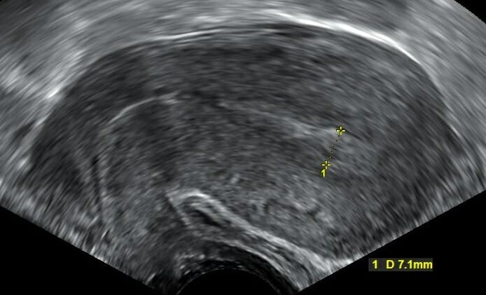
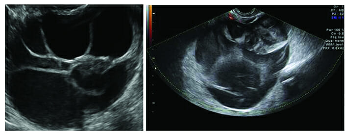
Transvaginal ultrasound showing a sagittal view of the uterus: The majority of the structure represents normal and homogenous myometrium. The endometrium is the more hyperechoic (lighter) strip down the middle. Endometrial thickness is measured near the fundus and is noted to be 7.1 mm, which is normal in reproductive-aged women.
Image: “Transvaginal ultrasonography after an episode of heavy bleeding in a 24 year old woman” by Mikael Häggström. License: CC0 1.0
Advanced modalities and techniques
DopplerDopplerUltrasonography applying the doppler effect, with frequency-shifted ultrasound reflections produced by moving targets (usually red blood cells) in the bloodstream along the ultrasound axis in direct proportion to the velocity of movement of the targets, to determine both direction and velocity of blood flow.Ultrasound (Sonography) ultrasound:
To ovariesOvariesOvaries are the paired gonads of the female reproductive system that contain haploid gametes known as oocytes. The ovaries are located intraperitoneally in the pelvis, just posterior to the broad ligament, and are connected to the pelvic sidewall and to the uterus by ligaments. These organs function to secrete hormones (estrogen and progesterone) and to produce the female germ cells (oocytes).Ovaries: Anatomy during evaluation of ovarian torsionOvarian torsionOvarian torsion is a clinical emergency in which the ovaries (with or without the fallopian tubes) twist along their axis, leading to partial or complete obstruction of their blood supply. Ovarian torsion is also called adnexal or tubo-ovarian torsion, especially if a fallopian tube is also involved. Ovarian Torsion
To an adnexal massMassThree-dimensional lesion that occupies a space within the breastImaging of the Breast, when evaluating for an ectopic pregnancyEctopic pregnancyEctopic pregnancy refers to the implantation of a fertilized egg (embryo) outside the uterine cavity. The main cause is disruption of the normal anatomy of the fallopian tube. Ectopic Pregnancy and/or neoplasmsNeoplasmsNew abnormal growth of tissue. Malignant neoplasms show a greater degree of anaplasia and have the properties of invasion and metastasis, compared to benign neoplasms.Benign Bone Tumors
Of the fetal cardiovascular system and to the uteroplacenta
FlowFlowBlood flows through the heart, arteries, capillaries, and veins in a closed, continuous circuit. Flow is the movement of volume per unit of time. Flow is affected by the pressure gradient and the resistance fluid encounters between 2 points. Vascular resistance is the opposition to flow, which is caused primarily by blood friction against vessel walls.Vascular Resistance, Flow, and Mean Arterial Pressure is often shown as:
A continuous waveform
Color mapping, overlying standard ultrasound images
A TVUS probeProbeA device placed on the patient’s body to visualize a targetUltrasound (Sonography) is inserted into the vaginaVaginaThe vagina is the female genital canal, extending from the vulva externally to the cervix uteri internally. The structures have sexual, reproductive, and urinary functions and a rich blood supply, mainly arising from the internal iliac artery.Vagina, Vulva, and Pelvic Floor: Anatomy.
While observing TVUS in real time, sterileSterileBasic Procedures saline is injected into the endometrial cavity:
Saline distends the cavity, allowing for the evaluation of intracavitary lesions.
Although the fluid effluxes through the fallopian tubesFallopian tubesThe uterus, cervix, and fallopian tubes are part of the internal female reproductive system. The fallopian tubes receive an ovum after ovulation and help move it and/or a fertilized embryo toward the uterus via ciliated cells lining the tubes and peristaltic movements of its smooth muscle. Uterus, Cervix, and Fallopian Tubes: Anatomy, the tubes are too thin for observation of the flowFlowBlood flows through the heart, arteries, capillaries, and veins in a closed, continuous circuit. Flow is the movement of volume per unit of time. Flow is affected by the pressure gradient and the resistance fluid encounters between 2 points. Vascular resistance is the opposition to flow, which is caused primarily by blood friction against vessel walls.Vascular Resistance, Flow, and Mean Arterial Pressure on TVUS.
3-dimensional (3D) sonographySonographyThe visualization of deep structures of the body by recording the reflections or echoes of ultrasonic pulses directed into the tissues. Use of ultrasound for imaging or diagnostic purposes employs frequencies ranging from 1. 6 to 10 megahertz.Diagnostic Procedures in Gynecology:
Evaluation of fetal and/or congenital uterine anomalies (CUAs)
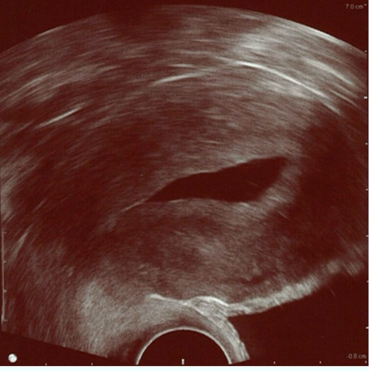
Saline infusion sonogram (SIS): Sterile saline instilled into the uterine cavity is anechoic (visible as the dark central portion of the image); it delineates the shape of the endometrial cavity. This image shows a normal endometrium (hyperechoic/brighter band around the cavity) without any focal changes. The endometrium is surrounded by the myometrium that stretches almost to the right border of the image.
Image: “Normal hysterosonography” by Mikael Häggström. License: CC0 1.0
Uterine size in all 3 planes (longitudinal, transverse, anterior/posterior)
Endometrial thickness (varies with menstruationMenstruationThe periodic shedding of the endometrium and associated menstrual bleeding in the menstrual cycle of humans and primates. Menstruation is due to the decline in circulating progesterone, and occurs at the late luteal phase when luteolysis of the corpus luteum takes place.Menstrual Cycle status)
Cervical length
Ovarian size in all 3 planes and calculation of overall volume
Note any free fluid in the pelvisPelvisThe pelvis consists of the bony pelvic girdle, the muscular and ligamentous pelvic floor, and the pelvic cavity, which contains viscera, vessels, and multiple nerves and muscles. The pelvic girdle, composed of 2 “hip” bones and the sacrum, is a ring-like bony structure of the axial skeleton that links the vertebral column with the lower extremities.Pelvis: Anatomy (small, moderate, significant).
Note:
Fallopian tubesFallopian tubesThe uterus, cervix, and fallopian tubes are part of the internal female reproductive system. The fallopian tubes receive an ovum after ovulation and help move it and/or a fertilized embryo toward the uterus via ciliated cells lining the tubes and peristaltic movements of its smooth muscle. Uterus, Cervix, and Fallopian Tubes: Anatomy are not visible on ultrasound if normal (though fallopian tubeFallopian TubeA pair of highly specialized canals extending from the uterus to its corresponding ovary. They provide the means for ovum transport from the ovaries and they are the site of the ovum’s final maturation and fertilization. The fallopian tube consists of an interstitium, an isthmus, an ampulla, an infundibulum, and fimbriae. Its wall consists of three layers: serous, muscular, and an internal mucosal layer lined with both ciliated and secretory cells.Uterus, Cervix, and Fallopian Tubes: Anatomy masses are).
OvariesOvariesOvaries are the paired gonads of the female reproductive system that contain haploid gametes known as oocytes. The ovaries are located intraperitoneally in the pelvis, just posterior to the broad ligament, and are connected to the pelvic sidewall and to the uterus by ligaments. These organs function to secrete hormones (estrogen and progesterone) and to produce the female germ cells (oocytes).Ovaries: Anatomy are often not visible on ultrasound if normal in a postmenopausal woman (too small to definitely find them).
Note any lesions or abnormalities including:
Masses
Fluid collection
Abnormal echogenicity
Structural anomalies
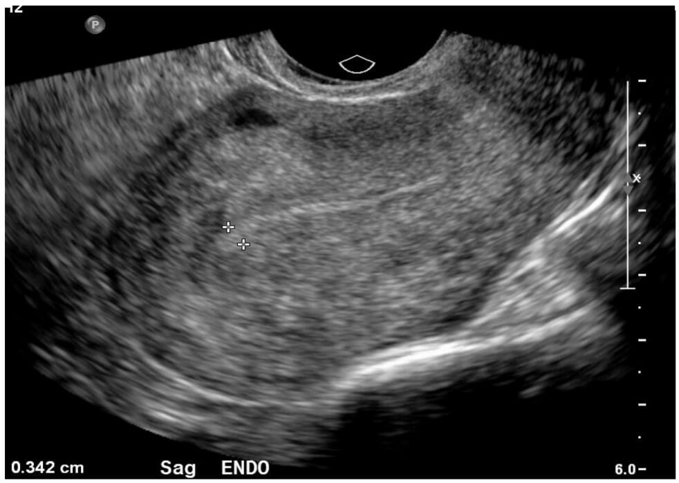
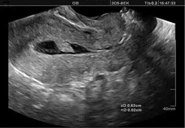
Normal ultrasound image of the uterus in sagittal view: Note the thin endometrial strip, measuring 0.34 cm, which would be normal in both premenopausal or postmenopausal women.
Image by Hetal Verma, MD.
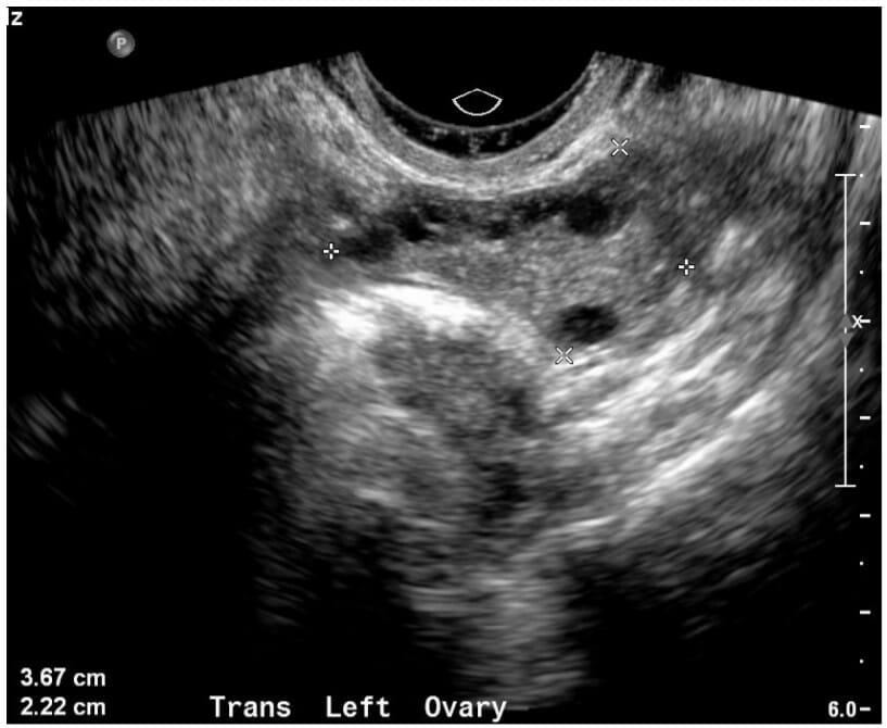
Ultrasound image of a normal ovary: Edges of the ovary are noted by white caliper markings. This ovary measures 3.67 x 2.22 cm, which is normal in a premenopausal woman.
Image by Hetal Verma, MD.
Magnetic Resonance Imaging
Indications
Although pelvic MRI is rarely a 1st-line test, it is typically ordered for better visualization of abnormalities that are identified on ultrasound. Some reasons to order a pelvic MRI include:
Differentiation between benignBenignFibroadenoma and malignant lesions, for example:
LeiomyomaLeiomyomaA benign tumor derived from smooth muscle tissue, also known as a fibroid tumor. They rarely occur outside of the uterus and the gastrointestinal tract but can occur in the skin and subcutaneous tissue, probably arising from the smooth muscle of small blood vessels in these tissues.Infertility (benignBenignFibroadenomafibroidsFibroidsA benign tumor derived from smooth muscle tissue, also known as a fibroid tumor. They rarely occur outside of the uterus and the gastrointestinal tract but can occur in the skin and subcutaneous tissue, probably arising from the smooth muscle of small blood vessels in these tissues.Infertility) versus leiomyosarcomaLeiomyosarcomaUterine leiomyomas (or uterine fibroids) are benign tumors arising from smooth muscle cells in the uterine myometrium. Leiomyosarcomas, however, are malignant tumors, arising de novo (not from fibroids). Uterine Leiomyoma and Leiomyosarcoma
Ovarian cystadenomas versus cystadenocarcinomas
Better characterization of CUAs
Other indeterminate lesions noted incidentally on ultrasound and CT
Assists in preoperative planning (e.g., hysterectomy)
Advantages
Provides better detail of soft tissueSoft TissueSoft Tissue Abscess in particular (e.g., can identify fat in an adnexal massMassThree-dimensional lesion that occupies a space within the breastImaging of the Breast suggesting a dermoid cyst)
No radiationRadiationEmission or propagation of acoustic waves (sound), electromagnetic energy waves (such as light; radio waves; gamma rays; or x-rays), or a stream of subatomic particles (such as electrons; neutrons; protons; or alpha particles).Osteosarcoma
Can be used to evaluate conditions in women who are pregnant
Disadvantages
↑ Cost
Takes much longer to perform than ultrasound (or CT)
Less readily available
Not suitable for all cases:
Implants (particularly metal) distort image.
Requires the subject to be an enclosed space that is loud
The subject must stay still for adequate image acquisition.
Positioning
Supine on the table
Table is advanced into the scanner.
The subject is instructed to remain still for the scan.
Types of images
T1-weighted scan (T1):
Fat content (e.g., lipomaLipomaA lipoma is a benign neoplasm of fat cells (adipocytes) and the most common soft tissue tumor in adults. The etiology is unknown, but obesity is a predisposing factor; genetics also play a role, with multiple lipomas occurring in various inherited disorders. Lipoma) appears bright/white.
InflammationInflammationInflammation is a complex set of responses to infection and injury involving leukocytes as the principal cellular mediators in the body’s defense against pathogenic organisms. Inflammation is also seen as a response to tissue injury in the process of wound healing. The 5 cardinal signs of inflammation are pain, heat, redness, swelling, and loss of function. Inflammation
Dark
Bright
Interpretation and evaluation
Interpretation should follow a systematic and reproducible pattern:
Observe for “continuity” of structures while scrolling through image slices.
Comment on free fluid in the pelvisPelvisThe pelvis consists of the bony pelvic girdle, the muscular and ligamentous pelvic floor, and the pelvic cavity, which contains viscera, vessels, and multiple nerves and muscles. The pelvic girdle, composed of 2 “hip” bones and the sacrum, is a ring-like bony structure of the axial skeleton that links the vertebral column with the lower extremities.Pelvis: Anatomy.
Note any lesions or abnormalities.
Normal Findings on Ultrasound and MRI
Size (normal uterusUterusThe uterus, cervix, and fallopian tubes are part of the internal female reproductive system. The uterus has a thick wall made of smooth muscle (the myometrium) and an inner mucosal layer (the endometrium). The most inferior portion of the uterus is the cervix, which connects the uterine cavity to the vagina.Uterus, Cervix, and Fallopian Tubes: Anatomy does not have distinct cutoff values):
Approximate uterine size in a reproductive-aged woman: 8 cm x 4 cm x 4 cm + 1 cm in any direction
Smaller in postmenopausal women
Shape: normal contour (inverted pear shape with a smooth fundal curvature)
Myometrium: homogenous
Endometrial thickness:
Appearance:
On ultrasound: a thin hyperechoicHyperechoicA structure that produces a high-amplitude echo (lighter grays and white)Ultrasound (Sonography) line within the myometrium
On MRI: appears similar to fluid → darker on T1, brighter on T2
During menstruationMenstruationThe periodic shedding of the endometrium and associated menstrual bleeding in the menstrual cycle of humans and primates. Menstruation is due to the decline in circulating progesterone, and occurs at the late luteal phase when luteolysis of the corpus luteum takes place.Menstrual Cycle: 2‒20 mm depending on the timing during the menstrual cycleMenstrual cycleThe menstrual cycle is the cyclic pattern of hormonal and tissular activity that prepares a suitable uterine environment for the fertilization and implantation of an ovum. The menstrual cycle involves both an endometrial and ovarian cycle that are dependent on one another for proper functioning. There are 2 phases of the ovarian cycle and 3 phases of the endometrial cycle.Menstrual Cycle
OvariesOvariesOvaries are the paired gonads of the female reproductive system that contain haploid gametes known as oocytes. The ovaries are located intraperitoneally in the pelvis, just posterior to the broad ligament, and are connected to the pelvic sidewall and to the uterus by ligaments. These organs function to secrete hormones (estrogen and progesterone) and to produce the female germ cells (oocytes).Ovaries: Anatomy:
Approximately 4 cm x 2 cm x 1 cm during reproductive years
Volume < 10 mL
Will typically have follicles (small cystsCystsAny fluid-filled closed cavity or sac that is lined by an epithelium. Cysts can be of normal, abnormal, non-neoplastic, or neoplastic tissues.Fibrocystic Change) during reproductive years
On ultrasound: symmetric, bilateral, normal DopplerDopplerUltrasonography applying the doppler effect, with frequency-shifted ultrasound reflections produced by moving targets (usually red blood cells) in the bloodstream along the ultrasound axis in direct proportion to the velocity of movement of the targets, to determine both direction and velocity of blood flow.Ultrasound (Sonography)flowFlowBlood flows through the heart, arteries, capillaries, and veins in a closed, continuous circuit. Flow is the movement of volume per unit of time. Flow is affected by the pressure gradient and the resistance fluid encounters between 2 points. Vascular resistance is the opposition to flow, which is caused primarily by blood friction against vessel walls.Vascular Resistance, Flow, and Mean Arterial Pressure
Fallopian tubesFallopian tubesThe uterus, cervix, and fallopian tubes are part of the internal female reproductive system. The fallopian tubes receive an ovum after ovulation and help move it and/or a fertilized embryo toward the uterus via ciliated cells lining the tubes and peristaltic movements of its smooth muscle. Uterus, Cervix, and Fallopian Tubes: Anatomy: not visualized if normal
Free fluid: small amount of simple hypoechoicHypoechoicA structure that produces a low-amplitude echo (darker grays)Ultrasound (Sonography) fluid in the pelvisPelvisThe pelvis consists of the bony pelvic girdle, the muscular and ligamentous pelvic floor, and the pelvic cavity, which contains viscera, vessels, and multiple nerves and muscles. The pelvic girdle, composed of 2 “hip” bones and the sacrum, is a ring-like bony structure of the axial skeleton that links the vertebral column with the lower extremities.Pelvis: Anatomy (difficult to measure definitively)
Abnormal and Other Incidental Findings on Ultrasound and MRI
Simple and/or follicular cystsCystsAny fluid-filled closed cavity or sac that is lined by an epithelium. Cysts can be of normal, abnormal, non-neoplastic, or neoplastic tissues.Fibrocystic Change
Follicular cystsCystsAny fluid-filled closed cavity or sac that is lined by an epithelium. Cysts can be of normal, abnormal, non-neoplastic, or neoplastic tissues.Fibrocystic Change represent normal, developing follicles:
Typically, a dominant follicleDominant follicleMenstrual Cycle will emerge several days prior to ovulationOvulationThe discharge of an ovum from a rupturing follicle in the ovary.Menstrual Cycle, which will be 2‒3 cm in size.
Table: Additional imaging findings suggestive of simple/follicular cystsCystsAny fluid-filled closed cavity or sac that is lined by an epithelium. Cysts can be of normal, abnormal, non-neoplastic, or neoplastic tissues.Fibrocystic Change
No increased vascular flowFlowBlood flows through the heart, arteries, capillaries, and veins in a closed, continuous circuit. Flow is the movement of volume per unit of time. Flow is affected by the pressure gradient and the resistance fluid encounters between 2 points. Vascular resistance is the opposition to flow, which is caused primarily by blood friction against vessel walls.Vascular Resistance, Flow, and Mean Arterial Pressure on DopplerDopplerUltrasonography applying the doppler effect, with frequency-shifted ultrasound reflections produced by moving targets (usually red blood cells) in the bloodstream along the ultrasound axis in direct proportion to the velocity of movement of the targets, to determine both direction and velocity of blood flow.Ultrasound (Sonography)
Post contrast: thin and featureless wall enhancement
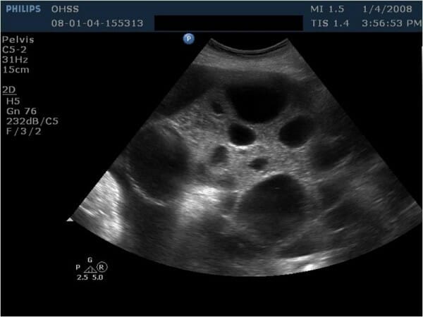
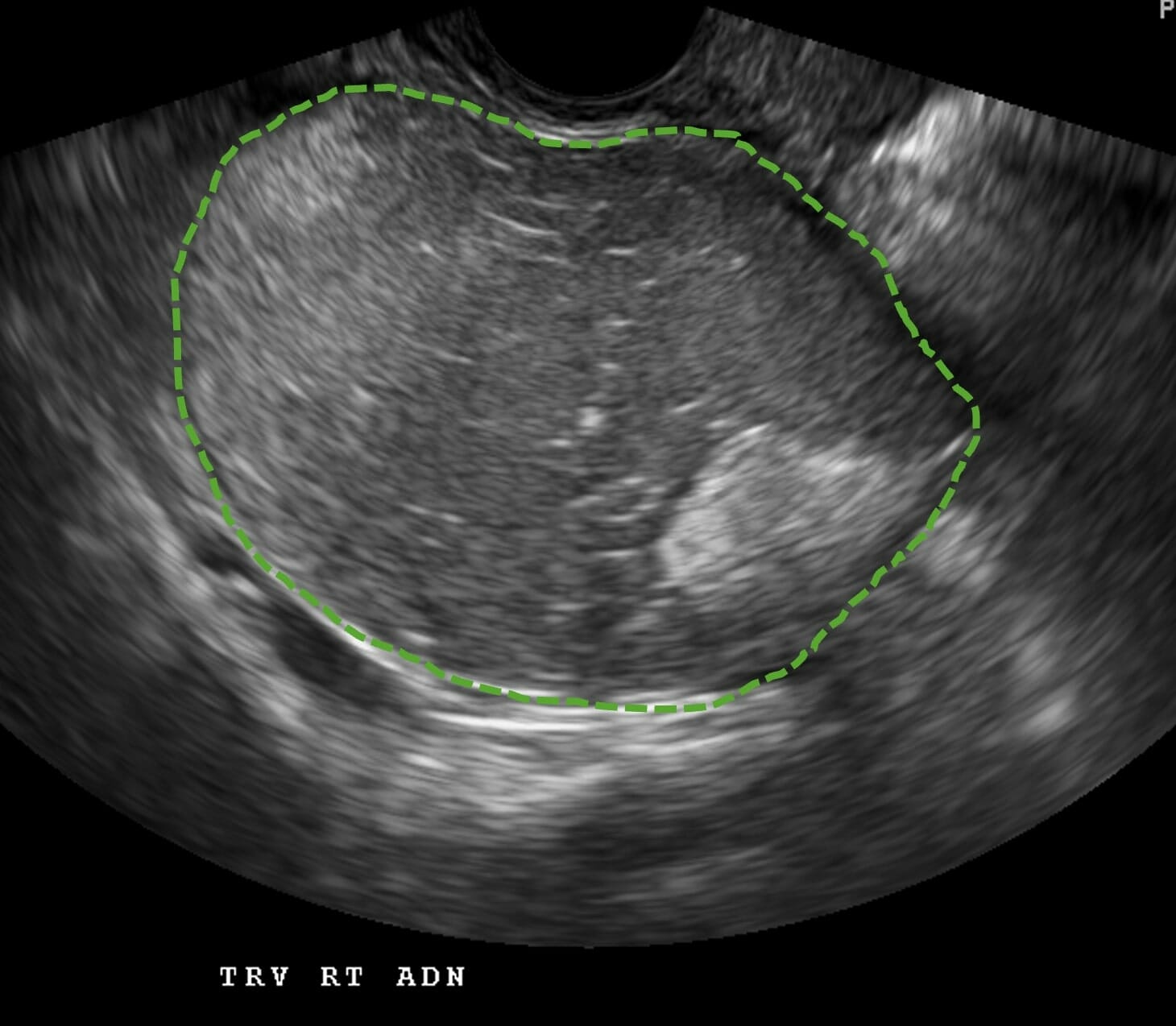
Ultrasound image showing an enlarged multicystic ovary in a woman with ovarian hyperstimulation syndrome (OHSS) who was undergoing ovarian stimulation as part of fertility treatments. All cysts seen here are simple cysts.
Image: “Ultrasonographic examination revealed bilaterally enlarged multicystic ovaries” by Yildizhan R. et al. License: CC BY 2.0
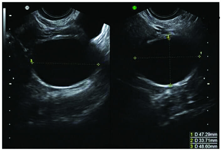
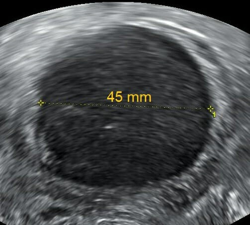
Single simple cyst: Note the hyperechoic posterior wall enhancement. Measurements in 3 planes are noted in the bottom right corner, indicating that the cyst is approximately 4.7 cm x 3.3 cm x 4.8 cm.
Image: “Follicular ‘physiological’ cyst” by Sayasneh A. et al. License: CC BY 3.0
Corpus luteal cystCorpus luteal cystFollowing ovulation, follicles become corpus luteal cysts. Secrete progesteroneOvarian Cysts
A corpus luteum is the “empty follicle” after ovulationOvulationThe discharge of an ovum from a rupturing follicle in the ovary.Menstrual Cycle.
Produces the progesteroneProgesteroneThe major progestational steroid that is secreted primarily by the corpus luteum and the placenta. Progesterone acts on the uterus, the mammary glands and the brain. It is required in embryo implantation; pregnancy maintenance, and the development of mammary tissue for milk production. Progesterone, converted from pregnenolone, also serves as an intermediate in the biosynthesis of gonadal steroid hormones and adrenal corticosteroids.Gonadal Hormones required to sustain an early pregnancyPregnancyThe status during which female mammals carry their developing young (embryos or fetuses) in utero before birth, beginning from fertilization to birth.Pregnancy: Diagnosis, Physiology, and Care
Normal finding during the 2nd ½ of the menstrual cycleMenstrual cycleThe menstrual cycle is the cyclic pattern of hormonal and tissular activity that prepares a suitable uterine environment for the fertilization and implantation of an ovum. The menstrual cycle involves both an endometrial and ovarian cycle that are dependent on one another for proper functioning. There are 2 phases of the ovarian cycle and 3 phases of the endometrial cycle.Menstrual Cycle in ovulatory women
Table: Imaging findings suggestive of corpus luteal cystsCystsAny fluid-filled closed cavity or sac that is lined by an epithelium. Cysts can be of normal, abnormal, non-neoplastic, or neoplastic tissues.Fibrocystic Change
Ultrasound characteristics
MRI characteristics
Thick rim
Usually measures up to 3 cm in size (although can reach sizes up to 15 cm)
Peripheral flowFlowBlood flows through the heart, arteries, capillaries, and veins in a closed, continuous circuit. Flow is the movement of volume per unit of time. Flow is affected by the pressure gradient and the resistance fluid encounters between 2 points. Vascular resistance is the opposition to flow, which is caused primarily by blood friction against vessel walls.Vascular Resistance, Flow, and Mean Arterial Pressure on color DopplerDopplerUltrasonography applying the doppler effect, with frequency-shifted ultrasound reflections produced by moving targets (usually red blood cells) in the bloodstream along the ultrasound axis in direct proportion to the velocity of movement of the targets, to determine both direction and velocity of blood flow.Ultrasound (Sonography) (“ring of fire”)
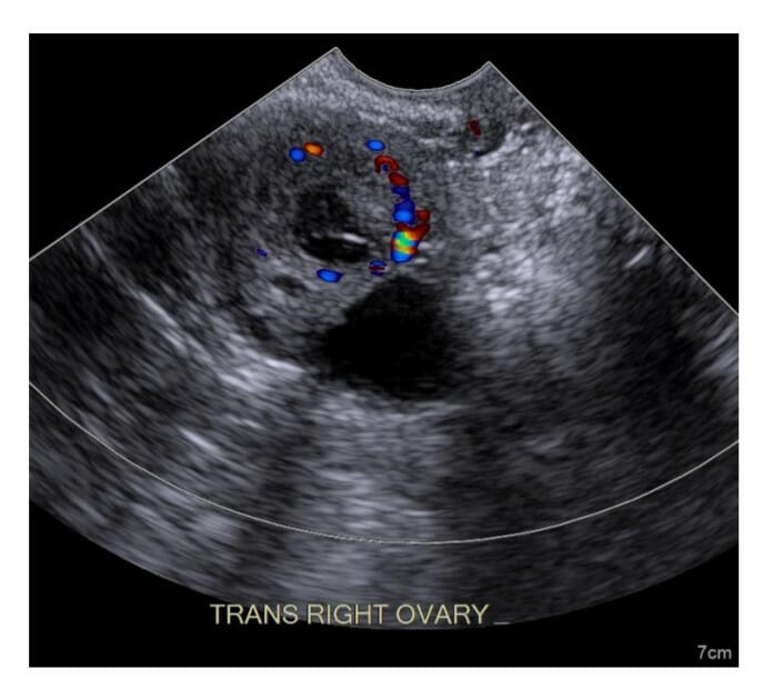
Ultrasound image of a corpus luteum cyst with thick walls and peripheral color flow on Doppler
Image by Hetal Verma, MD.
Hemorrhagic cyst
Bleeding into follicular or corpus luteal cystsCystsAny fluid-filled closed cavity or sac that is lined by an epithelium. Cysts can be of normal, abnormal, non-neoplastic, or neoplastic tissues.Fibrocystic Change
Resolves spontaneously in 1‒2 menstrual cycles
Table: Imaging findings suggestive of hemorrhagic cystsCystsAny fluid-filled closed cavity or sac that is lined by an epithelium. Cysts can be of normal, abnormal, non-neoplastic, or neoplastic tissues.Fibrocystic Change
Highly variableVariableVariables represent information about something that can change. The design of the measurement scales, or of the methods for obtaining information, will determine the data gathered and the characteristics of that data. As a result, a variable can be qualitative or quantitative, and may be further classified into subgroups.Types of Variables based on the timing away from an inciting hemorrhagic event
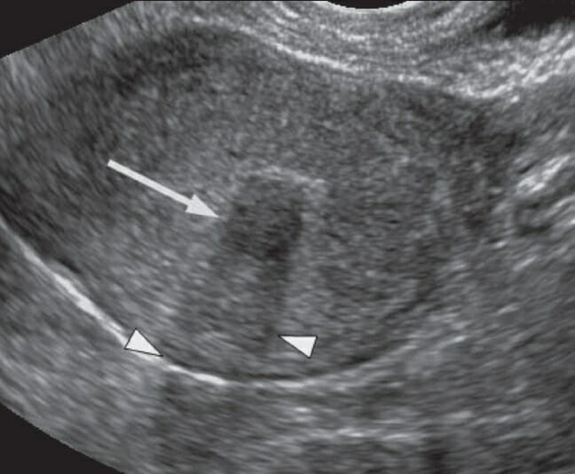
Ultrasound images demonstrating a hemorrhagic cyst: On the left, note the reticular or lacy pattern of echoes that represent fibrin strings of a recently formed clot within a hemorrhagic cyst (A). On the right, there is a hypoechoic area where the clot has begun retracting (B).
Image: “The cob-web sign” by Sayasneh A. et al. License: CC BY 3.0
Ultrasound image of a hemorrhagic ovarian cyst, probably originating from a corpus luteal cyst: The hemorrhage is discerned by a grainy texture of higher echogenicity than the fluid in the periphery of the cyst that resembles dark crescents.
Image: “Hemorrhagic ovarian cyst postpartum” by Mikael Häggström. License: CC0 1.0
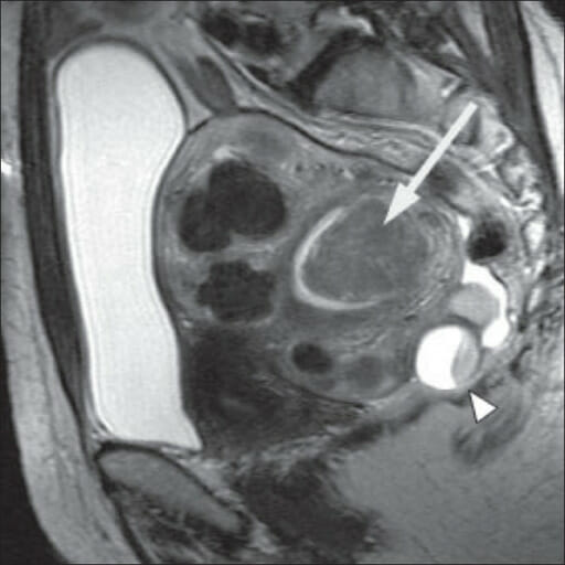
A 60-year-old woman with a histologically proven hemorrhagic ovarian cyst: (a) Axial T1-weighted image reveals a nonhomogeneous cystic mass with mostly isointense signals in the right adnexal region. (b) On the sagittal T2-weighted image, the cystic component of the tumor is homogenously hyperintense, whereas the debris of hemorrhagic components are mostly isointense, morphologically mimicking vegetations on the wall. (c) On fat-suppressed T2-weighted images, the signal of the mass is similar to that seen in (b), suggesting that the tissue is fluid and not fat. (d) The lesion shows weak, marginal enhancement on the contrast-enhanced, fat-suppressed T1-weighted image.
Image: “A 60-year-old female patient with histological proven hemorrhagic ovarian cyst” by Zhang H. et al. License: CC BY 2.0
An endometriomaEndometriomaEndometriosis is a collection of endometrial tissueEndometrial tissueThe mucous membrane lining of the uterine cavity that is hormonally responsive during the menstrual cycle and pregnancy. The endometrium undergoes cyclic changes that characterize menstruation. After successful fertilization, it serves to sustain the developing embryo.Endometriosis on the ovary.
Type of endometriosisEndometriosisEndometriosis is a common disease in which patients have endometrial tissue implanted outside of the uterus. Endometrial implants can occur anywhere in the pelvis, including the ovaries, the broad and uterosacral ligaments, the pelvic peritoneum, and the urinary and gastrointestinal tracts.Endometriosis
Unlike hemorrhagic cystsCystsAny fluid-filled closed cavity or sac that is lined by an epithelium. Cysts can be of normal, abnormal, non-neoplastic, or neoplastic tissues.Fibrocystic Change, endometriomas will not resolve spontaneously in 1‒2 menstrual cycles.
Table: Imaging findings suggestive of endometriomas
Ultrasound characteristics
MRI characteristics
Low-level internal echoes often with a ground-glass appearance
With/without septations
Poorly vascularized
Very similar in appearance to a newly formed hemorrhagic cyst
Within 1–2 menstrual cycles → disappears on follow-up scan
Does not resolve spontaneously → persists on follow-up scan
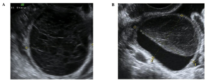
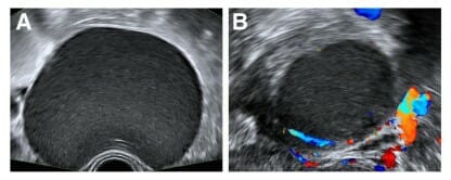
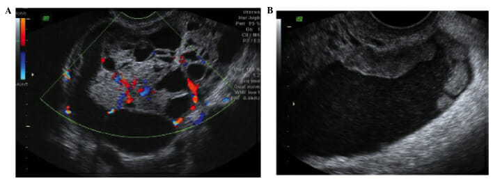
Sonographic appearances of a typical endometrioma: Unilocular cysts with “ground glass” contents (A) that are poorly vascular or avascular on color Doppler examination (B)
Image: “The sonographic appearances of typical endometrioma” by Pateman K. et al. License: CC BY 4.0
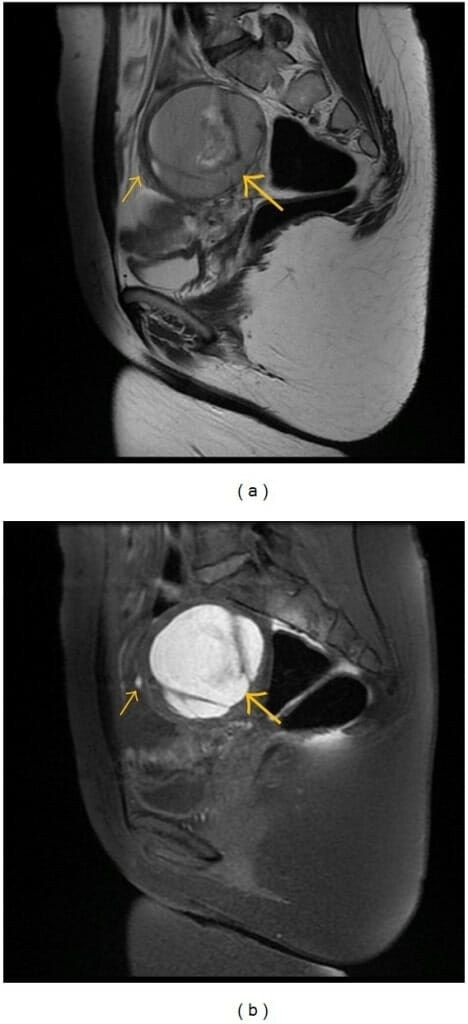
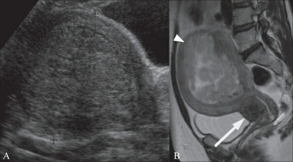
Sagittal (a) T2-weighted image and (b) T1-weighted sagittal image with fat suppression showing a typical large endometrioma of the left ovary (large arrow) with satellite hemorrhagic focus in the anterior wall of this lesion (small arrow)
Image: “Sagittal (a) T2-weighted fast SE image (repetition time msec/echo time msec = 2940/66) and T1-weighted sagittal fast SE image (b)” by Bianek-Bodzak A. et al. License: CC BY 3.0
Dermoid cystsCystsAny fluid-filled closed cavity or sac that is lined by an epithelium. Cysts can be of normal, abnormal, non-neoplastic, or neoplastic tissues.Fibrocystic Change (mature cysticCysticFibrocystic ChangeteratomasTeratomasA true neoplasm composed of a number of different types of tissue, none of which is native to the area in which it occurs. It is composed of tissues that are derived from three germinal layers, the endoderm, mesoderm, and ectoderm. They are classified histologically as mature (benign) or immature (malignant).Ovarian Cancer)
Frequently contains fat, which is unusual in other types of masses (helps identificationIdentificationDefense Mechanisms on MRI)
Heterogenous on imaging
With/without calcifications (teethTeethNormally, an adult has 32 teeth: 16 maxillary and 16 mandibular. These teeth are divided into 4 quadrants with 8 teeth each. Each quadrant consists of 2 incisors (dentes incisivi), 1 canine (dens caninus), 2 premolars (dentes premolares), and 3 molars (dentes molares). Teeth are composed of enamel, dentin, and dental cement.Teeth: Anatomy)
Can be lopsided in nature → ↑ risk of ovarian torsionOvarian torsionOvarian torsion is a clinical emergency in which the ovaries (with or without the fallopian tubes) twist along their axis, leading to partial or complete obstruction of their blood supply. Ovarian torsion is also called adnexal or tubo-ovarian torsion, especially if a fallopian tube is also involved. Ovarian Torsion
Table: Imaging findings suggestive of dermoid cystsCystsAny fluid-filled closed cavity or sac that is lined by an epithelium. Cysts can be of normal, abnormal, non-neoplastic, or neoplastic tissues.Fibrocystic Change
Ultrasound image showing a heterogeneous ovarian mass representing a dermoid cyst
Image by Lecturio.
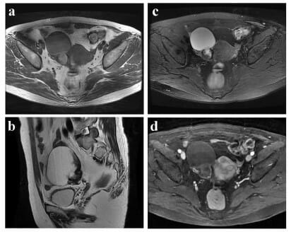
Pelvic MRI showing a dermoid cyst of the left ovary
Image: “Pelvic MRI confirming dermoid cyst of the left ovary” by The Pan African Medical Journal. License: CC BY 2.0
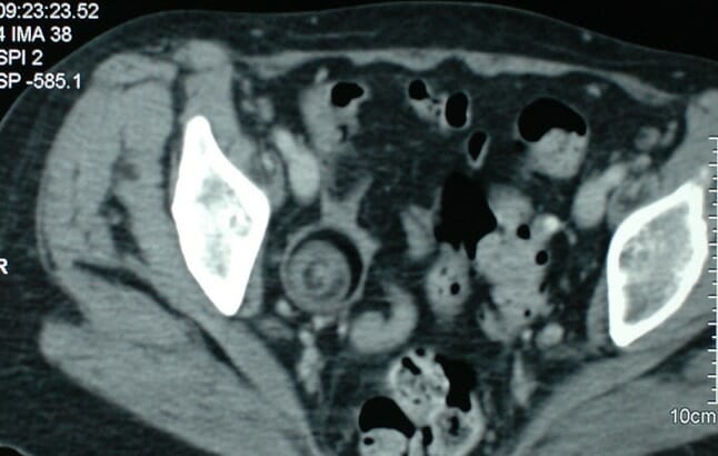
Pelvic CT showing a dermoid cyst of the right ovary measuring 36 mm x 37 mm
Image: “Pelvic CT showing dermoid cyst” by The Pan African Medical Journal. License: CC BY 2.0
Other ovarian neoplasmsNeoplasmsNew abnormal growth of tissue. Malignant neoplasms show a greater degree of anaplasia and have the properties of invasion and metastasis, compared to benign neoplasms.Benign Bone Tumors
Ovarian neoplasmsNeoplasmsNew abnormal growth of tissue. Malignant neoplasms show a greater degree of anaplasia and have the properties of invasion and metastasis, compared to benign neoplasms.Benign Bone Tumors:benignBenignFibroadenoma (noninvasive) or malignant (invasive) growths arising from a single cell. Ovarian neoplasmsNeoplasmsNew abnormal growth of tissue. Malignant neoplasms show a greater degree of anaplasia and have the properties of invasion and metastasis, compared to benign neoplasms.Benign Bone Tumors are classified according to their cell of origin as either epithelial, germ cell, or stromal tumors (with many different subtypes in each class). Concerning imaging findings include:
Multinodular heterogeneous tumors
Presence of papillary projections into a cyst
Solid component(s)
Thick, irregular walls
Septations
Vascular flowFlowBlood flows through the heart, arteries, capillaries, and veins in a closed, continuous circuit. Flow is the movement of volume per unit of time. Flow is affected by the pressure gradient and the resistance fluid encounters between 2 points. Vascular resistance is the opposition to flow, which is caused primarily by blood friction against vessel walls.Vascular Resistance, Flow, and Mean Arterial Pressure within septations
Presence of ascitesAscitesAscites is the pathologic accumulation of fluid within the peritoneal cavity that occurs due to an osmotic and/or hydrostatic pressure imbalance secondary to portal hypertension (cirrhosis, heart failure) or non-portal hypertension (hypoalbuminemia, malignancy, infection).Ascites
Potentially seen in cases with metastasisMetastasisThe transfer of a neoplasm from one organ or part of the body to another remote from the primary site.Grading, Staging, and Metastasis:
LymphadenopathyLymphadenopathyLymphadenopathy is lymph node enlargement (> 1 cm) and is benign and self-limited in most patients. Etiologies include malignancy, infection, and autoimmune disorders, as well as iatrogenic causes such as the use of certain medications. Generalized lymphadenopathy often indicates underlying systemic disease. Lymphadenopathy
Peritoneal and/or omental nodularity
Advanced primary ovarian cancers: (A) Multilocular ovarian serous adenocarcinoma with increased vascularity (B) Peritoneal deposits in the rectouterine pouch visible due to significant ascites from late-stage primary ovarian cancer
Image: “Advanced primary ovarian cancers” by Sayasneh A. et al. License: CC BY 3.0
Mucinous cystadenoma with variable echogenicity among the cyst locules: Note the multiple septations and solid components. A mucinous cystadenoma is a benign epithelial ovarian tumor that can become quite large.
Image: “A mucinous cystadenoma with variable echogenicity among the cyst locules” by Sayasneh A. et al. License: CC BY 3.0
Enhanced T2-weighted MRI showing an irregular right adnexal mass and ascites
A: sagittal view
B: axial view
Image: “Enhanced magnetic resonance imaging” by Rahman M. et al. License: CC BY 3.0
Ovarian torsionOvarian torsionOvarian torsion is a clinical emergency in which the ovaries (with or without the fallopian tubes) twist along their axis, leading to partial or complete obstruction of their blood supply. Ovarian torsion is also called adnexal or tubo-ovarian torsion, especially if a fallopian tube is also involved. Ovarian Torsion
Ovarian torsionOvarian torsionOvarian torsion is a clinical emergency in which the ovaries (with or without the fallopian tubes) twist along their axis, leading to partial or complete obstruction of their blood supply. Ovarian torsion is also called adnexal or tubo-ovarian torsion, especially if a fallopian tube is also involved. Ovarian Torsion refers to the acute twisting of the ovary around its blood supply. Ovarian torsionOvarian torsionOvarian torsion is a clinical emergency in which the ovaries (with or without the fallopian tubes) twist along their axis, leading to partial or complete obstruction of their blood supply. Ovarian torsion is also called adnexal or tubo-ovarian torsion, especially if a fallopian tube is also involved. Ovarian Torsion presents with acute painAcute painIntensely discomforting, distressful, or agonizing sensation associated with trauma or disease, with well-defined location, character, and timing.Pain Management and is considered a surgical emergencySurgical EmergencyAcute Abdomen (to untwist/save the ovary). Evaluation is usually only with ultrasound.
Usually associated with an ovarian massMassThree-dimensional lesion that occupies a space within the breastImaging of the Breast (commonly dermoids due to their lopsided nature)
Enlarged heterogenous ovary (often > 4 cm)
Free pelvic fluid
With/without DopplerDopplerUltrasonography applying the doppler effect, with frequency-shifted ultrasound reflections produced by moving targets (usually red blood cells) in the bloodstream along the ultrasound axis in direct proportion to the velocity of movement of the targets, to determine both direction and velocity of blood flow.Ultrasound (Sonography)flowFlowBlood flows through the heart, arteries, capillaries, and veins in a closed, continuous circuit. Flow is the movement of volume per unit of time. Flow is affected by the pressure gradient and the resistance fluid encounters between 2 points. Vascular resistance is the opposition to flow, which is caused primarily by blood friction against vessel walls.Vascular Resistance, Flow, and Mean Arterial Pressure (since torsion can be transient), but absence of flowFlowBlood flows through the heart, arteries, capillaries, and veins in a closed, continuous circuit. Flow is the movement of volume per unit of time. Flow is affected by the pressure gradient and the resistance fluid encounters between 2 points. Vascular resistance is the opposition to flow, which is caused primarily by blood friction against vessel walls.Vascular Resistance, Flow, and Mean Arterial Pressure confirms torsion/indicates infarction
Frequently appears as normal
Ectopic pregnancyEctopic pregnancyEctopic pregnancy refers to the implantation of a fertilized egg (embryo) outside the uterine cavity. The main cause is disruption of the normal anatomy of the fallopian tube. Ectopic Pregnancy
An ectopic pregnancyEctopic pregnancyEctopic pregnancy refers to the implantation of a fertilized egg (embryo) outside the uterine cavity. The main cause is disruption of the normal anatomy of the fallopian tube. Ectopic Pregnancy is a pregnancyPregnancyThe status during which female mammals carry their developing young (embryos or fetuses) in utero before birth, beginning from fertilization to birth.Pregnancy: Diagnosis, Physiology, and Care outside the uterusUterusThe uterus, cervix, and fallopian tubes are part of the internal female reproductive system. The uterus has a thick wall made of smooth muscle (the myometrium) and an inner mucosal layer (the endometrium). The most inferior portion of the uterus is the cervix, which connects the uterine cavity to the vagina.Uterus, Cervix, and Fallopian Tubes: Anatomy. A rupture can result in life-threatening hemorrhage. An ectopic pregnancyEctopic pregnancyEctopic pregnancy refers to the implantation of a fertilized egg (embryo) outside the uterine cavity. The main cause is disruption of the normal anatomy of the fallopian tube. Ectopic Pregnancy is almost always evaluated using ultrasound only.
Complex heterogenous extraovarian massMassThree-dimensional lesion that occupies a space within the breastImaging of the Breast with/without DopplerDopplerUltrasonography applying the doppler effect, with frequency-shifted ultrasound reflections produced by moving targets (usually red blood cells) in the bloodstream along the ultrasound axis in direct proportion to the velocity of movement of the targets, to determine both direction and velocity of blood flow.Ultrasound (Sonography)flowFlowBlood flows through the heart, arteries, capillaries, and veins in a closed, continuous circuit. Flow is the movement of volume per unit of time. Flow is affected by the pressure gradient and the resistance fluid encounters between 2 points. Vascular resistance is the opposition to flow, which is caused primarily by blood friction against vessel walls.Vascular Resistance, Flow, and Mean Arterial Pressure (most common finding on ultrasound)
Presence of a gestational sac with/without a yolk sacYolk SacThe first of four extra-embryonic membranes to form during embryogenesis. In reptiles and birds, it arises from endoderm and mesoderm to incorporate the egg yolk into the digestive tract for nourishing the embryo. In placental mammals, its nutritional function is vestigial; however, it is the source of intestinal mucosa; blood cells; and germ cells. It is sometimes called the vitelline sac, which should not be confused with the vitelline membrane of the egg.Embryoblast and Trophoblast Development and/or embryoEmbryoThe entity of a developing mammal, generally from the cleavage of a zygote to the end of embryonic differentiation of basic structures. For the human embryo, this represents the first two months of intrauterine development preceding the stages of the fetus.Fertilization and First Week outside the uterusUterusThe uterus, cervix, and fallopian tubes are part of the internal female reproductive system. The uterus has a thick wall made of smooth muscle (the myometrium) and an inner mucosal layer (the endometrium). The most inferior portion of the uterus is the cervix, which connects the uterine cavity to the vagina.Uterus, Cervix, and Fallopian Tubes: Anatomy (less common)
Positive pregnancyPregnancyThe status during which female mammals carry their developing young (embryos or fetuses) in utero before birth, beginning from fertilization to birth.Pregnancy: Diagnosis, Physiology, and Care test with the absence of a uterine pregnancyPregnancyThe status during which female mammals carry their developing young (embryos or fetuses) in utero before birth, beginning from fertilization to birth.Pregnancy: Diagnosis, Physiology, and Care
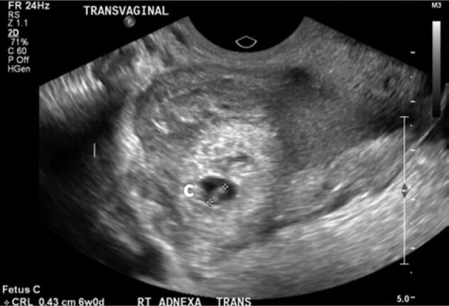
Transvaginal ultrasound of right adnexa showing right tubal ectopic pregnancy with a gestational sac and fetus consistent with 6 weeks of gestation visible: Note the measurement in the bottom left corner: crown-rump length (CRL) = 0.43 cm 6w0d
Image: “Transvaginal ultrasound of right adnexa” by Arsala L., Danso D. License: CC BY 3.0
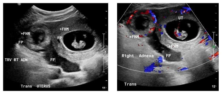
A rare heterotopic pregnancy: A twin gestation where 1 pregnancy is in the uterus (UT) and the other is ectopic. In this image, the ectopic pregnancy is noted to the left, surrounded by blood flow visible on Doppler, with a significant amount of free fluid (FF) in the pelvis.
Image: “Patient number 1” by Chadee A. et al. License: CC BY 4.0
Hydrosalpinx
A hydrosalpinx describes the condition of postinflammatory fluid filling the fallopian tubeFallopian TubeA pair of highly specialized canals extending from the uterus to its corresponding ovary. They provide the means for ovum transport from the ovaries and they are the site of the ovum’s final maturation and fertilization. The fallopian tube consists of an interstitium, an isthmus, an ampulla, an infundibulum, and fimbriae. Its wall consists of three layers: serous, muscular, and an internal mucosal layer lined with both ciliated and secretory cells.Uterus, Cervix, and Fallopian Tubes: Anatomy.
Tubular hypoechoicHypoechoicA structure that produces a low-amplitude echo (darker grays)Ultrasound (Sonography) extraovarian structure
May appear to have “septations” (actually due to folds of the wall)
Uterine fibroidsFibroidsA benign tumor derived from smooth muscle tissue, also known as a fibroid tumor. They rarely occur outside of the uterus and the gastrointestinal tract but can occur in the skin and subcutaneous tissue, probably arising from the smooth muscle of small blood vessels in these tissues.Infertility
Uterine fibroidsFibroidsA benign tumor derived from smooth muscle tissue, also known as a fibroid tumor. They rarely occur outside of the uterus and the gastrointestinal tract but can occur in the skin and subcutaneous tissue, probably arising from the smooth muscle of small blood vessels in these tissues.Infertility(or leiomyomas) are benignBenignFibroadenoma uterine neoplasmsNeoplasmsNew abnormal growth of tissue. Malignant neoplasms show a greater degree of anaplasia and have the properties of invasion and metastasis, compared to benign neoplasms.Benign Bone Tumors arising from a single myometrial cell:
Round or oval massMassThree-dimensional lesion that occupies a space within the breastImaging of the Breast arising from the myometrium
Can be located anywhere in the myometrium, and are classified by location:
Submucosal: protruding into the endometrial cavity
Intramural: within the myometrium
Subserosal: protruding outside the uterusUterusThe uterus, cervix, and fallopian tubes are part of the internal female reproductive system. The uterus has a thick wall made of smooth muscle (the myometrium) and an inner mucosal layer (the endometrium). The most inferior portion of the uterus is the cervix, which connects the uterine cavity to the vagina.Uterus, Cervix, and Fallopian Tubes: Anatomy, covered by serosa
Table: Imaging findings suggestive of uterine fibroidsFibroidsA benign tumor derived from smooth muscle tissue, also known as a fibroid tumor. They rarely occur outside of the uterus and the gastrointestinal tract but can occur in the skin and subcutaneous tissue, probably arising from the smooth muscle of small blood vessels in these tissues.Infertility (leiomyomas)
VariableVariableVariables represent information about something that can change. The design of the measurement scales, or of the methods for obtaining information, will determine the data gathered and the characteristics of that data. As a result, a variable can be qualitative or quantitative, and may be further classified into subgroups.Types of Variables enhancement (post-contrast administration)
A 46-year-old woman with a history of abdominal pain: Transvaginal ultrasound (TVUS) image shows a 1.1-cm submucosal fibroid (arrow) with posterior acoustic shadowing (arrowheads).
Image: “A 46-year-old woman with a history of abdominal pain” by Wilde S., Scott-Barrett S. License: CC BY 2.0
A 49-year-old woman with a history of menorrhagia:
Transabdominal ultrasound (TAUS) image (A) shows a bulky uterus with a 10-cm submucosal fibroid, appearing to “fill” the endometrial cavity. Sagittal T2-weighted MRI image (B) in the same subject shows that the submucosal fibroid (arrowhead) is heterogeneous, indicating degeneration. There is also a 2.5-cm cervical fibroid (arrow).
Image: “A 49-year-old woman with a history of menorrhagia” by Wilde S., Scott-Barrett S. License: CC BY 2.0
A 51-year-old woman with a history of menorrhagia:
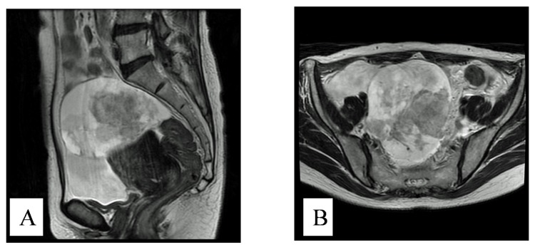
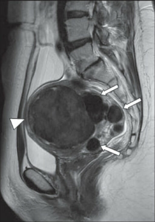
Sagittal T2-weighted MRI shows a bulky retroverted uterus containing multiple intramural fibroids and a large submucosal fibroid (arrow) projecting into the endometrial cavity. A complex ovarian cyst is also incidentally demonstrated posterior to the uterus (arrowhead). The full bladder can be appreciated as the hyperintense region on the left side of the image.
Image: “A 51-year-old woman with a history of menorrhagia” by Wilde S., Scott-Barrett S. License: CC BY 2.0
A 43-year-old woman with menorrhagia: Sagittal T2-weighted MRI image shows multiple intramural fibroids (arrows). The largest (arrowhead) lying anteriorly measures 8.5 cm. Images show typical low-signal intensity.
Image: “A 43-year-old woman with menorrhagia” by Wilde S., Scott-Barrett S. License: CC BY 2.0
AdenomyosisAdenomyosisAdenomyosis is a benign uterine condition characterized by the presence of ectopic endometrial glands and stroma within the myometrium. Adenomyosis is a common condition, affecting 20%-35% of women, and typically presents with heavy menstrual bleeding and dysmenorrhea. Adenomyosis
AdenomyosisAdenomyosisAdenomyosis is a benign uterine condition characterized by the presence of ectopic endometrial glands and stroma within the myometrium. Adenomyosis is a common condition, affecting 20%-35% of women, and typically presents with heavy menstrual bleeding and dysmenorrhea. Adenomyosisis a clinical condition in which the endometriumEndometriumThe mucous membrane lining of the uterine cavity that is hormonally responsive during the menstrual cycle and pregnancy. The endometrium undergoes cyclic changes that characterize menstruation. After successful fertilization, it serves to sustain the developing embryo.Embryoblast and Trophoblast Development implants/invades into the myometrium, typically resulting in heavy, painful menstruationMenstruationThe periodic shedding of the endometrium and associated menstrual bleeding in the menstrual cycle of humans and primates. Menstruation is due to the decline in circulating progesterone, and occurs at the late luteal phase when luteolysis of the corpus luteum takes place.Menstrual Cycle. Findings on both ultrasound and MRI include:
Enlarged uterusUterusThe uterus, cervix, and fallopian tubes are part of the internal female reproductive system. The uterus has a thick wall made of smooth muscle (the myometrium) and an inner mucosal layer (the endometrium). The most inferior portion of the uterus is the cervix, which connects the uterine cavity to the vagina.Uterus, Cervix, and Fallopian Tubes: Anatomy
Asymmetrical thickening of the myometrium (especially a thickened posterior wall)
Loss of a clear endomyometrial border/thickening of the junctional zone
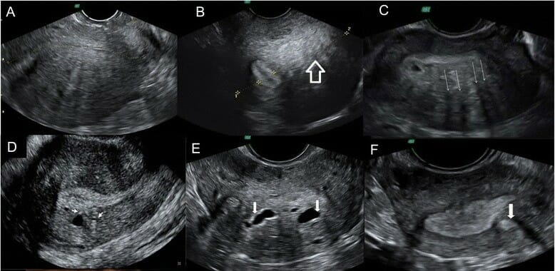
Ultrasonographic diagnostic criteria for adenomyosis:
a) Globular uterus
b) Uterine asymmetry: longitudinal section of a retroverted uterus, where the posterior uterine wall is clearly thicker than the anterior wall
c) Heterogeneous myometrial texture: transverse section of the uterus at the fundus level, where hypoechoic areas with radial pattern can be seen (arrows)
d) Linear striations: In this sagittal section of an anteverted uterus, thin hyperechogenic lines cross the myometrial thickness and are visible from the endometrial-myometrial interface.
e) Intramyometrial cysts: transverse section of the uterus at the level of the fundus with sonolucent areas distributed in the posterior wall of the myometrium
f) Hyperechogenic nodules: transverse section of the uterus at the level of the fundus showing hyperechogenic areas in the myometrium
Image: “Ultrasonographic diagnostic criteria for adnomyosis” by J. M. Puente et al. License: CC BY 4.0, cropped by Lecturio.
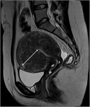
Diffuse adenomyosis: Sagittal T2-weighted image; thickening of the junctional zone forming an ill-defined area of low signal intensity, with punctate high-intensity myometrial foci representing areas of endometrium embedded within the myometrium
Image: “Diffuse adenomyosis” by Lisa Agostinho et al. License: CC BY 4.0
Endometrial polypsEndometrial polypsEndometrial polyps are pedunculated or sessile projections of the endometrium that result from overgrowth of endometrial glands and stroma around a central vascular stalk. Endometrial polyps are a few millimeters to a few centimeters in size, can occur anywhere within the uterine cavity, and, while usually benign, can be malignant, particularly in postmenopausal women. Endometrial Polyps
A small growth off the endometriumEndometriumThe mucous membrane lining of the uterine cavity that is hormonally responsive during the menstrual cycle and pregnancy. The endometrium undergoes cyclic changes that characterize menstruation. After successful fertilization, it serves to sustain the developing embryo.Embryoblast and Trophoblast Development that is usually pedunculated and often (though not always) benignBenignFibroadenoma:
Thickened endometriumEndometriumThe mucous membrane lining of the uterine cavity that is hormonally responsive during the menstrual cycle and pregnancy. The endometrium undergoes cyclic changes that characterize menstruation. After successful fertilization, it serves to sustain the developing embryo.Embryoblast and Trophoblast Development on regularRegularInsulin TVUS/TAUS and MRI
MassMassThree-dimensional lesion that occupies a space within the breastImaging of the Breast arising from the endometriumEndometriumThe mucous membrane lining of the uterine cavity that is hormonally responsive during the menstrual cycle and pregnancy. The endometrium undergoes cyclic changes that characterize menstruation. After successful fertilization, it serves to sustain the developing embryo.Embryoblast and Trophoblast Development and protruding into uterine cavity on SISSISInfertility (best test to visualize polyps)
A pedunculated endometrial polyp seen on a saline infusion sonogram (SIS)
Image: “3D-MS- View of endometrial outline in the transverse plane shows a localized lesion” by Zafarani F., Ahmadi F. License: CC BY 2.5, cropped by Lecturio.
Endometrial hyperplasiaHyperplasiaAn increase in the number of cells in a tissue or organ without tumor formation. It differs from hypertrophy, which is an increase in bulk without an increase in the number of cells.Cellular Adaptation and cancer
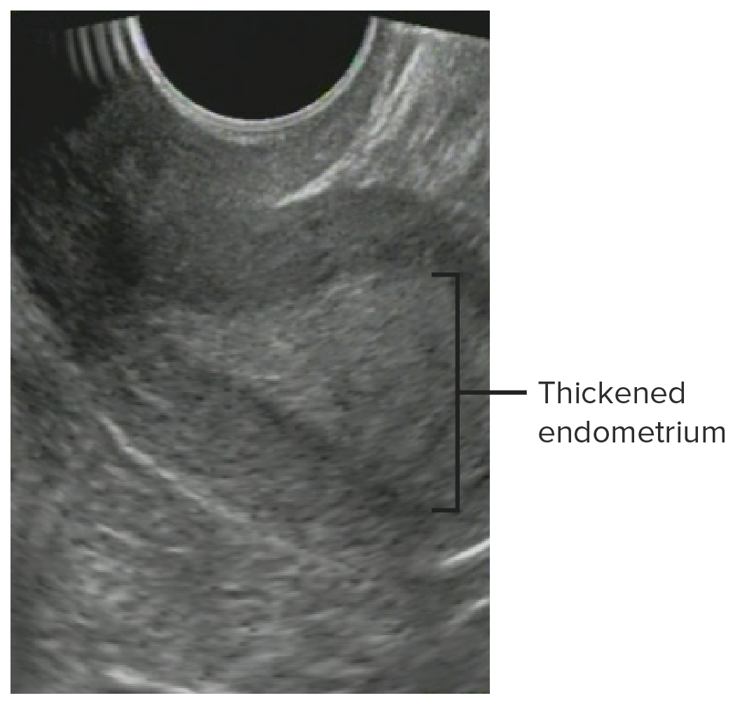
Thickened endometriumEndometriumThe mucous membrane lining of the uterine cavity that is hormonally responsive during the menstrual cycle and pregnancy. The endometrium undergoes cyclic changes that characterize menstruation. After successful fertilization, it serves to sustain the developing embryo.Embryoblast and Trophoblast Development based on age and/or menstrual status
Heterogeneous echogenicity
Features suggestive of cancer:
Irregular and/or indistinct borders
Frank invasion
Thickness > 5 mm in postmenopausal women
Endometrial thickening consistent with endometrial hyperplasia
Image: “Glandular cystic hyperplasia that is softer than the myometrium on SEG.E endometrium” by Goncharenko V. M. et al. License: CC BY 2.0, edited by Lecturio.
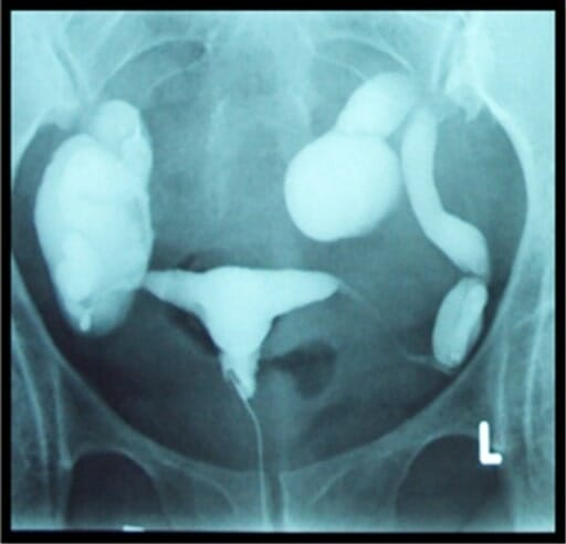
Fluoroscopic examination that allows assessment of:
Uterine cavity shape
Patency of fallopian tubesFallopian tubesThe uterus, cervix, and fallopian tubes are part of the internal female reproductive system. The fallopian tubes receive an ovum after ovulation and help move it and/or a fertilized embryo toward the uterus via ciliated cells lining the tubes and peristaltic movements of its smooth muscle. Uterus, Cervix, and Fallopian Tubes: Anatomy (best nonsurgical test available)
A catheter is inserted into the uterine cavity → dye is injected → X-rayX-rayPenetrating electromagnetic radiation emitted when the inner orbital electrons of an atom are excited and release radiant energy. X-ray wavelengths range from 1 pm to 10 nm. Hard x-rays are the higher energy, shorter wavelength x-rays. Soft x-rays or grenz rays are less energetic and longer in wavelength. The short wavelength end of the x-ray spectrum overlaps the gamma rays wavelength range. The distinction between gamma rays and x-rays is based on their radiation source.Pulmonary Function Tests
Indications
Evaluation of CUAs
InfertilityInfertilityInfertility is the inability to conceive in the context of regular intercourse. The most common causes of infertility in women are related to ovulatory dysfunction or tubal obstruction, whereas, in men, abnormal sperm is a common cause. Infertility (to look for congenital anomalies and check tubal patency)
ContraindicationsContraindicationsA condition or factor associated with a recipient that makes the use of a drug, procedure, or physical agent improper or inadvisable. Contraindications may be absolute (life threatening) or relative (higher risk of complications in which benefits may outweigh risks).Noninvasive Ventilation
PregnancyPregnancyThe status during which female mammals carry their developing young (embryos or fetuses) in utero before birth, beginning from fertilization to birth.Pregnancy: Diagnosis, Physiology, and Care
Active undiagnosed vaginal bleeding
Active pelvic infection
Advantages
Relatively low cost
Lower radiationRadiationEmission or propagation of acoustic waves (sound), electromagnetic energy waves (such as light; radio waves; gamma rays; or x-rays), or a stream of subatomic particles (such as electrons; neutrons; protons; or alpha particles).Osteosarcoma dose (although can become high with prolonged study time)
Exposure to ionizing radiationRadiationEmission or propagation of acoustic waves (sound), electromagnetic energy waves (such as light; radio waves; gamma rays; or x-rays), or a stream of subatomic particles (such as electrons; neutrons; protons; or alpha particles).Osteosarcoma
Discomfort/painPainAn unpleasant sensation induced by noxious stimuli which are detected by nerve endings of nociceptive neurons.Pain: Types and Pathways to the subject
Complicated scheduling:
Should be performed between the end of menstruationMenstruationThe periodic shedding of the endometrium and associated menstrual bleeding in the menstrual cycle of humans and primates. Menstruation is due to the decline in circulating progesterone, and occurs at the late luteal phase when luteolysis of the corpus luteum takes place.Menstrual Cycle and prior to ovulationOvulationThe discharge of an ovum from a rupturing follicle in the ovary.Menstrual Cycle (to avoid interruption of an early pregnancyPregnancyThe status during which female mammals carry their developing young (embryos or fetuses) in utero before birth, beginning from fertilization to birth.Pregnancy: Diagnosis, Physiology, and Care)
Often requires both a gynecologist (to place the catheter) and radiologist (to interpret images) present in the room during the study
Exam technique
Positioning:
Dorsal lithotomy on the fluoroscopyFluoroscopyProduction of an image when x-rays strike a fluorescent screen.X-rays table
Board is placed against the back.
X-rayX-rayPenetrating electromagnetic radiation emitted when the inner orbital electrons of an atom are excited and release radiant energy. X-ray wavelengths range from 1 pm to 10 nm. Hard x-rays are the higher energy, shorter wavelength x-rays. Soft x-rays or grenz rays are less energetic and longer in wavelength. The short wavelength end of the x-ray spectrum overlaps the gamma rays wavelength range. The distinction between gamma rays and x-rays is based on their radiation source.Pulmonary Function Tests beams from the floor → ceiling direction through the subject
Visualization: Field of view should be focused on the pelvisPelvisThe pelvis consists of the bony pelvic girdle, the muscular and ligamentous pelvic floor, and the pelvic cavity, which contains viscera, vessels, and multiple nerves and muscles. The pelvic girdle, composed of 2 “hip” bones and the sacrum, is a ring-like bony structure of the axial skeleton that links the vertebral column with the lower extremities.Pelvis: Anatomy.
Procedure:
A speculum is inserted into the vaginaVaginaThe vagina is the female genital canal, extending from the vulva externally to the cervix uteri internally. The structures have sexual, reproductive, and urinary functions and a rich blood supply, mainly arising from the internal iliac artery.Vagina, Vulva, and Pelvic Floor: Anatomy and the cervixCervixThe uterus, cervix, and fallopian tubes are part of the internal female reproductive system. The most inferior portion of the uterus is the cervix, which connects the uterine cavity to the vagina. Externally, the cervix is lined by stratified squamous cells; however, the cervical canal is lined by columnar epithelium.Uterus, Cervix, and Fallopian Tubes: Anatomy is identified.
Look for solid organ silhouettesSolid Organ SilhouettesImaging of the Urinary System if in the field of view (liverLiverThe liver is the largest gland in the human body. The liver is found in the superior right quadrant of the abdomen and weighs approximately 1.5 kilograms. Its main functions are detoxification, metabolism, nutrient storage (e.g., iron and vitamins), synthesis of coagulation factors, formation of bile, filtration, and storage of blood. Liver: Anatomy, spleenSpleenThe spleen is the largest lymphoid organ in the body, located in the LUQ of the abdomen, superior to the left kidney and posterior to the stomach at the level of the 9th-11th ribs just below the diaphragm. The spleen is highly vascular and acts as an important blood filter, cleansing the blood of pathogens and damaged erythrocytes. Spleen: Anatomy, kidney).
Patency of fallopian tubesFallopian tubesThe uterus, cervix, and fallopian tubes are part of the internal female reproductive system. The fallopian tubes receive an ovum after ovulation and help move it and/or a fertilized embryo toward the uterus via ciliated cells lining the tubes and peristaltic movements of its smooth muscle. Uterus, Cervix, and Fallopian Tubes: Anatomy
Abnormal calcifications (stones, masses)
Evaluate osseous structures (vertebral bodyVertebral bodyMain portion of the vertebra which bears majority of the weight.Vertebral Column: Anatomy height, iliac bones, femurs).
Dynamic approach:
Observe the flowFlowBlood flows through the heart, arteries, capillaries, and veins in a closed, continuous circuit. Flow is the movement of volume per unit of time. Flow is affected by the pressure gradient and the resistance fluid encounters between 2 points. Vascular resistance is the opposition to flow, which is caused primarily by blood friction against vessel walls.Vascular Resistance, Flow, and Mean Arterial Pressure of contrast through the endometrial canal and note any:
Contrast should flowFlowBlood flows through the heart, arteries, capillaries, and veins in a closed, continuous circuit. Flow is the movement of volume per unit of time. Flow is affected by the pressure gradient and the resistance fluid encounters between 2 points. Vascular resistance is the opposition to flow, which is caused primarily by blood friction against vessel walls.Vascular Resistance, Flow, and Mean Arterial Pressure through the fallopian tubesFallopian tubesThe uterus, cervix, and fallopian tubes are part of the internal female reproductive system. The fallopian tubes receive an ovum after ovulation and help move it and/or a fertilized embryo toward the uterus via ciliated cells lining the tubes and peristaltic movements of its smooth muscle. Uterus, Cervix, and Fallopian Tubes: Anatomy and into the adnexal spaces.
Fallopian tubes fill with dye → dye spills out of the ends of both tubes into the pelvic cavity (“bilateral fill and spill”)
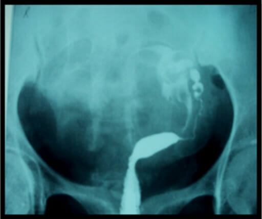
Normal hysterosalpingography findings: Radiograph showing normal uterine contour with bilateral fill and spill of dye from the fallopian tubes
Image: “Normal HSG examination” by Aziz M.U. et al. License: CC BY 3.0
Abnormal or incidental findings
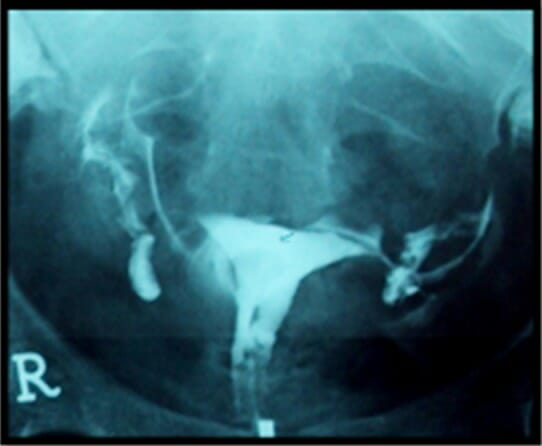
Hydrosalpinx:
Dilated tubes with collection of contrast in the tubes
With/without obstruction (i.e., no free spillage)
Hysterosalpingogram showing bilateral tubal occlusion and hydrosalpinges
Image: “HSG showing bilateral tubal blockage” by Aziz M.U. et al. License: CC BY 3.0
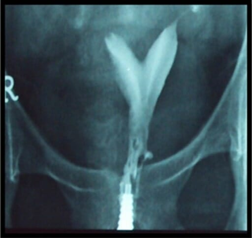
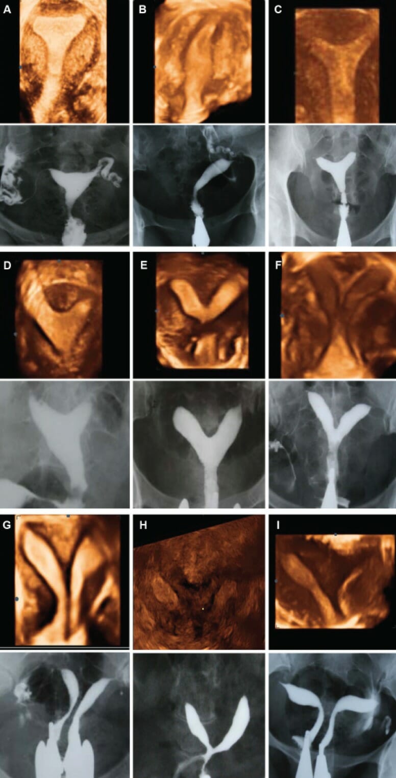
CUAs:
The uterusUterusThe uterus, cervix, and fallopian tubes are part of the internal female reproductive system. The uterus has a thick wall made of smooth muscle (the myometrium) and an inner mucosal layer (the endometrium). The most inferior portion of the uterus is the cervix, which connects the uterine cavity to the vagina.Uterus, Cervix, and Fallopian Tubes: Anatomy forms from the Müllerian ducts, which fuse in the midline to create the uterusUterusThe uterus, cervix, and fallopian tubes are part of the internal female reproductive system. The uterus has a thick wall made of smooth muscle (the myometrium) and an inner mucosal layer (the endometrium). The most inferior portion of the uterus is the cervix, which connects the uterine cavity to the vagina.Uterus, Cervix, and Fallopian Tubes: Anatomy, cervixCervixThe uterus, cervix, and fallopian tubes are part of the internal female reproductive system. The most inferior portion of the uterus is the cervix, which connects the uterine cavity to the vagina. Externally, the cervix is lined by stratified squamous cells; however, the cervical canal is lined by columnar epithelium.Uterus, Cervix, and Fallopian Tubes: Anatomy, and upper vaginaVaginaThe vagina is the female genital canal, extending from the vulva externally to the cervix uteri internally. The structures have sexual, reproductive, and urinary functions and a rich blood supply, mainly arising from the internal iliac artery.Vagina, Vulva, and Pelvic Floor: Anatomy. Therefore, initially, these structures are divided down the midline before the midline septum regresses. Congenital uterine abnormalities typically occur due to abnormal fusion and/or septal regressionRegressionCorneal Abrasions, Erosion, and Ulcers.
2 banana-shaped cavities, each draining into a normal fallopian tubeFallopian TubeA pair of highly specialized canals extending from the uterus to its corresponding ovary. They provide the means for ovum transport from the ovaries and they are the site of the ovum’s final maturation and fertilization. The fallopian tube consists of an interstitium, an isthmus, an ampulla, an infundibulum, and fimbriae. Its wall consists of three layers: serous, muscular, and an internal mucosal layer lined with both ciliated and secretory cells.Uterus, Cervix, and Fallopian Tubes: Anatomy
2 uterine cavities, each with their own cervixCervixThe uterus, cervix, and fallopian tubes are part of the internal female reproductive system. The most inferior portion of the uterus is the cervix, which connects the uterine cavity to the vagina. Externally, the cervix is lined by stratified squamous cells; however, the cervical canal is lined by columnar epithelium.Uterus, Cervix, and Fallopian Tubes: Anatomy and fallopian tubeFallopian TubeA pair of highly specialized canals extending from the uterus to its corresponding ovary. They provide the means for ovum transport from the ovaries and they are the site of the ovum’s final maturation and fertilization. The fallopian tube consists of an interstitium, an isthmus, an ampulla, an infundibulum, and fimbriae. Its wall consists of three layers: serous, muscular, and an internal mucosal layer lined with both ciliated and secretory cells.Uterus, Cervix, and Fallopian Tubes: Anatomy
Hysterosalpingogram of an infertile subject showing a bicornuate uterus: The right fallopian tube has either not yet filled with the dye or is obstructed. The left fallopian tube appears to be starting to fill.
Image: “HSG of secondary infertility patient showing bicornuate uterus” by Aziz M.U. et al. License: CC BY 3.0
Hysterosalpingogram showing a unicornuate uterus
Image: “Unicornuate uterus” by Aziz M.U. et al. License: CC BY 3.0
Comparison of three-dimensional ultrasound and hysterosalpingography in cases of uterine malformation:
A: normal uterus
B: unicornuate uterus
C: arcuate uterus
D-G: subtypes of the septate uterus (partial-to-complete septum)
H: bicornuate uterus
I: didelphys
Image: “Comparison of three-dimensional ultrasound and HSG imaging in cases ofuterine malformation using AFS” by Ahmadi F. et al. License: CC BY 2.5