Infertility is the inability to conceive in the context of regularRegularInsulin intercourse. The most common causes of infertility in women are related to ovulatory dysfunction or tubal obstruction, whereas, in men, abnormal sperm is a common cause. Diagnosis of infertility involves laboratory assessments for ovulatory function and a hysterosalpingogram to determine tubal patency in women, and semen analysis to assess the condition in men. Management involves treatment of the underlying pathology when possible, and may include ovulationOvulationThe discharge of an ovum from a rupturing follicle in the ovary.Menstrual Cycle induction with either timed intercourse or intrauterine insemination (IUI), in vitrofertilizationFertilizationTo undergo fertilization, the sperm enters the uterus, travels towards the ampulla of the fallopian tube, and encounters the oocyte. The zona pellucida (the outer layer of the oocyte) deteriorates along with the zygote, which travels towards the uterus and eventually forms a blastocyst, allowing for implantation to occur. Fertilization and First Week (IVF), and donor gametes, or by gestational surrogates or adoption.
Infertility is defined as the inability of a couple to conceive after 12 months of regular intercourse, in cases when the woman is < 35 years of age, or after 6 months of regular intercourse in couples when the woman is > 35 years of age.
Epidemiology
Normal fecundability (the probabilityProbabilityProbability is a mathematical tool used to study randomness and provide predictions about the likelihood of something happening. There are several basic rules of probability that can be used to help determine the probability of multiple events happening together, separately, or sequentially.Basics of Probability that a cycle will result in a pregnancyPregnancyThe status during which female mammals carry their developing young (embryos or fetuses) in utero before birth, beginning from fertilization to birth.Pregnancy: Diagnosis, Physiology, and Care):
25% in the 1st 3 months
15% in the 4th to 12th months
Approximately 80%–90% of healthy couples will conceive within 12 months.
PrevalencePrevalenceThe total number of cases of a given disease in a specified population at a designated time. It is differentiated from incidence, which refers to the number of new cases in the population at a given time.Measures of Disease Frequency of primary infertility in women:
To achieve pregnancyPregnancyThe status during which female mammals carry their developing young (embryos or fetuses) in utero before birth, beginning from fertilization to birth.Pregnancy: Diagnosis, Physiology, and Care, the female partner must be ovulatory with patent fallopian tubesFallopian tubesThe uterus, cervix, and fallopian tubes are part of the internal female reproductive system. The fallopian tubes receive an ovum after ovulation and help move it and/or a fertilized embryo toward the uterus via ciliated cells lining the tubes and peristaltic movements of its smooth muscle. Uterus, Cervix, and Fallopian Tubes: Anatomy and a receptive uterusUterusThe uterus, cervix, and fallopian tubes are part of the internal female reproductive system. The uterus has a thick wall made of smooth muscle (the myometrium) and an inner mucosal layer (the endometrium). The most inferior portion of the uterus is the cervix, which connects the uterine cavity to the vagina.Uterus, Cervix, and Fallopian Tubes: Anatomy, whereas the male partner must be able to produce sperm that is capable of fertilizing the oocyte.
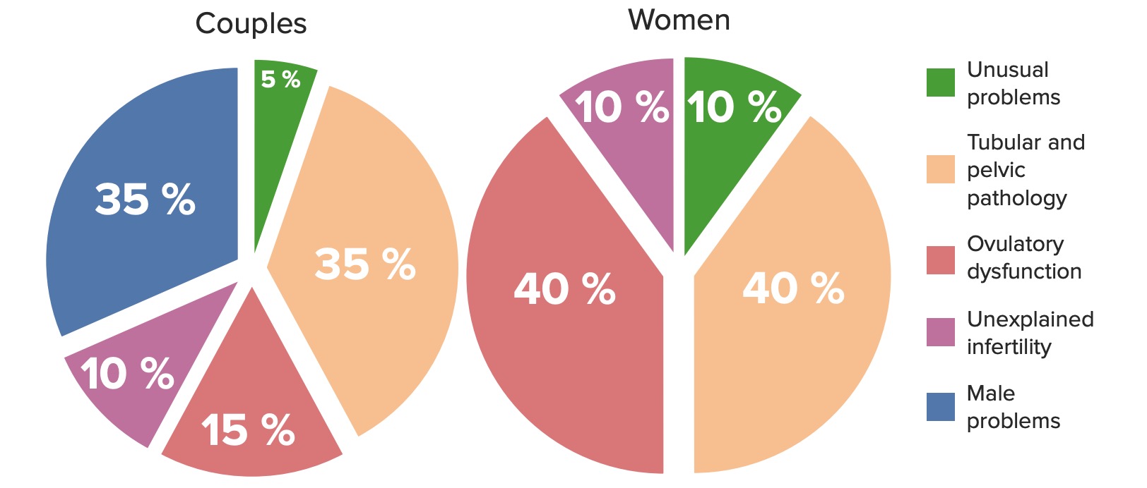
Etiologies of couples
Female factor alone: 37%
Male factor alone: 8%
Both female and male factors: 35%
Unexplained infertility: 5%
Exact etiologies often difficult to determine unless absolute infertility factors are present (e.g., bilateral tubal obstruction).
Couples often have more than 1 contributing etiology.
Etiologies of infertility in couples (left) and in women (right)
Oligoovulation: infrequent ovulationOvulationThe discharge of an ovum from a rupturing follicle in the ovary.Menstrual Cycle
AnovulationAnovulationSuspension or cessation of ovulation in animals or humans with follicle-containing ovaries (ovarian follicle). Depending on the etiology, ovulation may be induced with appropriate therapy.Polycystic Ovarian Syndrome: absence of ovulationOvulationThe discharge of an ovum from a rupturing follicle in the ovary.Menstrual Cycle
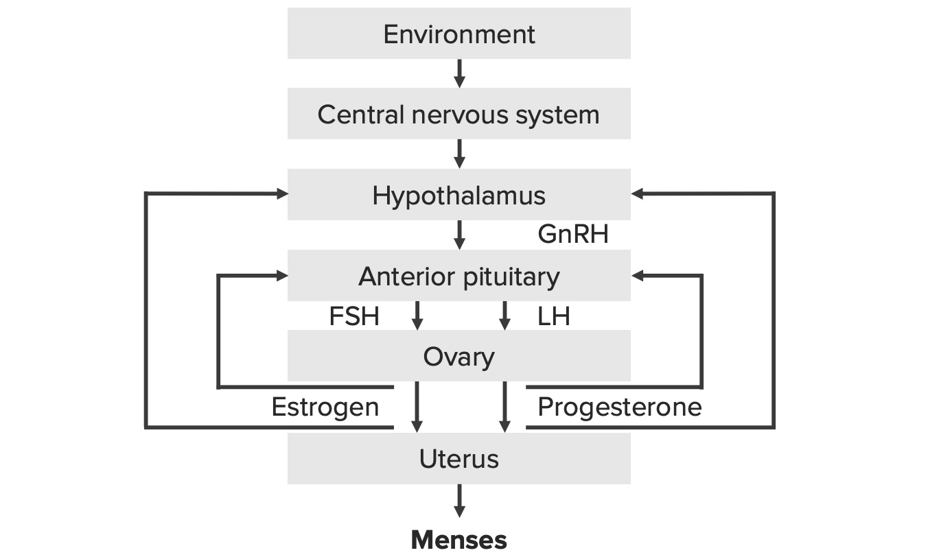
HypothalamusHypothalamusThe hypothalamus is a collection of various nuclei within the diencephalon in the center of the brain. The hypothalamus plays a vital role in endocrine regulation as the primary regulator of the pituitary gland, and it is the major point of integration between the central nervous and endocrine systems.Hypothalamus is not functioning properly.
↓ Gonadotropin-releasing hormoneGonadotropin-releasing hormoneA decapeptide that stimulates the synthesis and secretion of both pituitary gonadotropins, luteinizing hormone and follicle stimulating hormone. Gnrh is produced by neurons in the septum preoptic area of the hypothalamus and released into the pituitary portal blood, leading to stimulation of gonadotrophs in the anterior pituitary gland.Puberty (GnRH) → ↓ follicle-stimulating hormone (FSHFSHA major gonadotropin secreted by the adenohypophysis. Follicle-stimulating hormone stimulates gametogenesis and the supporting cells such as the ovarian granulosa cells, the testicular sertoli cells, and leydig cells. Fsh consists of two noncovalently linked subunits, alpha and beta. Within a species, the alpha subunit is common in the three pituitary glycoprotein hormones (TSH, LH, and FSH), but the beta subunit is unique and confers its biological specificity.Menstrual Cycle) → ↓ oocyte maturation → anovulationAnovulationSuspension or cessation of ovulation in animals or humans with follicle-containing ovaries (ovarian follicle). Depending on the etiology, ovulation may be induced with appropriate therapy.Polycystic Ovarian Syndrome
IdiopathicIdiopathicDermatomyositishypogonadotropic hypogonadismHypogonadotropic HypogonadismHypogonadism (IHHIHHPrimary Amenorrhea): congenital deficiency of GnRH, such as in Kallmann syndromeKallmann syndromeKallmann syndrome (KS), also called olfacto-genital syndrome, is a genetic condition that causes hypogonadotropic hypogonadism due to decreased secretion of gonadotropin-releasing hormone (GnRH) by the hypothalamus. The lack of sex hormones results in impaired pubertal development. Kallmann Syndrome (IHHIHHPrimary Amenorrhea associated with anosmiaAnosmiaComplete or severe loss of the subjective sense of smell. Loss of smell may be caused by many factors such as a cold, allergy, olfactory nerve diseases, viral respiratory tract infections (e.g., COVID-19), aging and various neurological disorders (e.g., Alzheimer disease).Cranial Nerve Palsies)
Sheehan syndrome: hypopituitarismHypopituitarismHypopituitarism is a condition characterized by pituitary hormone deficiency. This condition primarily results from a disease of the pituitary gland, but it may arise from hypothalamic dysfunction. Pituitary tumors are one of the most common causes. The majority of cases affect the anterior pituitary lobe (adenohypophysis), which accounts for 80% of the gland. Hypopituitarism caused by ischemiaIschemiaA hypoperfusion of the blood through an organ or tissue caused by a pathologic constriction or obstruction of its blood vessels, or an absence of blood circulation.Ischemic Cell Damage during postpartum hemorrhagePostpartum hemorrhagePostpartum hemorrhage is one of the most common and deadly obstetric complications. Since 2017, postpartum hemorrhage has been defined as blood loss greater than 1,000 mL for both cesarean and vaginal deliveries, or excessive blood loss with signs of hemodynamic instability. Postpartum Hemorrhage
Infiltrative disease (e.g., sarcoidosisSarcoidosisSarcoidosis is a multisystem inflammatory disease that causes noncaseating granulomas. The exact etiology is unknown. Sarcoidosis usually affects the lungs and thoracic lymph nodes, but it can also affect almost every system in the body, including the skin, heart, and eyes, most commonly. Sarcoidosis)
Sellar massMassThree-dimensional lesion that occupies a space within the breastImaging of the Breast
Normal GnRH and estrogens, but ↓ FSHFSHA major gonadotropin secreted by the adenohypophysis. Follicle-stimulating hormone stimulates gametogenesis and the supporting cells such as the ovarian granulosa cells, the testicular sertoli cells, and leydig cells. Fsh consists of two noncovalently linked subunits, alpha and beta. Within a species, the alpha subunit is common in the three pituitary glycoprotein hormones (TSH, LH, and FSH), but the beta subunit is unique and confers its biological specificity.Menstrual Cycle
Often oligomenorrheaOligomenorrheaPolycystic Ovarian Syndrome and ↑ androgensAndrogensAndrogens are naturally occurring steroid hormones responsible for development and maintenance of the male sex characteristics, including penile, scrotal, and clitoral growth, development of sexual hair, deepening of the voice, and musculoskeletal growth. Androgens and Antiandrogens
Examples:
Polycystic ovary syndrome(PCOSPCOSPolycystic ovarian syndrome (PCOS) is the most common endocrine disorder of reproductive-age women, affecting nearly 5%-10% of women in the age group. It is characterized by hyperandrogenism, chronic anovulation leading to oligomenorrhea (or amenorrhea), and metabolic dysfunction.Polycystic Ovarian Syndrome)
Cushing syndromeCushing syndromeA condition caused by prolonged exposure to excess levels of cortisol (hydrocortisone) or other glucocorticoids from endogenous or exogenous sources. It is characterized by upper body obesity; osteoporosis; hypertension; diabetes mellitus; hirsutism; amenorrhea; and excess body fluid. Endogenous Cushing syndrome or spontaneous hypercortisolism is divided into two groups, those due to an excess of adrenocorticotropin and those that are acth-independent.Paraneoplastic Syndromes
OvariesOvariesOvaries are the paired gonads of the female reproductive system that contain haploid gametes known as oocytes. The ovaries are located intraperitoneally in the pelvis, just posterior to the broad ligament, and are connected to the pelvic sidewall and to the uterus by ligaments. These organs function to secrete hormones (estrogen and progesterone) and to produce the female germ cells (oocytes).Ovaries: Anatomy not responsive to FSHFSHA major gonadotropin secreted by the adenohypophysis. Follicle-stimulating hormone stimulates gametogenesis and the supporting cells such as the ovarian granulosa cells, the testicular sertoli cells, and leydig cells. Fsh consists of two noncovalently linked subunits, alpha and beta. Within a species, the alpha subunit is common in the three pituitary glycoprotein hormones (TSH, LH, and FSH), but the beta subunit is unique and confers its biological specificity.Menstrual Cycle
↑ GnRH → ↑ FSHFSHA major gonadotropin secreted by the adenohypophysis. Follicle-stimulating hormone stimulates gametogenesis and the supporting cells such as the ovarian granulosa cells, the testicular sertoli cells, and leydig cells. Fsh consists of two noncovalently linked subunits, alpha and beta. Within a species, the alpha subunit is common in the three pituitary glycoprotein hormones (TSH, LH, and FSH), but the beta subunit is unique and confers its biological specificity.Menstrual Cycle → nonresponsive ovariesOvariesOvaries are the paired gonads of the female reproductive system that contain haploid gametes known as oocytes. The ovaries are located intraperitoneally in the pelvis, just posterior to the broad ligament, and are connected to the pelvic sidewall and to the uterus by ligaments. These organs function to secrete hormones (estrogen and progesterone) and to produce the female germ cells (oocytes).Ovaries: Anatomy → anovulationAnovulationSuspension or cessation of ovulation in animals or humans with follicle-containing ovaries (ovarian follicle). Depending on the etiology, ovulation may be induced with appropriate therapy.Polycystic Ovarian Syndrome
Example: primary ovarian insufficiencyPrimary ovarian insufficiencyCessation of ovarian function after menarche but before the age of 40, without or with ovarian follicle depletion. It is characterized by the presence of oligomenorrhea or amenorrhea, elevated gonadotropins, and low estradiol levels. It is a state of female hypergonadotropic hypogonadism. Etiologies include genetic defects, autoimmune processes, chemotherapy, radiation, and infections. The most commonly known genetic cause is the expansion of a cgg repeat to 55 to 199 copies in the 5′ untranslated region in the X-linked fmr1 gene.Primary Ovarian Insufficiency(POI)
Turner syndromeTurner syndromeTurner syndrome is a genetic condition affecting women, in which 1 X chromosome is partly or completely missing. The classic result is the karyotype 45,XO with a female phenotype. Turner syndrome is associated with decreased sex hormone levels and is the most common cause of primary amenorrhea.Turner Syndrome
RadiationRadiationEmission or propagation of acoustic waves (sound), electromagnetic energy waves (such as light; radio waves; gamma rays; or x-rays), or a stream of subatomic particles (such as electrons; neutrons; protons; or alpha particles).Osteosarcoma
Other etiologies of ovulatory dysfunction:
Oocyte aging
HyperprolactinemiaHyperprolactinemiaHyperprolactinemia is defined as a condition of elevated levels of prolactin (PRL) hormone in the blood. The PRL hormone is secreted by the anterior pituitary gland and is responsible for breast development and lactation. The most common cause is PRL-secreting pituitary adenomas (prolactinomas). Hyperprolactinemia
HypothyroidismHypothyroidismHypothyroidism is a condition characterized by a deficiency of thyroid hormones. Iodine deficiency is the most common cause worldwide, but Hashimoto’s disease (autoimmune thyroiditis) is the leading cause in non-iodine-deficient regions. Hypothyroidism
Estrogen- or androgen-secreting tumors:
SexSexThe totality of characteristics of reproductive structure, functions, phenotype, and genotype, differentiating the male from the female organism.Gender Dysphoria cord-stromal tumors
Adrenal tumors
↑ EstrogenEstrogenCompounds that interact with estrogen receptors in target tissues to bring about the effects similar to those of estradiol. Estrogens stimulate the female reproductive organs, and the development of secondary female sex characteristics. Estrogenic chemicals include natural, synthetic, steroidal, or non-steroidal compounds.Ovaries: Anatomy or androgensAndrogensAndrogens are naturally occurring steroid hormones responsible for development and maintenance of the male sex characteristics, including penile, scrotal, and clitoral growth, development of sexual hair, deepening of the voice, and musculoskeletal growth. Androgens and Antiandrogens → ↓ FSHFSHA major gonadotropin secreted by the adenohypophysis. Follicle-stimulating hormone stimulates gametogenesis and the supporting cells such as the ovarian granulosa cells, the testicular sertoli cells, and leydig cells. Fsh consists of two noncovalently linked subunits, alpha and beta. Within a species, the alpha subunit is common in the three pituitary glycoprotein hormones (TSH, LH, and FSH), but the beta subunit is unique and confers its biological specificity.Menstrual Cycle
Tubal factors:
Prevent sperm from reaching the egg due to:
Occlusion (usually from adhesions)
InflammationInflammationInflammation is a complex set of responses to infection and injury involving leukocytes as the principal cellular mediators in the body’s defense against pathogenic organisms. Inflammation is also seen as a response to tissue injury in the process of wound healing. The 5 cardinal signs of inflammation are pain, heat, redness, swelling, and loss of function. Inflammation
Causes:
Pelvic inflammatory diseasePelvic inflammatory diseasePelvic inflammatory disease (PID) is defined as a polymicrobial infection of the upper female reproductive system. The disease can affect the uterus, fallopian tubes, ovaries, and adjacent structures. Pelvic inflammatory disease is closely linked with sexually transmitted diseases, most commonly caused by Chlamydia trachomatis, Neisseria gonorrhoeae, and Gardnerella vaginalis. Pelvic Inflammatory Disease: caused by chlamydiaChlamydiaChlamydiae are obligate intracellular gram-negative bacteria. They lack a peptidoglycan layer and are best visualized using Giemsa stain. The family of Chlamydiaceae comprises 3 pathogens that can infect humans: Chlamydia trachomatis, Chlamydia psittaci, and Chlamydia pneumoniae.Chlamydia or gonorrheaGonorrheaGonorrhea is a sexually transmitted infection (STI) caused by the gram-negative bacteria Neisseria gonorrhoeae (N. gonorrhoeae). Gonorrhea may be asymptomatic but commonly manifests as cervicitis or urethritis with less common presentations such as proctitis, conjunctivitis, or pharyngitis. Gonorrhea
Hydrosalpinges
EndometriosisEndometriosisEndometriosis is a common disease in which patients have endometrial tissue implanted outside of the uterus. Endometrial implants can occur anywhere in the pelvis, including the ovaries, the broad and uterosacral ligaments, the pelvic peritoneum, and the urinary and gastrointestinal tracts.Endometriosis: fertility challenges due to both tubal adhesions and inflammationInflammationInflammation is a complex set of responses to infection and injury involving leukocytes as the principal cellular mediators in the body’s defense against pathogenic organisms. Inflammation is also seen as a response to tissue injury in the process of wound healing. The 5 cardinal signs of inflammation are pain, heat, redness, swelling, and loss of function. Inflammation
Prior tubal surgery
Prior ectopic pregnancyEctopic pregnancyEctopic pregnancy refers to the implantation of a fertilized egg (embryo) outside the uterine cavity. The main cause is disruption of the normal anatomy of the fallopian tube. Ectopic Pregnancy
Nontubal infectionsInfectionsInvasion of the host organism by microorganisms or their toxins or by parasites that can cause pathological conditions or diseases.Chronic Granulomatous Disease:
AppendicitisAppendicitisAppendicitis is the acute inflammation of the vermiform appendix and the most common abdominal surgical emergency globally. The condition has a lifetime risk of 8%. Characteristic features include periumbilical abdominal pain that migrates to the right lower quadrant, fever, anorexia, nausea, and vomiting.Appendicitis
Inflammatory bowel disease
Pelvic TBTBTuberculosis (TB) is an infectious disease caused by Mycobacterium tuberculosis complex bacteria. The bacteria usually attack the lungs but can also damage other parts of the body. Approximately 30% of people around the world are infected with this pathogen, with the majority harboring a latent infection. Tuberculosis spreads through the air when a person with active pulmonary infection coughs or sneezes. Tuberculosis
Endometrial polypsEndometrial polypsEndometrial polyps are pedunculated or sessile projections of the endometrium that result from overgrowth of endometrial glands and stroma around a central vascular stalk. Endometrial polyps are a few millimeters to a few centimeters in size, can occur anywhere within the uterine cavity, and, while usually benign, can be malignant, particularly in postmenopausal women. Endometrial Polyps
IHHIHHPrimary Amenorrhea/Kallmann syndromeKallmann syndromeKallmann syndrome (KS), also called olfacto-genital syndrome, is a genetic condition that causes hypogonadotropic hypogonadism due to decreased secretion of gonadotropin-releasing hormone (GnRH) by the hypothalamus. The lack of sex hormones results in impaired pubertal development. Kallmann Syndrome
Genetic defects affecting gonadotropins
Acquired conditions leading to hypothalamic or pituitaryPituitaryA small, unpaired gland situated in the sella turcica. It is connected to the hypothalamus by a short stalk which is called the infundibulum.Hormones: Overview and Types dysfunction:
Sellar masses
Infiltrative disease (e.g., sarcoidosisSarcoidosisSarcoidosis is a multisystem inflammatory disease that causes noncaseating granulomas. The exact etiology is unknown. Sarcoidosis usually affects the lungs and thoracic lymph nodes, but it can also affect almost every system in the body, including the skin, heart, and eyes, most commonly. Sarcoidosis)
HyperprolactinemiaHyperprolactinemiaHyperprolactinemia is defined as a condition of elevated levels of prolactin (PRL) hormone in the blood. The PRL hormone is secreted by the anterior pituitary gland and is responsible for breast development and lactation. The most common cause is PRL-secreting pituitary adenomas (prolactinomas). Hyperprolactinemia (e.g., medications)
ThyroidThyroidThe thyroid gland is one of the largest endocrine glands in the human body. The thyroid gland is a highly vascular, brownish-red gland located in the visceral compartment of the anterior region of the neck.Thyroid Gland: Anatomy disorders
Cushing syndromeCushing syndromeA condition caused by prolonged exposure to excess levels of cortisol (hydrocortisone) or other glucocorticoids from endogenous or exogenous sources. It is characterized by upper body obesity; osteoporosis; hypertension; diabetes mellitus; hirsutism; amenorrhea; and excess body fluid. Endogenous Cushing syndrome or spontaneous hypercortisolism is divided into two groups, those due to an excess of adrenocorticotropin and those that are acth-independent.Paraneoplastic Syndromes
Hormone-secreting tumors
Systemic illness
ObesityObesityObesity is a condition associated with excess body weight, specifically with the deposition of excessive adipose tissue. Obesity is considered a global epidemic. Major influences come from the western diet and sedentary lifestyles, but the exact mechanisms likely include a mixture of genetic and environmental factors. Obesity (can ↓ testosteroneTestosteroneA potent androgenic steroid and major product secreted by the leydig cells of the testis. Its production is stimulated by luteinizing hormone from the pituitary gland. In turn, testosterone exerts feedback control of the pituitary LH and FSH secretion. Depending on the tissues, testosterone can be further converted to dihydrotestosterone or estradiol.Androgens and Antiandrogens and testicular function)
Testicular defects in spermatogenesisSpermatogenesisThe process of germ cell development in the male from the primordial germ cells, through spermatogonia; spermatocytes; spermatids; to the mature haploid spermatozoa.Gametogenesis:
Teratozoospermia: ↑ number of sperm with abnormal morphology
Genetic causes:
Klinefelter syndromeKlinefelter syndromeKlinefelter syndrome is a chromosomal aneuploidy characterized by the presence of 1 or more extra X chromosomes in a male karyotype, most commonly leading to karyotype 47,XXY. Klinefelter syndrome is associated with decreased levels of testosterone and is the most common cause of congenital hypogonadism. Klinefelter Syndrome (47,XXYXXYKlinefelter syndrome is a chromosomal aneuploidy characterized by the presence of 1 or more extra X chromosomes in a male karyotype, most commonly leading to karyotype 47,XXY. Klinefelter syndrome is associated with decreased levels of testosterone and is the most common cause of congenital hypogonadism.Klinefelter Syndrome): one of the most common causes of primary hypogonadismPrimary HypogonadismMyotonic Dystrophies in men
Y microdeletions
CryptorchidismCryptorchidismCryptorchidism is one of the most common congenital anomalies in young boys. Typically, this asymptomatic condition presents during a routine well-child examination where 1 or both testicles are not palpable in the scrotum.Cryptorchidism: undescended testesTestesGonadal Hormones
Acquired causes:
VaricoceleVaricoceleA condition characterized by the dilated tortuous veins of the spermatic cord with a marked left-sided predominance. Adverse effect on male fertility occurs when varicocele leads to an increased scrotal (and testicular) temperature and reduced testicular volume.Varicocele, Hydrocele, and Spermatocele: dilation of the pampiniform plexus
Infection:
MumpsMumpsMumps is caused by a single-stranded, linear, negative-sense RNA virus of the family Paramyxoviridae. Mumps is typically a disease of childhood, which manifests initially with fever, muscle pain, headache, poor appetite, and a general feeling of malaise, and is classically followed by parotitis. Mumps Virus/Mumps
TBTBTuberculosis (TB) is an infectious disease caused by Mycobacterium tuberculosis complex bacteria. The bacteria usually attack the lungs but can also damage other parts of the body. Approximately 30% of people around the world are infected with this pathogen, with the majority harboring a latent infection. Tuberculosis spreads through the air when a person with active pulmonary infection coughs or sneezes. Tuberculosis
LeprosyLeprosyLeprosy, also known as Hansen’s disease, is a chronic bacterial infection caused by Mycobacterium leprae complex bacteria. Symptoms primarily affect the skin and peripheral nerves, resulting in cutaneous manifestations (e.g., hypopigmented macules) and neurologic manifestations (e.g., loss of sensation). Leprosy
GonorrheaGonorrheaGonorrhea is a sexually transmitted infection (STI) caused by the gram-negative bacteria Neisseria gonorrhoeae (N. gonorrhoeae). Gonorrhea may be asymptomatic but commonly manifests as cervicitis or urethritis with less common presentations such as proctitis, conjunctivitis, or pharyngitis. Gonorrhea and chlamydiaChlamydiaChlamydiae are obligate intracellular gram-negative bacteria. They lack a peptidoglycan layer and are best visualized using Giemsa stain. The family of Chlamydiaceae comprises 3 pathogens that can infect humans: Chlamydia trachomatis, Chlamydia psittaci, and Chlamydia pneumoniae.Chlamydia
RadiationRadiationEmission or propagation of acoustic waves (sound), electromagnetic energy waves (such as light; radio waves; gamma rays; or x-rays), or a stream of subatomic particles (such as electrons; neutrons; protons; or alpha particles).Osteosarcoma
Sperm transport and sexual dysfunctionSexual dysfunctionPhysiological disturbances in normal sexual performance in either the male or the female.Sexual Physiology disorders:
Congenital abnormalitiesCongenital AbnormalitiesMalformations of organs or body parts during development in utero.Omphalocele, dysfunction, or obstruction of:
EpididymisEpididymisThe convoluted cordlike structure attached to the posterior of the testis. Epididymis consists of the head (caput), the body (corpus), and the tail (cauda). A network of ducts leaving the testis joins into a common epididymal tubule proper which provides the transport, storage, and maturation of spermatozoa.Testicles: Anatomy
Vas differences
Ejaculatory ductsEjaculatory DuctsPaired ducts in the human male through which semen is ejaculated into the urethra.
Sexual dysfunctionSexual dysfunctionPhysiological disturbances in normal sexual performance in either the male or the female.Sexual Physiology:
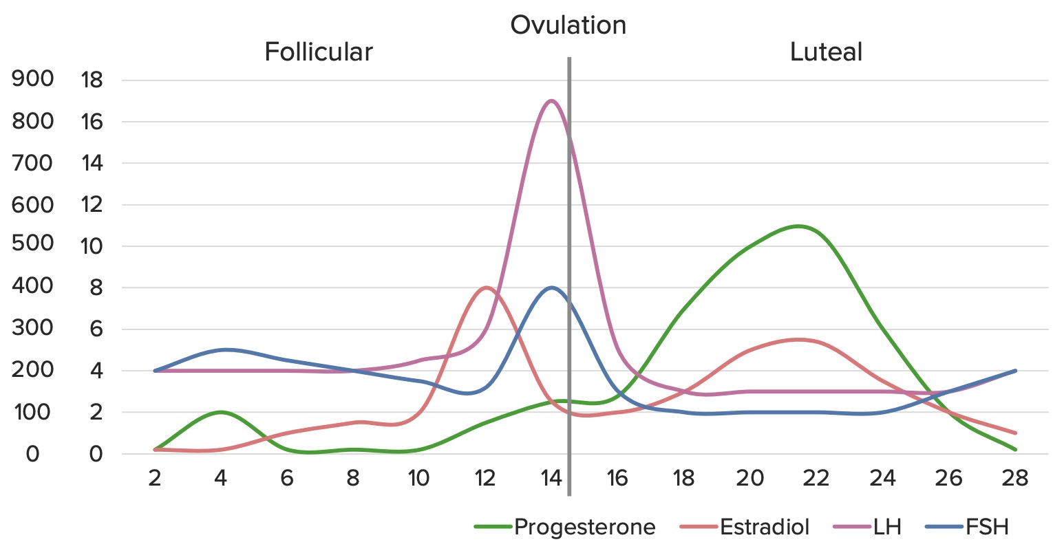
Cycle day 3 FSHFSHA major gonadotropin secreted by the adenohypophysis. Follicle-stimulating hormone stimulates gametogenesis and the supporting cells such as the ovarian granulosa cells, the testicular sertoli cells, and leydig cells. Fsh consists of two noncovalently linked subunits, alpha and beta. Within a species, the alpha subunit is common in the three pituitary glycoprotein hormones (TSH, LH, and FSH), but the beta subunit is unique and confers its biological specificity.Menstrual Cycle, LHLHA major gonadotropin secreted by the adenohypophysis. Luteinizing hormone regulates steroid production by the interstitial cells of the testis and the ovary. The preovulatory luteinizing hormone surge in females induces ovulation, and subsequent luteinization of the follicle. Luteinizing hormone consists of two noncovalently linked subunits, alpha and beta. Within a species, the alpha subunit is common in the three pituitary glycoprotein hormones (TSH, LH, and FSH), but the beta subunit is unique and confers its biological specificity.Menstrual Cycle, and estradiolEstradiolThe 17-beta-isomer of estradiol, an aromatized C18 steroid with hydroxyl group at 3-beta- and 17-beta-position. Estradiol-17-beta is the most potent form of mammalian estrogenic steroids.Noncontraceptive Estrogen and Progestins:
↓ FSHFSHA major gonadotropin secreted by the adenohypophysis. Follicle-stimulating hormone stimulates gametogenesis and the supporting cells such as the ovarian granulosa cells, the testicular sertoli cells, and leydig cells. Fsh consists of two noncovalently linked subunits, alpha and beta. Within a species, the alpha subunit is common in the three pituitary glycoprotein hormones (TSH, LH, and FSH), but the beta subunit is unique and confers its biological specificity.Menstrual Cycle with ↓ estrogenEstrogenCompounds that interact with estrogen receptors in target tissues to bring about the effects similar to those of estradiol. Estrogens stimulate the female reproductive organs, and the development of secondary female sex characteristics. Estrogenic chemicals include natural, synthetic, steroidal, or non-steroidal compounds.Ovaries: Anatomy → functional hypothalamic amenorrheaFunctional Hypothalamic AmenorrheaSecondary Amenorrhea
LHLHA major gonadotropin secreted by the adenohypophysis. Luteinizing hormone regulates steroid production by the interstitial cells of the testis and the ovary. The preovulatory luteinizing hormone surge in females induces ovulation, and subsequent luteinization of the follicle. Luteinizing hormone consists of two noncovalently linked subunits, alpha and beta. Within a species, the alpha subunit is common in the three pituitary glycoprotein hormones (TSH, LH, and FSH), but the beta subunit is unique and confers its biological specificity.Menstrual Cycle:FSHFSHA major gonadotropin secreted by the adenohypophysis. Follicle-stimulating hormone stimulates gametogenesis and the supporting cells such as the ovarian granulosa cells, the testicular sertoli cells, and leydig cells. Fsh consists of two noncovalently linked subunits, alpha and beta. Within a species, the alpha subunit is common in the three pituitary glycoprotein hormones (TSH, LH, and FSH), but the beta subunit is unique and confers its biological specificity.Menstrual Cycle ratio > 2 with normal estrogenEstrogenCompounds that interact with estrogen receptors in target tissues to bring about the effects similar to those of estradiol. Estrogens stimulate the female reproductive organs, and the development of secondary female sex characteristics. Estrogenic chemicals include natural, synthetic, steroidal, or non-steroidal compounds.Ovaries: Anatomy → PCOSPCOSPolycystic ovarian syndrome (PCOS) is the most common endocrine disorder of reproductive-age women, affecting nearly 5%-10% of women in the age group. It is characterized by hyperandrogenism, chronic anovulation leading to oligomenorrhea (or amenorrhea), and metabolic dysfunction.Polycystic Ovarian Syndrome
↑ FSHFSHA major gonadotropin secreted by the adenohypophysis. Follicle-stimulating hormone stimulates gametogenesis and the supporting cells such as the ovarian granulosa cells, the testicular sertoli cells, and leydig cells. Fsh consists of two noncovalently linked subunits, alpha and beta. Within a species, the alpha subunit is common in the three pituitary glycoprotein hormones (TSH, LH, and FSH), but the beta subunit is unique and confers its biological specificity.Menstrual Cycle with ↓ estrogenEstrogenCompounds that interact with estrogen receptors in target tissues to bring about the effects similar to those of estradiol. Estrogens stimulate the female reproductive organs, and the development of secondary female sex characteristics. Estrogenic chemicals include natural, synthetic, steroidal, or non-steroidal compounds.Ovaries: Anatomy → POI
↓ FSHFSHA major gonadotropin secreted by the adenohypophysis. Follicle-stimulating hormone stimulates gametogenesis and the supporting cells such as the ovarian granulosa cells, the testicular sertoli cells, and leydig cells. Fsh consists of two noncovalently linked subunits, alpha and beta. Within a species, the alpha subunit is common in the three pituitary glycoprotein hormones (TSH, LH, and FSH), but the beta subunit is unique and confers its biological specificity.Menstrual Cycle with ↑ estrogenEstrogenCompounds that interact with estrogen receptors in target tissues to bring about the effects similar to those of estradiol. Estrogens stimulate the female reproductive organs, and the development of secondary female sex characteristics. Estrogenic chemicals include natural, synthetic, steroidal, or non-steroidal compounds.Ovaries: Anatomy → estrogen-secreting tumorTumorInflammation
Cycle day 21 progesteroneProgesteroneThe major progestational steroid that is secreted primarily by the corpus luteum and the placenta. Progesterone acts on the uterus, the mammary glands and the brain. It is required in embryo implantation; pregnancy maintenance, and the development of mammary tissue for milk production. Progesterone, converted from pregnenolone, also serves as an intermediate in the biosynthesis of gonadal steroid hormones and adrenal corticosteroids.Gonadal Hormones: ↑ progesteroneProgesteroneThe major progestational steroid that is secreted primarily by the corpus luteum and the placenta. Progesterone acts on the uterus, the mammary glands and the brain. It is required in embryo implantation; pregnancy maintenance, and the development of mammary tissue for milk production. Progesterone, converted from pregnenolone, also serves as an intermediate in the biosynthesis of gonadal steroid hormones and adrenal corticosteroids.Gonadal Hormones in the luteal phaseLuteal phaseThe period in the menstrual cycle that follows ovulation, characterized by the development of corpus luteum, increase in progesterone production by the ovary and secretion by the glandular epithelium of the endometrium. The luteal phase begins with ovulation and ends with the onset of menstruation.Menstrual Cycle confirms ovulationOvulationThe discharge of an ovum from a rupturing follicle in the ovary.Menstrual Cycle
Anti-Müllerian hormone (AMHAMHA glycoprotein that causes regression of mullerian ducts. It is produced by sertoli cells of the testes. In the absence of this hormone, the mullerian ducts develop into structures of the female reproductive tract. In males, defects of this hormone result in persistent mullerian duct, a form of male pseudohermaphroditism.Primary Amenorrhea):
Ovarian reserve test
↓ AMHAMHA glycoprotein that causes regression of mullerian ducts. It is produced by sertoli cells of the testes. In the absence of this hormone, the mullerian ducts develop into structures of the female reproductive tract. In males, defects of this hormone result in persistent mullerian duct, a form of male pseudohermaphroditism.Primary Amenorrhea → POI
Other hormonal abnormalities that contribute to ovulatory dysfunction:
↑ ProlactinProlactinA lactogenic hormone secreted by the adenohypophysis. It is a polypeptide of approximately 23 kd. Besides its major action on lactation, in some species prolactin exerts effects on reproduction, maternal behavior, fat metabolism, immunomodulation and osmoregulation.Breasts: Anatomy → hyperprolactinemiaHyperprolactinemiaHyperprolactinemia is defined as a condition of elevated levels of prolactin (PRL) hormone in the blood. The PRL hormone is secreted by the anterior pituitary gland and is responsible for breast development and lactation. The most common cause is PRL-secreting pituitary adenomas (prolactinomas). Hyperprolactinemia
↑ Thyroid-stimulating hormoneThyroid-stimulating hormoneA glycoprotein hormone secreted by the adenohypophysis. Thyrotropin stimulates thyroid gland by increasing the iodide transport, synthesis and release of thyroid hormones (thyroxine and triiodothyronine).Thyroid Hormones → hypothyroidismHypothyroidismHypothyroidism is a condition characterized by a deficiency of thyroid hormones. Iodine deficiency is the most common cause worldwide, but Hashimoto’s disease (autoimmune thyroiditis) is the leading cause in non-iodine-deficient regions. Hypothyroidism
↑ TestosteroneTestosteroneA potent androgenic steroid and major product secreted by the leydig cells of the testis. Its production is stimulated by luteinizing hormone from the pituitary gland. In turn, testosterone exerts feedback control of the pituitary LH and FSH secretion. Depending on the tissues, testosterone can be further converted to dihydrotestosterone or estradiol.Androgens and Antiandrogens → PCOSPCOSPolycystic ovarian syndrome (PCOS) is the most common endocrine disorder of reproductive-age women, affecting nearly 5%-10% of women in the age group. It is characterized by hyperandrogenism, chronic anovulation leading to oligomenorrhea (or amenorrhea), and metabolic dysfunction.Polycystic Ovarian Syndrome
Imaging:
Ultrasound:
Antral follicle count (assessment of ovarian reserve)
Leiomyomas
Polycystic-appearing ovariesOvariesOvaries are the paired gonads of the female reproductive system that contain haploid gametes known as oocytes. The ovaries are located intraperitoneally in the pelvis, just posterior to the broad ligament, and are connected to the pelvic sidewall and to the uterus by ligaments. These organs function to secrete hormones (estrogen and progesterone) and to produce the female germ cells (oocytes).Ovaries: Anatomy
Injection of saline into the uterine cavity to distend it during sonographySonographyThe visualization of deep structures of the body by recording the reflections or echoes of ultrasonic pulses directed into the tissues. Use of ultrasound for imaging or diagnostic purposes employs frequencies ranging from 1. 6 to 10 megahertz.Diagnostic Procedures in Gynecology
To diagnose polyps, uterine septa, synechiae
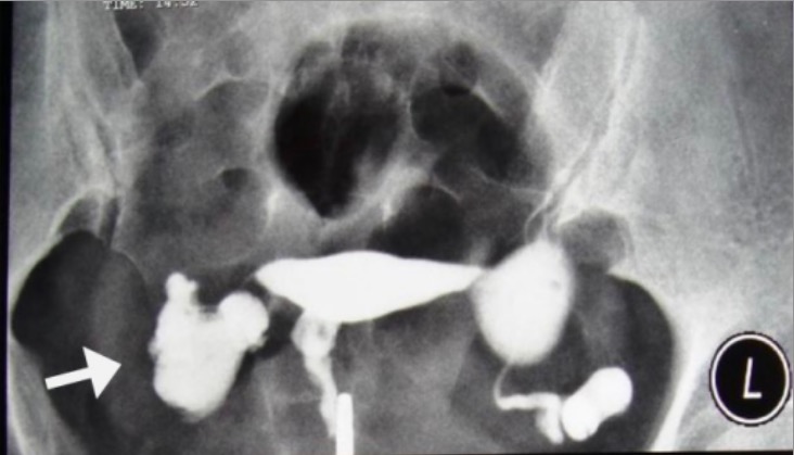
Hysterosalpingogram:
Inject dye into the uterine cavity under fluoroscopyFluoroscopyProduction of an image when x-rays strike a fluorescent screen.X-rays.
Bilateral “fill and spill” of dye confirms tubal patency.
Some uterine anomalies may be visible.
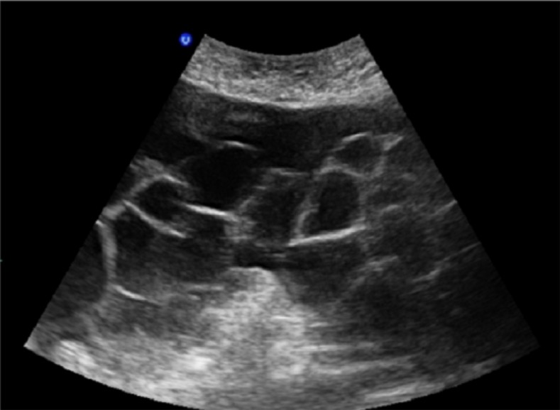
A 38-year-old woman with genital TB: Hysterosalpingogram shows terminal saculation and occlusion of both fallopian tubes causing hydrosalpinx (arrows). Uterine cavity has a normal appearance.
Image: “Genital tuberculosis” by Surgical Oncology Research Center, Mashhad University of Medical Sciences, Mashhad, Iran. License: CC BY 3.0
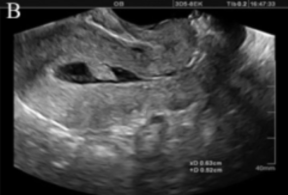
Saline infusion sonogram demonstrating a pedunculated lesion (likely a polyp) in the uterine cavity: On normal transvaginal ultrasonography without the infusion of saline, this finding would simply appear as an area of thickened endometrial lining.
Image: “A:B ratio less than 1 indicating pendunculated lesion” by Department of Reproductive Imaging at Reproductive Biomedicine Research Center, Royan Institute for Reproductive Biomedicine, ACECR, Tehran, Iran. License: CC BY 2.5, cropped by Lecturio.
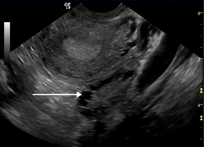
Transvaginal ultrasound showing a polycystic ovary: Note the multiple cysts on the periphery of the ovary (white arrow)
Image: “Transvaginal ultrasound scan of polycystic ovary” by Schomynv. License: CC0 1.0
LaparoscopyLaparoscopyLaparoscopy is surgical exploration and interventions performed through small incisions with a camera and long instruments. Laparotomy and Laparoscopy with chromopertubation
Inject dye through tubes to assess patency.
Allows for assessment and treatment of endometriosisEndometriosisEndometriosis is a common disease in which patients have endometrial tissue implanted outside of the uterus. Endometrial implants can occur anywhere in the pelvis, including the ovaries, the broad and uterosacral ligaments, the pelvic peritoneum, and the urinary and gastrointestinal tracts.Endometriosis and some pelvic adhesions
Evaluation of men
Semen analysis:
Volume
pHpHThe quantitative measurement of the acidity or basicity of a solution.Acid-Base Balance
Laboratory and imaging if semen analysis is abnormal:
FSHFSHA major gonadotropin secreted by the adenohypophysis. Follicle-stimulating hormone stimulates gametogenesis and the supporting cells such as the ovarian granulosa cells, the testicular sertoli cells, and leydig cells. Fsh consists of two noncovalently linked subunits, alpha and beta. Within a species, the alpha subunit is common in the three pituitary glycoprotein hormones (TSH, LH, and FSH), but the beta subunit is unique and confers its biological specificity.Menstrual Cycle, LHLHA major gonadotropin secreted by the adenohypophysis. Luteinizing hormone regulates steroid production by the interstitial cells of the testis and the ovary. The preovulatory luteinizing hormone surge in females induces ovulation, and subsequent luteinization of the follicle. Luteinizing hormone consists of two noncovalently linked subunits, alpha and beta. Within a species, the alpha subunit is common in the three pituitary glycoprotein hormones (TSH, LH, and FSH), but the beta subunit is unique and confers its biological specificity.Menstrual Cycle, and morning total testosteroneTestosteroneA potent androgenic steroid and major product secreted by the leydig cells of the testis. Its production is stimulated by luteinizing hormone from the pituitary gland. In turn, testosterone exerts feedback control of the pituitary LH and FSH secretion. Depending on the tissues, testosterone can be further converted to dihydrotestosterone or estradiol.Androgens and Antiandrogens:
↑ FSHFSHA major gonadotropin secreted by the adenohypophysis. Follicle-stimulating hormone stimulates gametogenesis and the supporting cells such as the ovarian granulosa cells, the testicular sertoli cells, and leydig cells. Fsh consists of two noncovalently linked subunits, alpha and beta. Within a species, the alpha subunit is common in the three pituitary glycoprotein hormones (TSH, LH, and FSH), but the beta subunit is unique and confers its biological specificity.Menstrual Cycle and LHLHA major gonadotropin secreted by the adenohypophysis. Luteinizing hormone regulates steroid production by the interstitial cells of the testis and the ovary. The preovulatory luteinizing hormone surge in females induces ovulation, and subsequent luteinization of the follicle. Luteinizing hormone consists of two noncovalently linked subunits, alpha and beta. Within a species, the alpha subunit is common in the three pituitary glycoprotein hormones (TSH, LH, and FSH), but the beta subunit is unique and confers its biological specificity.Menstrual Cycle with ↓ testosteroneTestosteroneA potent androgenic steroid and major product secreted by the leydig cells of the testis. Its production is stimulated by luteinizing hormone from the pituitary gland. In turn, testosterone exerts feedback control of the pituitary LH and FSH secretion. Depending on the tissues, testosterone can be further converted to dihydrotestosterone or estradiol.Androgens and Antiandrogens → hypergonadotropic hypogonadismHypergonadotropic HypogonadismHypogonadism (testicular defects)
Normal or ↓ FSHFSHA major gonadotropin secreted by the adenohypophysis. Follicle-stimulating hormone stimulates gametogenesis and the supporting cells such as the ovarian granulosa cells, the testicular sertoli cells, and leydig cells. Fsh consists of two noncovalently linked subunits, alpha and beta. Within a species, the alpha subunit is common in the three pituitary glycoprotein hormones (TSH, LH, and FSH), but the beta subunit is unique and confers its biological specificity.Menstrual Cycle and LHLHA major gonadotropin secreted by the adenohypophysis. Luteinizing hormone regulates steroid production by the interstitial cells of the testis and the ovary. The preovulatory luteinizing hormone surge in females induces ovulation, and subsequent luteinization of the follicle. Luteinizing hormone consists of two noncovalently linked subunits, alpha and beta. Within a species, the alpha subunit is common in the three pituitary glycoprotein hormones (TSH, LH, and FSH), but the beta subunit is unique and confers its biological specificity.Menstrual Cycle with ↓ testosteroneTestosteroneA potent androgenic steroid and major product secreted by the leydig cells of the testis. Its production is stimulated by luteinizing hormone from the pituitary gland. In turn, testosterone exerts feedback control of the pituitary LH and FSH secretion. Depending on the tissues, testosterone can be further converted to dihydrotestosterone or estradiol.Androgens and Antiandrogens → hypogonadotropic hypogonadismHypogonadotropic HypogonadismHypogonadism (hypothalamic or pituitaryPituitaryA small, unpaired gland situated in the sella turcica. It is connected to the hypothalamus by a short stalk which is called the infundibulum.Hormones: Overview and Types defects)
Normal FSHFSHA major gonadotropin secreted by the adenohypophysis. Follicle-stimulating hormone stimulates gametogenesis and the supporting cells such as the ovarian granulosa cells, the testicular sertoli cells, and leydig cells. Fsh consists of two noncovalently linked subunits, alpha and beta. Within a species, the alpha subunit is common in the three pituitary glycoprotein hormones (TSH, LH, and FSH), but the beta subunit is unique and confers its biological specificity.Menstrual Cycle with ↑ LHLHA major gonadotropin secreted by the adenohypophysis. Luteinizing hormone regulates steroid production by the interstitial cells of the testis and the ovary. The preovulatory luteinizing hormone surge in females induces ovulation, and subsequent luteinization of the follicle. Luteinizing hormone consists of two noncovalently linked subunits, alpha and beta. Within a species, the alpha subunit is common in the three pituitary glycoprotein hormones (TSH, LH, and FSH), but the beta subunit is unique and confers its biological specificity.Menstrual Cycle and testosteroneTestosteroneA potent androgenic steroid and major product secreted by the leydig cells of the testis. Its production is stimulated by luteinizing hormone from the pituitary gland. In turn, testosterone exerts feedback control of the pituitary LH and FSH secretion. Depending on the tissues, testosterone can be further converted to dihydrotestosterone or estradiol.Androgens and Antiandrogens → partial androgen resistanceResistancePhysiologically, the opposition to flow of air caused by the forces of friction. As a part of pulmonary function testing, it is the ratio of driving pressure to the rate of air flow.Ventilation: Mechanics of Breathing
↓↓ LHLHA major gonadotropin secreted by the adenohypophysis. Luteinizing hormone regulates steroid production by the interstitial cells of the testis and the ovary. The preovulatory luteinizing hormone surge in females induces ovulation, and subsequent luteinization of the follicle. Luteinizing hormone consists of two noncovalently linked subunits, alpha and beta. Within a species, the alpha subunit is common in the three pituitary glycoprotein hormones (TSH, LH, and FSH), but the beta subunit is unique and confers its biological specificity.Menstrual Cycle with ↑ muscle massMassThree-dimensional lesion that occupies a space within the breastImaging of the Breast → suspect androgen abuse
Genetic testingGenetic TestingDetection of a mutation; genotype; karyotype; or specific alleles associated with genetic traits, heritable diseases, or predisposition to a disease, or that may lead to the disease in descendants. It includes prenatal genetic testing.Myotonic Dystrophies (if abnormalities are suspected):
KaryotypeKaryotypeThe full set of chromosomes presented as a systematized array of metaphase chromosomes from a photomicrograph of a single cell nucleus arranged in pairs in descending order of size and according to the position of the centromere.Congenital Malformations of the Female Reproductive System → Klinefelter syndromeKlinefelter syndromeKlinefelter syndrome is a chromosomal aneuploidy characterized by the presence of 1 or more extra X chromosomes in a male karyotype, most commonly leading to karyotype 47,XXY. Klinefelter syndrome is associated with decreased levels of testosterone and is the most common cause of congenital hypogonadism. Klinefelter Syndrome
Y-chromosome microdeletions
CysticCysticFibrocystic ChangefibrosisFibrosisAny pathological condition where fibrous connective tissue invades any organ, usually as a consequence of inflammation or other injury.Bronchiolitis Obliterans transmembrane conductance regulator (CFTR) mutations → cysticCysticFibrocystic ChangefibrosisFibrosisAny pathological condition where fibrous connective tissue invades any organ, usually as a consequence of inflammation or other injury.Bronchiolitis Obliterans
Scrotal and transrectal ultrasound: dilated seminal vesiclesVesiclesFemale Genitourinary Examination → obstruction of ejaculatory ductsEjaculatory DuctsPaired ducts in the human male through which semen is ejaculated into the urethra.
Management
Lifestyle factors
Coital frequency of every 1–2 days around ovulationOvulationThe discharge of an ovum from a rupturing follicle in the ovary.Menstrual Cycle
SmokingSmokingWillful or deliberate act of inhaling and exhaling smoke from burning substances or agents held by hand.Interstitial Lung Diseases cessation
LimitLimitA value (e.g., pressure or time) that should not be exceeded and which is specified by the operator to protect the lungInvasive Mechanical Ventilation excessive alcohol and caffeineCaffeineA methylxanthine naturally occurring in some beverages and also used as a pharmacological agent. Caffeine’s most notable pharmacological effect is as a central nervous system stimulant, increasing alertness and producing agitation. Several cellular actions of caffeine have been observed, but it is not entirely clear how each contributes to its pharmacological profile. Among the most important are inhibition of cyclic nucleotide phosphodiesterases, antagonism of adenosine receptors, and modulation of intracellular calcium handling.Stimulants intake.
Fertility-friendly lubricants (many common brands inhibit sperm motilityMotilityThe motor activity of the gastrointestinal tract.Gastrointestinal Motility)
Weight lossWeight lossDecrease in existing body weight.Bariatric Surgery in the case of obesityObesityObesity is a condition associated with excess body weight, specifically with the deposition of excessive adipose tissue. Obesity is considered a global epidemic. Major influences come from the western diet and sedentary lifestyles, but the exact mechanisms likely include a mixture of genetic and environmental factors. Obesity or in overweight women
Weight gain for women who are underweight
Reduce environmental toxins: pesticides, cleaning solvents, and heavy metals
Surgical correction of uterine anomalies
Fibroids
Polyps
Synechiae
Septa
OvulationOvulationThe discharge of an ovum from a rupturing follicle in the ovary.Menstrual Cycle induction, ovarian hyperstimulation, and insemination
Requirements:
OvariesOvariesOvaries are the paired gonads of the female reproductive system that contain haploid gametes known as oocytes. The ovaries are located intraperitoneally in the pelvis, just posterior to the broad ligament, and are connected to the pelvic sidewall and to the uterus by ligaments. These organs function to secrete hormones (estrogen and progesterone) and to produce the female germ cells (oocytes).Ovaries: Anatomy capable of normal function
Patent tubes
Sperm
LetrozoleLetrozoleA triazole and benzonitrile derivative that is a selective non-steroidal aromatase inhibitor, similar to anastrozole. It is used in the treatment of metastatic or locally advanced breast cancer in postmenopausal women.Antiestrogens:
AromataseAromataseAn enzyme that catalyzes the desaturation (aromatization) of the ring a of C19 androgens and converts them to C18 estrogens. In this process, the 19-methyl is removed. This enzyme is membrane-bound, located in the endoplasmic reticulum of estrogen-producing cells of ovaries, placenta, testes, adipose, and brain tissues. Aromatase is encoded by the cyp19 gene, and functions in complex with NADPH-ferrihemoprotein reductase in the cytochrome p450 system.Adipose Tissue: Histology inhibitor that ↓ estrogenEstrogenCompounds that interact with estrogen receptors in target tissues to bring about the effects similar to those of estradiol. Estrogens stimulate the female reproductive organs, and the development of secondary female sex characteristics. Estrogenic chemicals include natural, synthetic, steroidal, or non-steroidal compounds.Ovaries: Anatomy → ↓ pituitaryPituitaryA small, unpaired gland situated in the sella turcica. It is connected to the hypothalamus by a short stalk which is called the infundibulum.Hormones: Overview and Types inhibition → ↑ FSHFSHA major gonadotropin secreted by the adenohypophysis. Follicle-stimulating hormone stimulates gametogenesis and the supporting cells such as the ovarian granulosa cells, the testicular sertoli cells, and leydig cells. Fsh consists of two noncovalently linked subunits, alpha and beta. Within a species, the alpha subunit is common in the three pituitary glycoprotein hormones (TSH, LH, and FSH), but the beta subunit is unique and confers its biological specificity.Menstrual Cycle
Used in normogonadotropic normoestrogenic ovulatory dysfunction
Not FDA approved, but considered as 1st-line therapy
↓ Rate of twins compared with clomiphene citrateClomiphene citrateA triphenyl ethylene stilbene derivative which is an estrogen agonist or antagonist depending on the target tissue.Antiestrogens
Clomiphene citrateClomiphene citrateA triphenyl ethylene stilbene derivative which is an estrogen agonist or antagonist depending on the target tissue.Antiestrogens:
Selective estrogenEstrogenCompounds that interact with estrogen receptors in target tissues to bring about the effects similar to those of estradiol. Estrogens stimulate the female reproductive organs, and the development of secondary female sex characteristics. Estrogenic chemicals include natural, synthetic, steroidal, or non-steroidal compounds.Ovaries: AnatomyreceptorReceptorReceptors are proteins located either on the surface of or within a cell that can bind to signaling molecules known as ligands (e.g., hormones) and cause some type of response within the cell.Receptors modulator → ↓ pituitaryPituitaryA small, unpaired gland situated in the sella turcica. It is connected to the hypothalamus by a short stalk which is called the infundibulum.Hormones: Overview and Types inhibition → ↑ FSHFSHA major gonadotropin secreted by the adenohypophysis. Follicle-stimulating hormone stimulates gametogenesis and the supporting cells such as the ovarian granulosa cells, the testicular sertoli cells, and leydig cells. Fsh consists of two noncovalently linked subunits, alpha and beta. Within a species, the alpha subunit is common in the three pituitary glycoprotein hormones (TSH, LH, and FSH), but the beta subunit is unique and confers its biological specificity.Menstrual Cycle
Used in normogonadotropic normoestrogenic ovulatory dysfunction
Classic treatment still used, but no longer 1st line
Injectable gonadotropins (e.g., FSHFSHA major gonadotropin secreted by the adenohypophysis. Follicle-stimulating hormone stimulates gametogenesis and the supporting cells such as the ovarian granulosa cells, the testicular sertoli cells, and leydig cells. Fsh consists of two noncovalently linked subunits, alpha and beta. Within a species, the alpha subunit is common in the three pituitary glycoprotein hormones (TSH, LH, and FSH), but the beta subunit is unique and confers its biological specificity.Menstrual Cycle):
High risk of multiples, including higher-order multiples
Used in egg retrieval prior to in vitrofertilizationFertilizationTo undergo fertilization, the sperm enters the uterus, travels towards the ampulla of the fallopian tube, and encounters the oocyte. The zona pellucida (the outer layer of the oocyte) deteriorates along with the zygote, which travels towards the uterus and eventually forms a blastocyst, allowing for implantation to occur. Fertilization and First Week (IVF) and hypothalamic hypogonadismHypogonadismHypogonadism is a condition characterized by reduced or no sex hormone production by the testes or ovaries. Hypogonadism can result from primary (hypergonadotropic) or secondary (hypogonadotropic) failure. Symptoms include infertility, increased risk of osteoporosis, erectile dysfunction, decreased libido, and regression (or absence) of secondary sexual characteristics.Hypogonadism
Requires monitoring with frequent ultrasound procedures
Other medical treatments:
Insulin-sensitizing agents:
MetforminMetforminA biguanide hypoglycemic agent used in the treatment of non-insulin-dependent diabetes mellitus not responding to dietary modification. Metformin improves glycemic control by improving insulin sensitivity and decreasing intestinal absorption of glucose.Non-insulinotropic Diabetes Drugs
Used in overweight patientsPatientsIndividuals participating in the health care system for the purpose of receiving therapeutic, diagnostic, or preventive procedures.Clinician–Patient Relationship with insulin resistanceInsulin resistanceDiminished effectiveness of insulin in lowering blood sugar levels: requiring the use of 200 units or more of insulin per day to prevent hyperglycemia or ketosis.Diabetes Mellitus and PCOSPCOSPolycystic ovarian syndrome (PCOS) is the most common endocrine disorder of reproductive-age women, affecting nearly 5%-10% of women in the age group. It is characterized by hyperandrogenism, chronic anovulation leading to oligomenorrhea (or amenorrhea), and metabolic dysfunction.Polycystic Ovarian Syndrome in combination with other ovulation-inducing agents
DopamineDopamineOne of the catecholamine neurotransmitters in the brain. It is derived from tyrosine and is the precursor to norepinephrine and epinephrine. Dopamine is a major transmitter in the extrapyramidal system of the brain, and important in regulating movement.Receptors and Neurotransmitters of the CNS agonists:
BromocriptineBromocriptineA semisynthetic ergotamine alkaloid that is a dopamine D2 agonist. It suppresses prolactin secretion.Parkinson’s Disease Drugs
Cabergoline
Used to ↓ prolactinProlactinA lactogenic hormone secreted by the adenohypophysis. It is a polypeptide of approximately 23 kd. Besides its major action on lactation, in some species prolactin exerts effects on reproduction, maternal behavior, fat metabolism, immunomodulation and osmoregulation.Breasts: Anatomy levels in hyperprolactinemiaHyperprolactinemiaHyperprolactinemia is defined as a condition of elevated levels of prolactin (PRL) hormone in the blood. The PRL hormone is secreted by the anterior pituitary gland and is responsible for breast development and lactation. The most common cause is PRL-secreting pituitary adenomas (prolactinomas). Hyperprolactinemia
Intrauterine insemination (IUI)
Injection of a processed semen sample into the uterusUterusThe uterus, cervix, and fallopian tubes are part of the internal female reproductive system. The uterus has a thick wall made of smooth muscle (the myometrium) and an inner mucosal layer (the endometrium). The most inferior portion of the uterus is the cervix, which connects the uterine cavity to the vagina.Uterus, Cervix, and Fallopian Tubes: Anatomy
Often combined with ovulationOvulationThe discharge of an ovum from a rupturing follicle in the ovary.Menstrual Cycle induction to ↑ pregnancyPregnancyThe status during which female mammals carry their developing young (embryos or fetuses) in utero before birth, beginning from fertilization to birth.Pregnancy: Diagnosis, Physiology, and Care rates
Intrauterine insemination
Image: “Assisted reproductive technology process” by BruceBlaus. License: CC BY 3.0
In vitrofertilizationFertilizationTo undergo fertilization, the sperm enters the uterus, travels towards the ampulla of the fallopian tube, and encounters the oocyte. The zona pellucida (the outer layer of the oocyte) deteriorates along with the zygote, which travels towards the uterus and eventually forms a blastocyst, allowing for implantation to occur. Fertilization and First Week
For patientsPatientsIndividuals participating in the health care system for the purpose of receiving therapeutic, diagnostic, or preventive procedures.Clinician–Patient Relationship with:
Failed ovulationOvulationThe discharge of an ovum from a rupturing follicle in the ovary.Menstrual Cycle induction/IUI
Severe tubal disease
Advanced age
Procedure:
OocytesOocytesFemale germ cells derived from oogonia and termed oocytes when they enter meiosis. The primary oocytes begin meiosis but are arrested at the diplotene state until ovulation at puberty to give rise to haploid secondary oocytes or ova (ovum).Ovaries: Anatomy are surgically retrieved using ultrasound guidance.
FertilizationFertilizationTo undergo fertilization, the sperm enters the uterus, travels towards the ampulla of the fallopian tube, and encounters the oocyte. The zona pellucida (the outer layer of the oocyte) deteriorates along with the zygote, which travels towards the uterus and eventually forms a blastocyst, allowing for implantation to occur. Fertilization and First Week occurs via:
Introduction of sperm from a semen sample
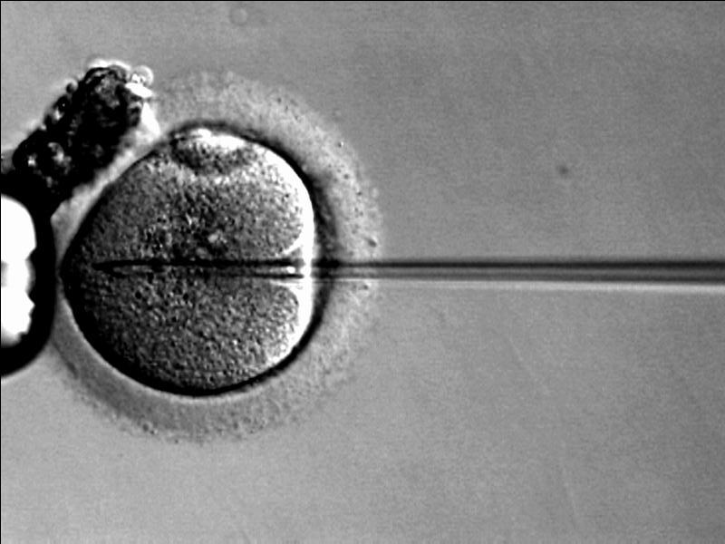
Intracytoplasmic sperm injection (ICSI)
Embryos are cultivated in a Petri dish and reimplanted into the endometriumEndometriumThe mucous membrane lining of the uterine cavity that is hormonally responsive during the menstrual cycle and pregnancy. The endometrium undergoes cyclic changes that characterize menstruation. After successful fertilization, it serves to sustain the developing embryo.Embryoblast and Trophoblast Development.
Additional options often combined with IVF:
Preimplantation genetic diagnosisPreimplantation genetic diagnosisDetermination of the nature of a pathological condition or disease in the ovum; zygote; or blastocyst prior to implantation. Cytogenetic analysis is performed to determine the presence or absence of genetic disease.Reproductive Ethical Issues (PGDPGDDetermination of the nature of a pathological condition or disease in the ovum; zygote; or blastocyst prior to implantation. Cytogenetic analysis is performed to determine the presence or absence of genetic disease.Reproductive Ethical Issues): screeningScreeningPreoperative Care of blastocysts for euploidy or a specific genetic defectGenetic DefectIon Channel Myopathy prior to reimplantation
ICSI:
Injection of a single sperm into a retrieved oocyte
Used in patientsPatientsIndividuals participating in the health care system for the purpose of receiving therapeutic, diagnostic, or preventive procedures.Clinician–Patient Relationship with abnormal sperm motilityMotilityThe motor activity of the gastrointestinal tract.Gastrointestinal Motility or morphology
Bilateral multilocular cystic masses in a patient with ovarian hyperstimulation syndrome in a spontaneous pregnancy with invasive mole
Image: “Bilateral multilocular cystic masses” by Myriam Rachad et al. License: CC BY 4.0
Intracytoplasmic sperm injection (ICSI): A technique used in couples who have low motility on semen analysis or multiple failed attempts in in vitro fertilization
Schorge, J.O., Schaffer, J.I., et al. (2008). Williams Gynecology (1st ed. pp. 426-467).
Practice Committee of the American Society for Reproductive Medicine. (2008). Definitions of infertility and recurrent pregnancy loss. Fertil Steril. 90(5 Suppl), S60 https://pubmed.ncbi.nlm.nih.gov/19007647/