Achieve Mastery of Medical Concepts

Study for medical school and boards with Lecturio

Klinefelter Syndrome

Klinefelter syndrome is a chromosomal aneuploidy characterized by the presence of 1 or more extra X chromosomes Chromosomes In a prokaryotic cell or in the nucleus of a eukaryotic cell, a structure consisting of or containing DNA which carries the genetic information essential to the cell. DNA Types and Structure in a male karyotype Karyotype The full set of chromosomes presented as a systematized array of metaphase chromosomes from a photomicrograph of a single cell nucleus arranged in pairs in descending order of size and according to the position of the centromere. Congenital Malformations of the Female Reproductive System, most commonly leading to karyotype Karyotype The full set of chromosomes presented as a systematized array of metaphase chromosomes from a photomicrograph of a single cell nucleus arranged in pairs in descending order of size and according to the position of the centromere. Congenital Malformations of the Female Reproductive System 47,XXY. Klinefelter syndrome is associated with decreased levels of testosterone Testosterone A potent androgenic steroid and major product secreted by the leydig cells of the testis. Its production is stimulated by luteinizing hormone from the pituitary gland. In turn, testosterone exerts feedback control of the pituitary LH and FSH secretion. Depending on the tissues, testosterone can be further converted to dihydrotestosterone or estradiol. Androgens and Antiandrogens and is the most common cause of congenital Congenital Chorioretinitis hypogonadism Hypogonadism Hypogonadism is a condition characterized by reduced or no sex hormone production by the testes or ovaries. Hypogonadism can result from primary (hypergonadotropic) or secondary (hypogonadotropic) failure. Symptoms include infertility, increased risk of osteoporosis, erectile dysfunction, decreased libido, and regression (or absence) of secondary sexual characteristics. Hypogonadism. Symptoms are often not noticed until adolescence or adulthood. Individuals with this condition tend to present as tall, phenotypic men with small testes Testes Gonadal Hormones, decreased body hair, gynecomastia Gynecomastia Gynecomastia is a benign proliferation of male breast glandular ductal tissue, usually bilateral, caused by increased estrogen activity, decreased testosterone activity, or medications. The condition is common and physiological in neonates, adolescent boys, and elderly men. Gynecomastia, and infertility Infertility Infertility is the inability to conceive in the context of regular intercourse. The most common causes of infertility in women are related to ovulatory dysfunction or tubal obstruction, whereas, in men, abnormal sperm is a common cause. Infertility. Treatment consists of life-long testosterone Testosterone A potent androgenic steroid and major product secreted by the leydig cells of the testis. Its production is stimulated by luteinizing hormone from the pituitary gland. In turn, testosterone exerts feedback control of the pituitary LH and FSH secretion. Depending on the tissues, testosterone can be further converted to dihydrotestosterone or estradiol. Androgens and Antiandrogens replacement therapy.

Last updated: Dec 13, 2022

Editorial responsibility: Stanley Oiseth, Lindsay Jones, Evelin Maza

Epidemiology and Genetics

Epidemiology

  • Incidence Incidence The number of new cases of a given disease during a given period in a specified population. It also is used for the rate at which new events occur in a defined population. It is differentiated from prevalence, which refers to all cases in the population at a given time. Measures of Disease Frequency: approximately 1 in 600 male births
  • Only diagnosed in approximately 25% of adult men with the condition

Genetics Genetics Genetics is the study of genes and their functions and behaviors. Basic Terms of Genetics

  • Karyotype Karyotype The full set of chromosomes presented as a systematized array of metaphase chromosomes from a photomicrograph of a single cell nucleus arranged in pairs in descending order of size and according to the position of the centromere. Congenital Malformations of the Female Reproductive System: 2 or more X chromosomes Chromosomes In a prokaryotic cell or in the nucleus of a eukaryotic cell, a structure consisting of or containing DNA which carries the genetic information essential to the cell. DNA Types and Structure with at least 1 predominantly inactive X chromosome X chromosome The female sex chromosome, being the differential sex chromosome carried by half the male gametes and all female gametes in human and other male-heterogametic species. Basic Terms of Genetics ( Barr body Barr body In the interphase nucleus, a condensed mass of chromatin representing an inactivated X chromosome. Each X chromosome, in excess of one, forms sex chromatin (Barr body) in the mammalian nucleus. Basic Terms of Genetics) present
    • 47,XXY: most common, karyotype Karyotype The full set of chromosomes presented as a systematized array of metaphase chromosomes from a photomicrograph of a single cell nucleus arranged in pairs in descending order of size and according to the position of the centromere. Congenital Malformations of the Female Reproductive System in about 80% of cases
    • 48,XXXY: less common
    • 48,XXYY: less common (additional Y chromosome Y chromosome The male sex chromosome, being the differential sex chromosome carried by half the male gametes and none of the female gametes in humans and in some other male-heterogametic species in which the homologue of the X chromosome has been retained. Basic Terms of Genetics)
  • Etiology:
    • Most commonly due to meiotic nondisjunction Nondisjunction The failure of homologous chromosomes or chromatids to segregate during mitosis or meiosis with the result that one daughter cell has both of a pair of parental chromosomes or chromatids and the other has none. Basic Terms of Genetics of the sex chromosomes Sex chromosomes The homologous chromosomes that are dissimilar in the heterogametic sex. There are the X chromosome, the Y chromosome, and the w, z chromosomes (in animals in which the female is the heterogametic sex (the silkworm moth bombyx mori, for example)). In such cases the w chromosome is the female-determining and the male is zz. Basic Terms of Genetics in parental germ cells Germ Cells The reproductive cells in multicellular organisms at various stages during gametogenesis. Gametogenesis
      • In meiosis I Meiosis I Following DNA replication, meiosis I creates 2 daughter cells containing half the genetic information of the mother cell (1n) but the same number of chromosomes (2c) by segregating sister chromatids into the same daughter cell Meiosis: Paired homologous chromosomes Homologous chromosomes Basic Terms of Genetics fail to separate.
      • In meiosis II Meiosis II Meiosis II is a cellular division event wherein the number of chromosomes in the daughter cells is halved from that of the mother cell. Meiosis II: similar to meiosis I but not preceded by interphase (DNA replication) Meiosis: Paired sister chromatids fail to separate.
    • May also occur due to mitotic nondisjunction Nondisjunction The failure of homologous chromosomes or chromatids to segregate during mitosis or meiosis with the result that one daughter cell has both of a pair of parental chromosomes or chromatids and the other has none. Basic Terms of Genetics in the developing zygote Zygote The fertilized ovum resulting from the fusion of a male and a female gamete. Fertilization and First Week
    • Origin of nondisjunction Nondisjunction The failure of homologous chromosomes or chromatids to segregate during mitosis or meiosis with the result that one daughter cell has both of a pair of parental chromosomes or chromatids and the other has none. Basic Terms of Genetics may be either maternal or paternal.
  • Risk factor: advanced maternal age
Human chromosomes xxy

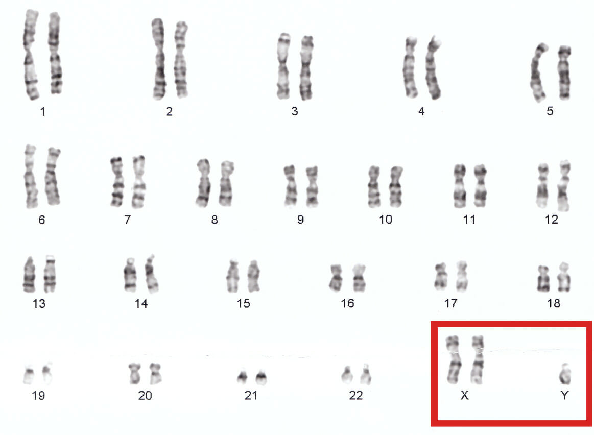
The most common karyotype in Klinefelter syndrome is 47, XXY

Image: “Human chromosomes XXY” by Nami-ja. License: Public Domain

Clinical Presentation

Clinical phenotype Phenotype The complete genetic complement contained in the DNA of a set of chromosomes in a human. The length of the human genome is about 3 billion base pairs. Basic Terms of Genetics

  • Outer appearance:
    • Male phenotype Phenotype The complete genetic complement contained in the DNA of a set of chromosomes in a human. The length of the human genome is about 3 billion base pairs. Basic Terms of Genetics
    • Tall with disproportionately long extremities
    • Eunuchoid features (both male and female features)
    • Classically slender build, but may have abdominal obesity with “pear-shaped” body habitus
    • Gynecomastia Gynecomastia Gynecomastia is a benign proliferation of male breast glandular ductal tissue, usually bilateral, caused by increased estrogen activity, decreased testosterone activity, or medications. The condition is common and physiological in neonates, adolescent boys, and elderly men. Gynecomastia (due to ↓ levels of testosterone Testosterone A potent androgenic steroid and major product secreted by the leydig cells of the testis. Its production is stimulated by luteinizing hormone from the pituitary gland. In turn, testosterone exerts feedback control of the pituitary LH and FSH secretion. Depending on the tissues, testosterone can be further converted to dihydrotestosterone or estradiol. Androgens and Antiandrogens and ↑ estrogen Estrogen Compounds that interact with estrogen receptors in target tissues to bring about the effects similar to those of estradiol. Estrogens stimulate the female reproductive organs, and the development of secondary female sex characteristics. Estrogenic chemicals include natural, synthetic, steroidal, or non-steroidal compounds. Ovaries: Anatomy)
    • Decreased facial/body/pubic hair
    • Decreased muscle mass Mass Three-dimensional lesion that occupies a space within the breast Imaging of the Breast/growth
    • Small, firm testes Testes Gonadal Hormones
  • Cognition/intelligence:
    • IQ (intelligence quotient) may be normal or mildly reduced.
    • Educational difficulties, especially with language-related functions
    • Symptoms may be more severe with increasing number of X chromosomes Chromosomes In a prokaryotic cell or in the nucleus of a eukaryotic cell, a structure consisting of or containing DNA which carries the genetic information essential to the cell. DNA Types and Structure present in karyotype Karyotype The full set of chromosomes presented as a systematized array of metaphase chromosomes from a photomicrograph of a single cell nucleus arranged in pairs in descending order of size and according to the position of the centromere. Congenital Malformations of the Female Reproductive System.
  • Behavioral abnormalities:
    • Increased risk of psychological disorders
    • Tendency toward poor social adjustments, but normal adult relationships
  • Sex Sex The totality of characteristics of reproductive structure, functions, phenotype, and genotype, differentiating the male from the female organism. Gender Dysphoria development/fertility:
    • Delayed puberty Delayed Puberty Delayed puberty (DP) is defined as the lack of testicular growth in boys past the age of 14 and the lack of thelarche in girls past the age of 13. Delayed puberty affects up to 5% of healthy boys and girls, and half of all cases are due to constitutional growth delay. Delayed Puberty
    • Hypergonadotropic hypogonadism Hypergonadotropic Hypogonadism Hypogonadism
      • Primary testicular failure → ↓ testosterone Testosterone A potent androgenic steroid and major product secreted by the leydig cells of the testis. Its production is stimulated by luteinizing hormone from the pituitary gland. In turn, testosterone exerts feedback control of the pituitary LH and FSH secretion. Depending on the tissues, testosterone can be further converted to dihydrotestosterone or estradiol. Androgens and Antiandrogens
      • Fibrosis Fibrosis Any pathological condition where fibrous connective tissue invades any organ, usually as a consequence of inflammation or other injury. Bronchiolitis Obliterans of seminiferous tubules Seminiferous Tubules The convoluted tubules in the testis where sperm are produced (spermatogenesis) and conveyed to the rete testis. Spermatogenic tubules are composed of developing germ cells and the supporting sertoli cells. Testicles: Anatomy within the testes Testes Gonadal Hormones and loss of function Loss of Function Inflammation of Sertoli cells Sertoli Cells Supporting cells projecting inward from the basement membrane of seminiferous tubules. They surround and nourish the developing male germ cells and secrete androgen-binding protein and hormones such as anti-mullerian hormone. The tight junctions of sertoli cells with the spermatogonia and spermatocytes provide a blood-testis barrier. Testicles: Anatomy (site of spermatogenesis Spermatogenesis The process of germ cell development in the male from the primordial germ cells, through spermatogonia; spermatocytes; spermatids; to the mature haploid spermatozoa. Gametogenesis)
      • Testosterone Testosterone A potent androgenic steroid and major product secreted by the leydig cells of the testis. Its production is stimulated by luteinizing hormone from the pituitary gland. In turn, testosterone exerts feedback control of the pituitary LH and FSH secretion. Depending on the tissues, testosterone can be further converted to dihydrotestosterone or estradiol. Androgens and Antiandrogens and lack of testes Testes Gonadal Hormones‘ ability to respond to luteinizing hormone ( LH LH A major gonadotropin secreted by the adenohypophysis. Luteinizing hormone regulates steroid production by the interstitial cells of the testis and the ovary. The preovulatory luteinizing hormone surge in females induces ovulation, and subsequent luteinization of the follicle. Luteinizing hormone consists of two noncovalently linked subunits, alpha and beta. Within a species, the alpha subunit is common in the three pituitary glycoprotein hormones (TSH, LH, and FSH), but the beta subunit is unique and confers its biological specificity. Menstrual Cycle) and follicle-stimulating hormone ( FSH FSH A major gonadotropin secreted by the adenohypophysis. Follicle-stimulating hormone stimulates gametogenesis and the supporting cells such as the ovarian granulosa cells, the testicular sertoli cells, and leydig cells. Fsh consists of two noncovalently linked subunits, alpha and beta. Within a species, the alpha subunit is common in the three pituitary glycoprotein hormones (TSH, LH, and FSH), but the beta subunit is unique and confers its biological specificity. Menstrual Cycle) → lack of feedback inhibition in pituitary Pituitary A small, unpaired gland situated in the sella turcica. It is connected to the hypothalamus by a short stalk which is called the infundibulum. Hormones: Overview and Types gland → ↑ LH LH A major gonadotropin secreted by the adenohypophysis. Luteinizing hormone regulates steroid production by the interstitial cells of the testis and the ovary. The preovulatory luteinizing hormone surge in females induces ovulation, and subsequent luteinization of the follicle. Luteinizing hormone consists of two noncovalently linked subunits, alpha and beta. Within a species, the alpha subunit is common in the three pituitary glycoprotein hormones (TSH, LH, and FSH), but the beta subunit is unique and confers its biological specificity. Menstrual Cycle and FSH FSH A major gonadotropin secreted by the adenohypophysis. Follicle-stimulating hormone stimulates gametogenesis and the supporting cells such as the ovarian granulosa cells, the testicular sertoli cells, and leydig cells. Fsh consists of two noncovalently linked subunits, alpha and beta. Within a species, the alpha subunit is common in the three pituitary glycoprotein hormones (TSH, LH, and FSH), but the beta subunit is unique and confers its biological specificity. Menstrual Cycle levels 
    • Infertility Infertility Infertility is the inability to conceive in the context of regular intercourse. The most common causes of infertility in women are related to ovulatory dysfunction or tubal obstruction, whereas, in men, abnormal sperm is a common cause. Infertility due to oligospermia (reduced number of sperm in semen) or azoospermia Azoospermia A condition of having no sperm present in the ejaculate (semen). Infertility (absence of sperm in semen)

Associated conditions

  • Cardiac abnormalities: mitral valve Mitral valve The valve between the left atrium and left ventricle of the heart. Heart: Anatomy prolapse
  • Malignancies:
    • Increased risk of breast cancer Breast cancer Breast cancer is a disease characterized by malignant transformation of the epithelial cells of the breast. Breast cancer is the most common form of cancer and 2nd most common cause of cancer-related death among women. Breast Cancer (due to ↑ estrogen Estrogen Compounds that interact with estrogen receptors in target tissues to bring about the effects similar to those of estradiol. Estrogens stimulate the female reproductive organs, and the development of secondary female sex characteristics. Estrogenic chemicals include natural, synthetic, steroidal, or non-steroidal compounds. Ovaries: Anatomy and ↓ levels of testosterone Testosterone A potent androgenic steroid and major product secreted by the leydig cells of the testis. Its production is stimulated by luteinizing hormone from the pituitary gland. In turn, testosterone exerts feedback control of the pituitary LH and FSH secretion. Depending on the tissues, testosterone can be further converted to dihydrotestosterone or estradiol. Androgens and Antiandrogens)
    • Increased risk of testicular cancer Testicular cancer Testicular cancer is the most common solid malignancy affecting men 15-35 years of age. Most of the testicular cancers are of the germ cell tumor type, and they can be classified as seminomas and nonseminomas. The most common presentation of testicular cancer is a painless testicular mass. Testicular Cancer
  • Osteoporosis Osteoporosis Osteoporosis refers to a decrease in bone mass and density leading to an increased number of fractures. There are 2 forms of osteoporosis: primary, which is commonly postmenopausal or senile; and secondary, which is a manifestation of immobilization, underlying medical disorders, or long-term use of certain medications. Osteoporosis in adulthood

Diagnosis and Management

Diagnosis

  • History and clinical examination
  • Genetic testing Genetic Testing Detection of a mutation; genotype; karyotype; or specific alleles associated with genetic traits, heritable diseases, or predisposition to a disease, or that may lead to the disease in descendants. It includes prenatal genetic testing. Myotonic Dystrophies: Karyotyping Karyotyping Mapping of the karyotype of a cell. Chromosome Testing confirms the diagnosis.
  • Labs show: ↓ testosterone Testosterone A potent androgenic steroid and major product secreted by the leydig cells of the testis. Its production is stimulated by luteinizing hormone from the pituitary gland. In turn, testosterone exerts feedback control of the pituitary LH and FSH secretion. Depending on the tissues, testosterone can be further converted to dihydrotestosterone or estradiol. Androgens and Antiandrogens level and ↑ LH LH A major gonadotropin secreted by the adenohypophysis. Luteinizing hormone regulates steroid production by the interstitial cells of the testis and the ovary. The preovulatory luteinizing hormone surge in females induces ovulation, and subsequent luteinization of the follicle. Luteinizing hormone consists of two noncovalently linked subunits, alpha and beta. Within a species, the alpha subunit is common in the three pituitary glycoprotein hormones (TSH, LH, and FSH), but the beta subunit is unique and confers its biological specificity. Menstrual Cycle and FSH FSH A major gonadotropin secreted by the adenohypophysis. Follicle-stimulating hormone stimulates gametogenesis and the supporting cells such as the ovarian granulosa cells, the testicular sertoli cells, and leydig cells. Fsh consists of two noncovalently linked subunits, alpha and beta. Within a species, the alpha subunit is common in the three pituitary glycoprotein hormones (TSH, LH, and FSH), but the beta subunit is unique and confers its biological specificity. Menstrual Cycle levels
  • Infertility Infertility Infertility is the inability to conceive in the context of regular intercourse. The most common causes of infertility in women are related to ovulatory dysfunction or tubal obstruction, whereas, in men, abnormal sperm is a common cause. Infertility screening Screening Preoperative Care should also be done; typically shows low sperm count.

Management

  • Testosterone Testosterone A potent androgenic steroid and major product secreted by the leydig cells of the testis. Its production is stimulated by luteinizing hormone from the pituitary gland. In turn, testosterone exerts feedback control of the pituitary LH and FSH secretion. Depending on the tissues, testosterone can be further converted to dihydrotestosterone or estradiol. Androgens and Antiandrogens replacement therapy (life-long): improves facial/body hair growth, strength/muscle size, libido, and energy level
  • Fertility treatment if children desired: Genetic counseling Genetic Counseling An educational process that provides information and advice to individuals or families about a genetic condition that may affect them. The purpose is to help individuals make informed decisions about marriage, reproduction, and other health management issues based on information about the genetic disease, the available diagnostic tests, and management programs. Psychosocial support is usually offered. Myotonic Dystrophies is warranted as there is a slightly increased risk of chromosomal abnormalities in offspring.

Clinical Relevance

Differential diagnoses

The following conditions are differential diagnoses of Klinefelter syndrome:

  • Kallmann syndrome Kallmann syndrome Kallmann syndrome (KS), also called olfacto-genital syndrome, is a genetic condition that causes hypogonadotropic hypogonadism due to decreased secretion of gonadotropin-releasing hormone (GnRH) by the hypothalamus. The lack of sex hormones results in impaired pubertal development. Kallmann Syndrome: a genetic condition that causes hypogonadotropic hypogonadism Hypogonadotropic Hypogonadism Hypogonadism due to gonadotropin-releasing hormone Gonadotropin-releasing hormone A decapeptide that stimulates the synthesis and secretion of both pituitary gonadotropins, luteinizing hormone and follicle stimulating hormone. Gnrh is produced by neurons in the septum preoptic area of the hypothalamus and released into the pituitary portal blood, leading to stimulation of gonadotrophs in the anterior pituitary gland. Puberty (GnRH) deficiency. Men and women are both affected. Men may present with delayed puberty Delayed Puberty Delayed puberty (DP) is defined as the lack of testicular growth in boys past the age of 14 and the lack of thelarche in girls past the age of 13. Delayed puberty affects up to 5% of healthy boys and girls, and half of all cases are due to constitutional growth delay. Delayed Puberty, decreased testosterone Testosterone A potent androgenic steroid and major product secreted by the leydig cells of the testis. Its production is stimulated by luteinizing hormone from the pituitary gland. In turn, testosterone exerts feedback control of the pituitary LH and FSH secretion. Depending on the tissues, testosterone can be further converted to dihydrotestosterone or estradiol. Androgens and Antiandrogens levels, and small testes Testes Gonadal Hormones. However, an absent or decreased sense of smell Smell The sense of smell, or olfaction, begins in a small area on the roof of the nasal cavity, which is covered in specialized mucosa. From there, the olfactory nerve transmits the sensory perception of smell via the olfactory pathway. This pathway is composed of the olfactory cells and bulb, the tractus and striae olfactoriae, and the primary olfactory cortex and amygdala. Olfaction: Anatomy is pathognomonic. Unlike in Klinefelter syndrome, LH LH A major gonadotropin secreted by the adenohypophysis. Luteinizing hormone regulates steroid production by the interstitial cells of the testis and the ovary. The preovulatory luteinizing hormone surge in females induces ovulation, and subsequent luteinization of the follicle. Luteinizing hormone consists of two noncovalently linked subunits, alpha and beta. Within a species, the alpha subunit is common in the three pituitary glycoprotein hormones (TSH, LH, and FSH), but the beta subunit is unique and confers its biological specificity. Menstrual Cycle and FSH FSH A major gonadotropin secreted by the adenohypophysis. Follicle-stimulating hormone stimulates gametogenesis and the supporting cells such as the ovarian granulosa cells, the testicular sertoli cells, and leydig cells. Fsh consists of two noncovalently linked subunits, alpha and beta. Within a species, the alpha subunit is common in the three pituitary glycoprotein hormones (TSH, LH, and FSH), but the beta subunit is unique and confers its biological specificity. Menstrual Cycle levels are elevated. Genetic testing Genetic Testing Detection of a mutation; genotype; karyotype; or specific alleles associated with genetic traits, heritable diseases, or predisposition to a disease, or that may lead to the disease in descendants. It includes prenatal genetic testing. Myotonic Dystrophies confirms the diagnosis.
  • Fragile X syndrome Fragile X syndrome Fragile X syndrome (FXS), also known as Martin-Bell syndrome, is a genetic condition with X-linked inheritance. Both boys and girls may be affected, but the severity is much worse in boys. Characteristic features include a long face, prominent forehead and chin, large ears, flat feet, and large testes post-puberty for boys. Fragile X Syndrome: an X-linked X-linked Genetic diseases that are linked to gene mutations on the X chromosome in humans or the X chromosome in other species. Included here are animal models of human X-linked diseases. Common Variable Immunodeficiency (CVID) dominant condition affecting men more commonly than women. Affected persons are usually tall with long, narrow faces and large ears. Abnormally large testes Testes Gonadal Hormones are noted after puberty Puberty Puberty is a complex series of physical, psychosocial, and cognitive transitions usually experienced by adolescents (11-19 years of age). Puberty is marked by a growth in stature and the development of secondary sexual characteristics, achievement of fertility, and changes in most body systems. Puberty, unlike the small testes Testes Gonadal Hormones seen in Klinefelter syndrome. Developmental delays are common. Genetic testing Genetic Testing Detection of a mutation; genotype; karyotype; or specific alleles associated with genetic traits, heritable diseases, or predisposition to a disease, or that may lead to the disease in descendants. It includes prenatal genetic testing. Myotonic Dystrophies confirms the diagnosis. 
  • Marfan syndrome Marfan syndrome Marfan syndrome is a genetic condition with autosomal dominant inheritance. Marfan syndrome affects the elasticity of connective tissues throughout the body, most notably in the cardiovascular, ocular, and musculoskeletal systems. Marfan Syndrome: an autosomal dominant Autosomal dominant Autosomal inheritance, both dominant and recessive, refers to the transmission of genes from the 22 autosomal chromosomes. Autosomal dominant diseases are expressed when only 1 copy of the dominant allele is inherited. Autosomal Recessive and Autosomal Dominant Inheritance connective tissue Connective tissue Connective tissues originate from embryonic mesenchyme and are present throughout the body except inside the brain and spinal cord. The main function of connective tissues is to provide structural support to organs. Connective tissues consist of cells and an extracellular matrix. Connective Tissue: Histology disorder that affects both men and women. Affected persons are usually tall with long arms and legs and arachnodactyly Arachnodactyly An abnormal bone development that is characterized by extra long and slender hands and fingers, such that the clenched thumb extends beyond the ulnar side of the hand. Arachnodactyly can include feet and toes. Arachnodactyly has been associated with several gene mutations and syndromes. Marfan Syndrome (abnormally long, slender fingers and toes). Associated with cardiovascular complications, musculoskeletal abnormalities, and lens Lens A transparent, biconvex structure of the eye, enclosed in a capsule and situated behind the iris and in front of the vitreous humor (vitreous body). It is slightly overlapped at its margin by the ciliary processes. Adaptation by the ciliary body is crucial for ocular accommodation. Eye: Anatomy dislocation/ vision Vision Ophthalmic Exam impairment. Marfan syndrome Marfan syndrome Marfan syndrome is a genetic condition with autosomal dominant inheritance. Marfan syndrome affects the elasticity of connective tissues throughout the body, most notably in the cardiovascular, ocular, and musculoskeletal systems. Marfan Syndrome does not cause hypogonadism Hypogonadism Hypogonadism is a condition characterized by reduced or no sex hormone production by the testes or ovaries. Hypogonadism can result from primary (hypergonadotropic) or secondary (hypogonadotropic) failure. Symptoms include infertility, increased risk of osteoporosis, erectile dysfunction, decreased libido, and regression (or absence) of secondary sexual characteristics. Hypogonadism or cognitive disabilities. Genetic testing Genetic Testing Detection of a mutation; genotype; karyotype; or specific alleles associated with genetic traits, heritable diseases, or predisposition to a disease, or that may lead to the disease in descendants. It includes prenatal genetic testing. Myotonic Dystrophies confirms the diagnosis.

Related condition

The following condition is related to Klinefelter syndrome as it also causes gonadal insufficiency due to a chromosomal abnormality:

Turner syndrome Turner syndrome Turner syndrome is a genetic condition affecting women, in which 1 X chromosome is partly or completely missing. The classic result is the karyotype 45,XO with a female phenotype. Turner syndrome is associated with decreased sex hormone levels and is the most common cause of primary amenorrhea. Turner Syndrome: a chromosomal abnormality disorder affecting only women. Karyotype Karyotype The full set of chromosomes presented as a systematized array of metaphase chromosomes from a photomicrograph of a single cell nucleus arranged in pairs in descending order of size and according to the position of the centromere. Congenital Malformations of the Female Reproductive System is 45,X0 instead of XX. Causes delayed puberty Delayed Puberty Delayed puberty (DP) is defined as the lack of testicular growth in boys past the age of 14 and the lack of thelarche in girls past the age of 13. Delayed puberty affects up to 5% of healthy boys and girls, and half of all cases are due to constitutional growth delay. Delayed Puberty and primary hypergonadotropic hypogonadism Hypergonadotropic Hypogonadism Hypogonadism (ovarian failure) in women. Affected women are characterized by short stature, widely spaced nipples, and webbed neck Webbed neck Turner Syndrome. Genetic testing Genetic Testing Detection of a mutation; genotype; karyotype; or specific alleles associated with genetic traits, heritable diseases, or predisposition to a disease, or that may lead to the disease in descendants. It includes prenatal genetic testing. Myotonic Dystrophies confirms the diagnosis. Management includes hormone replacement therapy Hormone Replacement Therapy Hormone replacement therapy (HRT) is used to treat symptoms associated with female menopause and in combination to suppress ovulation. Risks and side effects include uterine bleeding, predisposition to cancer, breast tenderness, hyperpigmentation, migraine headaches, hypertension, bloating, and mood changes. Noncontraceptive Estrogen and Progestins and fertility treatments if desired.

References

  1. Wattendorf, Daniel J., & Muenke, Maximilian. (2005). Klinefelter Syndrome. Am Fam Physician.72(11):2259–2262.
  2. Smith, C.M., & Bremner, W.J.(1998). Klinefelter Syndrome. Arch Intern Med. 158(12):1309–14. doi: 10.1001/archinte.158.12.1309.
  3. Defendi, Germaine L. Klinefelter Syndrome. Medscape. Retrieved November 23, 2020, from https://img.medscape.com/pi/iphone/medscapeapp/html/A945649-business.html

Create your free account or log in to continue reading!

Sign up now and get free access to Lecturio with concept pages, medical videos, and questions for your medical education.

User Reviews

Details