Advertisement

Advertisement

Advertisement

Advertisement

Meiosis

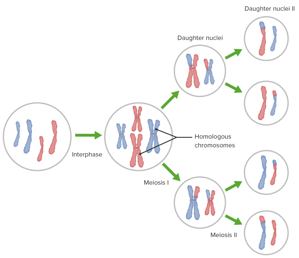
The creation of eukaryotic Eukaryotic Eukaryotes can be single-celled or multicellular organisms and include plants, animals, fungi, and protozoa. Eukaryotic cells contain a well-organized nucleus contained by a membrane, along with other membrane-bound organelles. Cell Types: Eukaryotic versus Prokaryotic gametes involves a DNA replication DNA replication The entire DNA of a cell is replicated during the S (synthesis) phase of the cell cycle. The principle of replication is based on complementary nucleotide base pairing: adenine forms hydrogen bonds with thymine (or uracil in RNA) and guanine forms hydrogen bonds with cytosine. DNA Replication phase followed by 2 cellular division stages: meiosis I and meiosis II. Meiosis I separates homologous chromosomes Homologous chromosomes Basic Terms of Genetics into separate cells (1n, 2c), while meiosis II separates sister chromatids into gametes (1n, 1c). Unique combinations of gametes via sexual reproduction are a major driver of evolutionary fitness in complex organisms.

Last updated: Dec 5, 2024

Editorial responsibility: Stanley Oiseth, Lindsay Jones, Evelin Maza

Advertisement

Advertisement

Advertisement

Advertisement

Advertisement

Advertisement

Overview of Meiosis

  • Meiosis: a cellular division process that creates aneuploid gametes in sexually reproducing species
  • Occurs in ovaries Ovaries Ovaries are the paired gonads of the female reproductive system that contain haploid gametes known as oocytes. The ovaries are located intraperitoneally in the pelvis, just posterior to the broad ligament, and are connected to the pelvic sidewall and to the uterus by ligaments. These organs function to secrete hormones (estrogen and progesterone) and to produce the female germ cells (oocytes). Ovaries: Anatomy and testes Testes Gonadal Hormones
  • Prior to mitosis Mitosis A type of cell nucleus division by means of which the two daughter nuclei normally receive identical complements of the number of chromosomes of the somatic cells of the species. Cell Cycle, the cell undergoes a growth and DNA replication DNA replication The entire DNA of a cell is replicated during the S (synthesis) phase of the cell cycle. The principle of replication is based on complementary nucleotide base pairing: adenine forms hydrogen bonds with thymine (or uracil in RNA) and guanine forms hydrogen bonds with cytosine. DNA Replication cycle called interphase Interphase The interval between two successive cell divisions during which the chromosomes are not individually distinguishable. It is composed of the g phases (g1 phase; g0 phase; g2 phase) and s phase (when DNA replication occurs). Cell Cycle.
  • 1 round of DNA replication DNA replication The entire DNA of a cell is replicated during the S (synthesis) phase of the cell cycle. The principle of replication is based on complementary nucleotide base pairing: adenine forms hydrogen bonds with thymine (or uracil in RNA) and guanine forms hydrogen bonds with cytosine. DNA Replication ( interphase Interphase The interval between two successive cell divisions during which the chromosomes are not individually distinguishable. It is composed of the g phases (g1 phase; g0 phase; g2 phase) and s phase (when DNA replication occurs). Cell Cycle), followed by 2 rounds of cellular division
  • 2 rounds of cell division Cell Division A type of cell nucleus division by means of which the two daughter nuclei normally receive identical complements of the number of chromosomes of the somatic cells of the species. Cell Cycle (meiosis I and meiosis II) divided into phases:
    • Prophase Prophase The first phase of cell nucleus division, in which the chromosomes become visible, the cell nucleus starts to lose its identity, the spindle apparatus appears, and the centrioles migrate toward opposite poles. Cell Cycle
    • Metaphase Metaphase The phase of cell nucleus division following prometaphase, in which the chromosomes line up across the equatorial plane of the spindle apparatus prior to separation. Cell Cycle
    • Anaphase Anaphase The phase of cell nucleus division following metaphase, in which the chromatids separate and migrate to opposite poles of the spindle. Cell Cycle
    • Telophase Telophase The final phase of cell nucleus division following anaphase, in which two daughter nuclei are formed, the cytoplasm completes division, and the chromosomes lose their distinctness and are transformed into chromatin threads. Cell Cycle
    • Cytokinesis
  • The resulting daughter cells have ½ the number of chromosomes Chromosomes In a prokaryotic cell or in the nucleus of a eukaryotic cell, a structure consisting of or containing DNA which carries the genetic information essential to the cell. DNA Types and Structure as the mother cells.
  • Nomenclature: n = number of homologous chromosomes Homologous chromosomes Basic Terms of Genetics, c = total number of chromosomes Chromosomes In a prokaryotic cell or in the nucleus of a eukaryotic cell, a structure consisting of or containing DNA which carries the genetic information essential to the cell. DNA Types and Structure (homologous and sister)
Overview of meiosis i and ii

Overview of meiosis I and II, crossing over (homologous recombination), and independent assortment

Image by Lecturio.

Meiosis I

Following DNA replication DNA replication The entire DNA of a cell is replicated during the S (synthesis) phase of the cell cycle. The principle of replication is based on complementary nucleotide base pairing: adenine forms hydrogen bonds with thymine (or uracil in RNA) and guanine forms hydrogen bonds with cytosine. DNA Replication, meiosis I creates 2 daughter cells containing half the genetic information of the mother cell (1n) but the same number of chromosomes Chromosomes In a prokaryotic cell or in the nucleus of a eukaryotic cell, a structure consisting of or containing DNA which carries the genetic information essential to the cell. DNA Types and Structure (2c) by segregating sister chromatids into the same daughter cell.
  • Prophase Prophase The first phase of cell nucleus division, in which the chromosomes become visible, the cell nucleus starts to lose its identity, the spindle apparatus appears, and the centrioles migrate toward opposite poles. Cell Cycle I: longest phase of meiosis
    • Chromosomes Chromosomes In a prokaryotic cell or in the nucleus of a eukaryotic cell, a structure consisting of or containing DNA which carries the genetic information essential to the cell. DNA Types and Structure start to condense.
    • Homologous chromosomes Homologous chromosomes Basic Terms of Genetics align and a synapse Synapse The junction between 2 neurons is called a synapse. The synapse allows a neuron to pass an electrical or chemical signal to another neuron or target effector cell. Synapses and Neurotransmission forms.
    • Crossing over Crossing over The reciprocal exchange of segments at corresponding positions along pairs of homologous chromosomes by symmetrical breakage and crosswise rejoining forming cross-over sites (holliday junctions) that are resolved during chromosome segregation. Crossing-over typically occurs during meiosis but it may also occur in the absence of meiosis, for example, with bacterial chromosomes, organelle chromosomes, or somatic cell nuclear chromosomes. Basic Terms of Genetics (overlap and fusion of homologous areas of aligned chromosomes Chromosomes In a prokaryotic cell or in the nucleus of a eukaryotic cell, a structure consisting of or containing DNA which carries the genetic information essential to the cell. DNA Types and Structure, causing an exchange of genetic material) occurs and is visible because of the chiasmata (point of crossing over Crossing over The reciprocal exchange of segments at corresponding positions along pairs of homologous chromosomes by symmetrical breakage and crosswise rejoining forming cross-over sites (holliday junctions) that are resolved during chromosome segregation. Crossing-over typically occurs during meiosis but it may also occur in the absence of meiosis, for example, with bacterial chromosomes, organelle chromosomes, or somatic cell nuclear chromosomes. Basic Terms of Genetics).
    • Crossing over Crossing over The reciprocal exchange of segments at corresponding positions along pairs of homologous chromosomes by symmetrical breakage and crosswise rejoining forming cross-over sites (holliday junctions) that are resolved during chromosome segregation. Crossing-over typically occurs during meiosis but it may also occur in the absence of meiosis, for example, with bacterial chromosomes, organelle chromosomes, or somatic cell nuclear chromosomes. Basic Terms of Genetics occurs between non-sister chromatid segments (homologous recombination Recombination Production of new arrangements of DNA by various mechanisms such as assortment and segregation, crossing over; gene conversion; genetic transformation; genetic conjugation; genetic transduction; or mixed infection of viruses. Virology).
  • Metaphase Metaphase The phase of cell nucleus division following prometaphase, in which the chromosomes line up across the equatorial plane of the spindle apparatus prior to separation. Cell Cycle I
    • The nuclear membrane disassembles.
    • Chromosomes Chromosomes In a prokaryotic cell or in the nucleus of a eukaryotic cell, a structure consisting of or containing DNA which carries the genetic information essential to the cell. DNA Types and Structure align themselves on the equatorial plane and attach to spindle fibers.
    • Spindle fibers push the chromosome Chromosome In a prokaryotic cell or in the nucleus of a eukaryotic cell, a structure consisting of or containing DNA which carries the genetic information essential to the cell. Basic Terms of Genetics pairs to stay in the middle of the cell.
    • Cohesins hold sister chromatids together from replication to anaphase Anaphase The phase of cell nucleus division following metaphase, in which the chromatids separate and migrate to opposite poles of the spindle. Cell Cycle.
  • Anaphase Anaphase The phase of cell nucleus division following metaphase, in which the chromatids separate and migrate to opposite poles of the spindle. Cell Cycle I
    • Spindle fibers shorten.
    • Separation of the chromosome Chromosome In a prokaryotic cell or in the nucleus of a eukaryotic cell, a structure consisting of or containing DNA which carries the genetic information essential to the cell. Basic Terms of Genetics pairs
    • The cell becomes oblong in preparation for division.
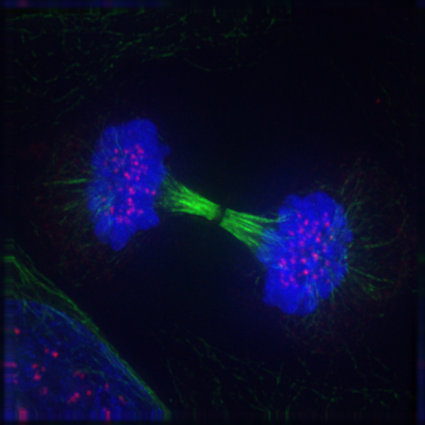
  • Telophase Telophase The final phase of cell nucleus division following anaphase, in which two daughter nuclei are formed, the cytoplasm completes division, and the chromosomes lose their distinctness and are transformed into chromatin threads. Cell Cycle I and cytokinesis
    • Spindle fibers disassemble.
    • Chromosomes Chromosomes In a prokaryotic cell or in the nucleus of a eukaryotic cell, a structure consisting of or containing DNA which carries the genetic information essential to the cell. DNA Types and Structure become less condensed, and the nuclear envelope Envelope Bilayer lipid membrane acquired by viral particles during viral morphogenesis. Although the lipids of the viral envelope are host derived, various virus-encoded integral membrane proteins, i.e. Viral envelope proteins are incorporated there. Virology may start to re-form.
    • Cytokinesis divides the cell into 2 daughter cells, each of which contains only 1 set of chromosomes Chromosomes In a prokaryotic cell or in the nucleus of a eukaryotic cell, a structure consisting of or containing DNA which carries the genetic information essential to the cell. DNA Types and Structure and is considered haploid Haploid The chromosomal constitution of cells, in which each type of chromosome is represented once. Symbol: n. Basic Terms of Genetics.
Telophase reappearance of the nuclear membrane and nucleolus

Reappearance of the nuclear membrane and nucleolus: the telophase

Image: “Reappearance of the nuclear membrane and nucleolus: the telophase” by Roy van Heesbeen. License: Public Domain

Meiosis II

Meiosis II is a cellular division event wherein the number of chromosomes Chromosomes In a prokaryotic cell or in the nucleus of a eukaryotic cell, a structure consisting of or containing DNA which carries the genetic information essential to the cell. DNA Types and Structure in the daughter cells is halved from that of the mother cell.

  • Meiosis II: similar to meiosis I but not preceded by interphase Interphase The interval between two successive cell divisions during which the chromosomes are not individually distinguishable. It is composed of the g phases (g1 phase; g0 phase; g2 phase) and s phase (when DNA replication occurs). Cell Cycle ( DNA replication DNA replication The entire DNA of a cell is replicated during the S (synthesis) phase of the cell cycle. The principle of replication is based on complementary nucleotide base pairing: adenine forms hydrogen bonds with thymine (or uracil in RNA) and guanine forms hydrogen bonds with cytosine. DNA Replication)
  • Stages of meiosis II:
    • Prophase Prophase The first phase of cell nucleus division, in which the chromosomes become visible, the cell nucleus starts to lose its identity, the spindle apparatus appears, and the centrioles migrate toward opposite poles. Cell Cycle II
      • The nuclear envelope Envelope Bilayer lipid membrane acquired by viral particles during viral morphogenesis. Although the lipids of the viral envelope are host derived, various virus-encoded integral membrane proteins, i.e. Viral envelope proteins are incorporated there. Virology disassembles.
      • Spindle fibers again form and chromosomes Chromosomes In a prokaryotic cell or in the nucleus of a eukaryotic cell, a structure consisting of or containing DNA which carries the genetic information essential to the cell. DNA Types and Structure start to condense.
    • Metaphase Metaphase The phase of cell nucleus division following prometaphase, in which the chromosomes line up across the equatorial plane of the spindle apparatus prior to separation. Cell Cycle II
      • Spindle fibers push the chromosomes Chromosomes In a prokaryotic cell or in the nucleus of a eukaryotic cell, a structure consisting of or containing DNA which carries the genetic information essential to the cell. DNA Types and Structure to the middle of the cell.
      • The cell elongates.
    • Anaphase Anaphase The phase of cell nucleus division following metaphase, in which the chromatids separate and migrate to opposite poles of the spindle. Cell Cycle II
      • Spindle fibers shorten.
      • Sister chromatids are separated.
    • Telophase Telophase The final phase of cell nucleus division following anaphase, in which two daughter nuclei are formed, the cytoplasm completes division, and the chromosomes lose their distinctness and are transformed into chromatin threads. Cell Cycle II
      • Chromosomes Chromosomes In a prokaryotic cell or in the nucleus of a eukaryotic cell, a structure consisting of or containing DNA which carries the genetic information essential to the cell. DNA Types and Structure become less condense.
      • Spindle fibers disassemble.
      • Cytokinesis: Cells divide into 2 daughter cells that are haploid Haploid The chromosomal constitution of cells, in which each type of chromosome is represented once. Symbol: n. Basic Terms of Genetics.

Meiosis in Humans

The human karyotype Karyotype The full set of chromosomes presented as a systematized array of metaphase chromosomes from a photomicrograph of a single cell nucleus arranged in pairs in descending order of size and according to the position of the centromere. Congenital Malformations of the Female Reproductive System normally contains 23 pairs of chromosomes Chromosomes In a prokaryotic cell or in the nucleus of a eukaryotic cell, a structure consisting of or containing DNA which carries the genetic information essential to the cell. DNA Types and Structure.

  • 22 pairs of autosomes
  • 1 pair of allosomes ( sex chromosomes Sex chromosomes The homologous chromosomes that are dissimilar in the heterogametic sex. There are the X chromosome, the Y chromosome, and the w, z chromosomes (in animals in which the female is the heterogametic sex (the silkworm moth bombyx mori, for example)). In such cases the w chromosome is the female-determining and the male is zz. Basic Terms of Genetics)
  • 23 chromosomes Chromosomes In a prokaryotic cell or in the nucleus of a eukaryotic cell, a structure consisting of or containing DNA which carries the genetic information essential to the cell. DNA Types and Structure from each parent
  • Total of 46 chromosomes Chromosomes In a prokaryotic cell or in the nucleus of a eukaryotic cell, a structure consisting of or containing DNA which carries the genetic information essential to the cell. DNA Types and Structure

Meiosis is important in the production of haploid Haploid The chromosomal constitution of cells, in which each type of chromosome is represented once. Symbol: n. Basic Terms of Genetics cells (gametes).

  • Spermatocytes and oocytes Oocytes Female germ cells derived from oogonia and termed oocytes when they enter meiosis. The primary oocytes begin meiosis but are arrested at the diplotene state until ovulation at puberty to give rise to haploid secondary oocytes or ova (ovum). Ovaries: Anatomy are the 2 types of haploid Haploid The chromosomal constitution of cells, in which each type of chromosome is represented once. Symbol: n. Basic Terms of Genetics cells (gametes) produced during meiosis.
  • Sexual reproduction: 2 haploid Haploid The chromosomal constitution of cells, in which each type of chromosome is represented once. Symbol: n. Basic Terms of Genetics cells combine to make 1 diploid Diploid The chromosomal constitution of cells, in which each type of chromosome is represented twice. Symbol: 2n or 2x. Basic Terms of Genetics cell.
  • 1 haploid Haploid The chromosomal constitution of cells, in which each type of chromosome is represented once. Symbol: n. Basic Terms of Genetics from the mother, 1 haploid Haploid The chromosomal constitution of cells, in which each type of chromosome is represented once. Symbol: n. Basic Terms of Genetics from the father
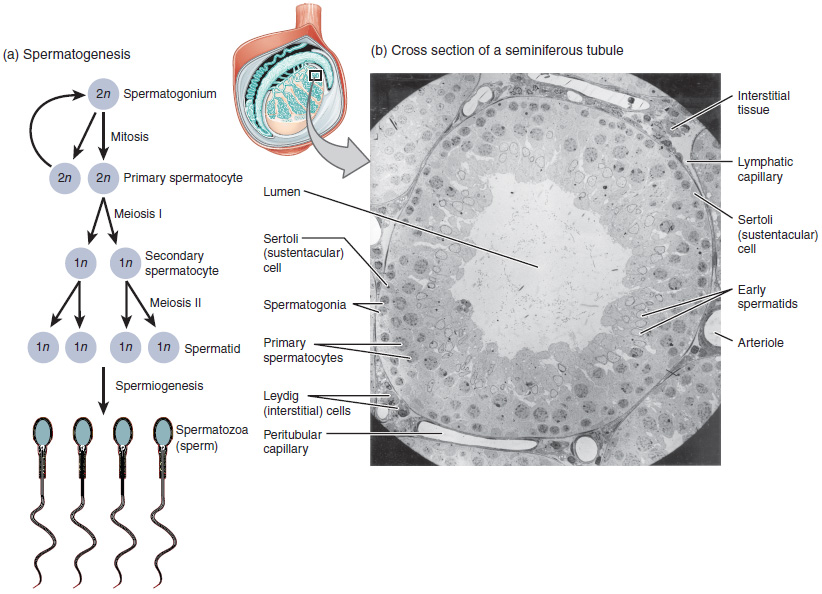
  • Spermatocytes are produced during spermatogenesis Spermatogenesis The process of germ cell development in the male from the primordial germ cells, through spermatogonia; spermatocytes; spermatids; to the mature haploid spermatozoa. Gametogenesis.
  • Oocytes Oocytes Female germ cells derived from oogonia and termed oocytes when they enter meiosis. The primary oocytes begin meiosis but are arrested at the diplotene state until ovulation at puberty to give rise to haploid secondary oocytes or ova (ovum). Ovaries: Anatomy are produced during oogenesis Oogenesis The process of germ cell development in the female from the primordial germ cells through oogonia to the mature haploid ova (ovum). Gametogenesis.

Spermatogenesis Spermatogenesis The process of germ cell development in the male from the primordial germ cells, through spermatogonia; spermatocytes; spermatids; to the mature haploid spermatozoa. Gametogenesis

  • Occurs in the seminiferous tubules Seminiferous Tubules The convoluted tubules in the testis where sperm are produced (spermatogenesis) and conveyed to the rete testis. Spermatogenic tubules are composed of developing germ cells and the supporting sertoli cells. Testicles: Anatomy of the testes Testes Gonadal Hormones
  • Spermatogonial stage
    • Mitotic clonal expansion Clonal Expansion Seborrheic Keratosis occurs.
    • Spermatogonia = precursor cells for spermatocytes
    • Type A spermatogonia from the primordial germ cell divide to maintain the stem cell pool population.
    • Some of the type A spermatogonia return or stay at the resting pool while some proliferate and undergo differentiation.
    • Type A spermatogonia may be converted to type B spermatogonia.
    • Type B spermatogonia then enter the preleptotene stage and become primary spermatocytes.
  • Meiotic stage
    • Primary spermatocytes undergo 2 stages of meiosis.
    • Haploid Haploid The chromosomal constitution of cells, in which each type of chromosome is represented once. Symbol: n. Basic Terms of Genetics daughter cells are called round spermatids.
  • Spermiogenesis stage
    • Round spermatids enter the spermatogenesis Spermatogenesis The process of germ cell development in the male from the primordial germ cells, through spermatogonia; spermatocytes; spermatids; to the mature haploid spermatozoa. Gametogenesis stage.
    • Spermatids undergo nuclear and cytoplasmic changes, leading to the formation of spermatozoa:
      • Formation of the acrosome (a modified lysosome)
      • The nucleus Nucleus Within a eukaryotic cell, a membrane-limited body which contains chromosomes and one or more nucleoli (cell nucleolus). The nuclear membrane consists of a double unit-type membrane which is perforated by a number of pores; the outermost membrane is continuous with the endoplasmic reticulum. A cell may contain more than one nucleus. The Cell: Organelles becomes condensed and is moved to the periphery of the cell.
      • Microtubules Microtubules Slender, cylindrical filaments found in the cytoskeleton of plant and animal cells. They are composed of the protein tubulin and are influenced by tubulin modulators. The Cell: Cytosol and Cytoskeleton then form, producing flagella Flagella A whiplike motility appendage present on the surface cells. Prokaryote flagella are composed of a protein called flagellin. Bacteria can have a single flagellum, a tuft at one pole, or multiple flagella covering the entire surface. In eukaryotes, flagella are threadlike protoplasmic extensions used to propel flagellates and sperm. Flagella have the same basic structure as cilia but are longer in proportion to the cell bearing them and present in much smaller numbers. Helicobacter.
      • A large part of the cytoplasm is removed as a residual body.
      • Sertoli cells Sertoli Cells Supporting cells projecting inward from the basement membrane of seminiferous tubules. They surround and nourish the developing male germ cells and secrete androgen-binding protein and hormones such as anti-mullerian hormone. The tight junctions of sertoli cells with the spermatogonia and spermatocytes provide a blood-testis barrier. Testicles: Anatomy phagocytose these bodies.
    • Spermatozoa are stored to mature before being released into the epididymis Epididymis The convoluted cordlike structure attached to the posterior of the testis. Epididymis consists of the head (caput), the body (corpus), and the tail (cauda). A network of ducts leaving the testis joins into a common epididymal tubule proper which provides the transport, storage, and maturation of spermatozoa. Testicles: Anatomy.

Oogenesis Oogenesis The process of germ cell development in the female from the primordial germ cells through oogonia to the mature haploid ova (ovum). Gametogenesis
  • Birth to puberty Puberty Puberty is a complex series of physical, psychosocial, and cognitive transitions usually experienced by adolescents (11-19 years of age). Puberty is marked by a growth in stature and the development of secondary sexual characteristics, achievement of fertility, and changes in most body systems. Puberty
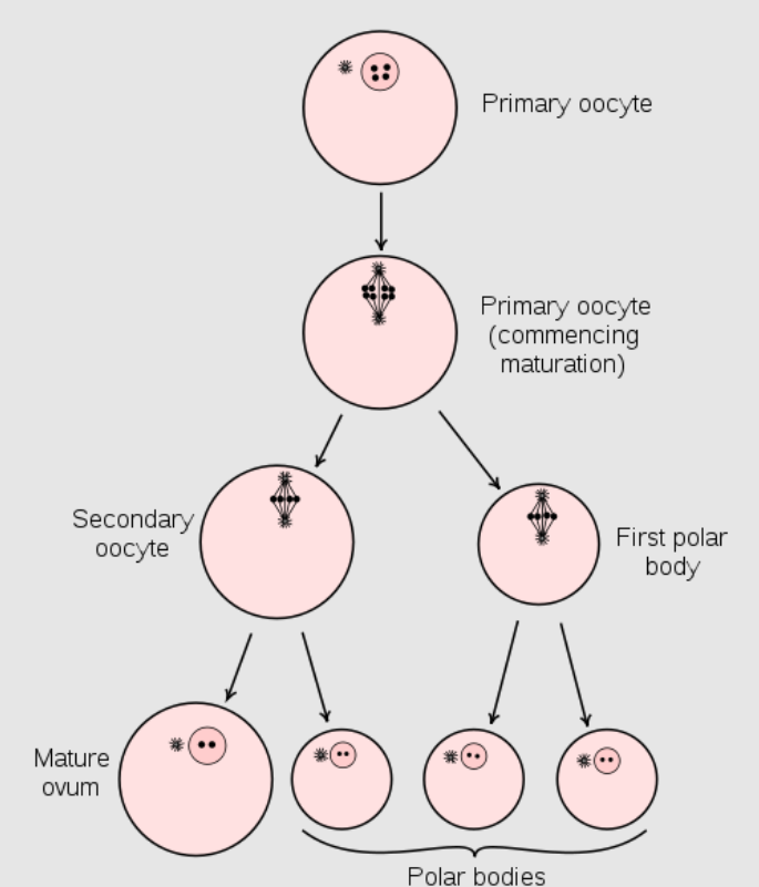
    • Before birth, oogonium Oogonium Euploid female germ cells of an early stage of oogenesis, derived from primordial germ cells during ovarian differentiation. Oogonia undergo meiosis and give rise to haploid oocytes. Gametogenesis, which also arise from germs cells, undergo mitosis Mitosis A type of cell nucleus division by means of which the two daughter nuclei normally receive identical complements of the number of chromosomes of the somatic cells of the species. Cell Cycle to produce primary oocytes Oocytes Female germ cells derived from oogonia and termed oocytes when they enter meiosis. The primary oocytes begin meiosis but are arrested at the diplotene state until ovulation at puberty to give rise to haploid secondary oocytes or ova (ovum). Ovaries: Anatomy.
    • Undergoes meiosis I, but process is halted at the prophase Prophase The first phase of cell nucleus division, in which the chromosomes become visible, the cell nucleus starts to lose its identity, the spindle apparatus appears, and the centrioles migrate toward opposite poles. Cell Cycle I
  • After puberty Puberty Puberty is a complex series of physical, psychosocial, and cognitive transitions usually experienced by adolescents (11-19 years of age). Puberty is marked by a growth in stature and the development of secondary sexual characteristics, achievement of fertility, and changes in most body systems. Puberty
    • Primary oocytes Oocytes Female germ cells derived from oogonia and termed oocytes when they enter meiosis. The primary oocytes begin meiosis but are arrested at the diplotene state until ovulation at puberty to give rise to haploid secondary oocytes or ova (ovum). Ovaries: Anatomy complete meiosis I forming a secondary oocyte and the 1st polar body
    • The secondary oocytes Oocytes Female germ cells derived from oogonia and termed oocytes when they enter meiosis. The primary oocytes begin meiosis but are arrested at the diplotene state until ovulation at puberty to give rise to haploid secondary oocytes or ova (ovum). Ovaries: Anatomy then begin meiosis II and are ovulated.
    • The secondary oocyte are arrested in metaphase Metaphase The phase of cell nucleus division following prometaphase, in which the chromosomes line up across the equatorial plane of the spindle apparatus prior to separation. Cell Cycle II.
  • Fertilization Fertilization To undergo fertilization, the sperm enters the uterus, travels towards the ampulla of the fallopian tube, and encounters the oocyte. The zona pellucida (the outer layer of the oocyte) deteriorates along with the zygote, which travels towards the uterus and eventually forms a blastocyst, allowing for implantation to occur. Fertilization and First Week
    • A secondary oocyte and a sperm cell fuse.
    • Meiosis II proceeds and is completed when the sperm cell completely penetrates the secondary oocyte.
    • A 2nd polar body and the ovum are produced after meiosis II.
    • When the nuclei of the sperm cells and the ovum unite, a diploid Diploid The chromosomal constitution of cells, in which each type of chromosome is represented twice. Symbol: 2n or 2x. Basic Terms of Genetics zygote Zygote The fertilized ovum resulting from the fusion of a male and a female gamete. Fertilization and First Week is produced.

Related videos

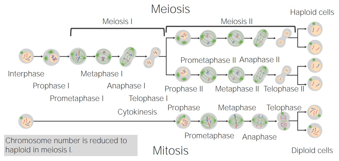
Meiosis vs. Mitosis

  • The major difference between these 2 processes is the number of steps needed to complete cell division Cell Division A type of cell nucleus division by means of which the two daughter nuclei normally receive identical complements of the number of chromosomes of the somatic cells of the species. Cell Cycle.
  • Mitosis Mitosis A type of cell nucleus division by means of which the two daughter nuclei normally receive identical complements of the number of chromosomes of the somatic cells of the species. Cell Cycle involves a single division producing 2 daughter cells similar to the parent cell.
  • Meiosis occurs as a 2-cell division process that produces 4 daughter cells that are completely distinct from both each other and the parent cells.
  • The final product of meiosis has half the number of chromosomes Chromosomes In a prokaryotic cell or in the nucleus of a eukaryotic cell, a structure consisting of or containing DNA which carries the genetic information essential to the cell. DNA Types and Structure compared to the parent cell, while mitosis Mitosis A type of cell nucleus division by means of which the two daughter nuclei normally receive identical complements of the number of chromosomes of the somatic cells of the species. Cell Cycle produces daughter cells with the same number of chromosomes Chromosomes In a prokaryotic cell or in the nucleus of a eukaryotic cell, a structure consisting of or containing DNA which carries the genetic information essential to the cell. DNA Types and Structure.
  • Mitosis Mitosis A type of cell nucleus division by means of which the two daughter nuclei normally receive identical complements of the number of chromosomes of the somatic cells of the species. Cell Cycle occurs in somatic cells and during early cell division Cell Division A type of cell nucleus division by means of which the two daughter nuclei normally receive identical complements of the number of chromosomes of the somatic cells of the species. Cell Cycle in gamete Gamete Gametogenesis formation, while meiosis occurs only at the final division of gamete Gamete Gametogenesis maturation.
Comparing-mitosis-meisosis

Comparing mitosis and meiosis

Image by Lecturio.

Clinical Relevance

  • Down syndrome Down syndrome Down syndrome, or trisomy 21, is the most common chromosomal aberration and the most frequent genetic cause of developmental delay. Both boys and girls are affected and have characteristic craniofacial and musculoskeletal features, as well as multiple medical anomalies involving the cardiac, gastrointestinal, ocular, and auditory systems. Down syndrome (Trisomy 21) ((DS) trisomy 21 Trisomy 21 Down syndrome, or trisomy 21, is the most common chromosomal aberration and the most frequent genetic cause of developmental delay. Both boys and girls are affected and have characteristic craniofacial and musculoskeletal features, as well as multiple medical anomalies involving the cardiac, gastrointestinal, ocular, and auditory systems. Down syndrome (Trisomy 21)): the most common chromosomal aberration and the most frequent genetic cause of developmental delay. Down syndrome Down syndrome Down syndrome, or trisomy 21, is the most common chromosomal aberration and the most frequent genetic cause of developmental delay. Both boys and girls are affected and have characteristic craniofacial and musculoskeletal features, as well as multiple medical anomalies involving the cardiac, gastrointestinal, ocular, and auditory systems. Down syndrome (Trisomy 21) results from a failure of meiosis. Characteristic traits include upslanting, almond-shaped eyes with skin Skin The skin, also referred to as the integumentary system, is the largest organ of the body. The skin is primarily composed of the epidermis (outer layer) and dermis (deep layer). The epidermis is primarily composed of keratinocytes that undergo rapid turnover, while the dermis contains dense layers of connective tissue. Skin: Structure and Functions covering the inner aspects, a broad flattened nasal bridge, small rounded ears, and a small mouth with a large tongue Tongue The tongue, on the other hand, is a complex muscular structure that permits tasting and facilitates the process of mastication and communication. The blood supply of the tongue originates from the external carotid artery, and the innervation is through cranial nerves. Lips and Tongue: Anatomy
  • Patau’s syndrome ( trisomy Trisomy The possession of a third chromosome of any one type in an otherwise diploid cell. Types of Mutations 13): a genetic syndrome caused by the presence of 3 copies of chromosome Chromosome In a prokaryotic cell or in the nucleus of a eukaryotic cell, a structure consisting of or containing DNA which carries the genetic information essential to the cell. Basic Terms of Genetics 13. Findings include craniofacial and cardiac malformations, severe intellectual disability Disability Determination of the degree of a physical, mental, or emotional handicap. The diagnosis is applied to legal qualification for benefits and income under disability insurance and to eligibility for social security and workman’s compensation benefits. ABCDE Assessment, and greatly reduced life expectancy Life expectancy Based on known statistical data, the number of years which any person of a given age may reasonably expected to live. Population Pyramids. Most infants do not survive past 3 months. 
  • Edwards syndrome Edwards syndrome Edwards syndrome, or trisomy 18, is a genetic syndrome caused by the presence of an extra chromosome 18. The extra chromosome is either from 3 full copies of chromosome 18 or an additional segment of chromosome 18. As the 2nd most common trisomy, Edwards syndrome is seen in 1 out of every 5,500 live births. Edwards Syndrome (Trisomy 18) ( trisomy Trisomy The possession of a third chromosome of any one type in an otherwise diploid cell. Types of Mutations 18): a genetic syndrome caused by the presence of an extra chromosome Chromosome In a prokaryotic cell or in the nucleus of a eukaryotic cell, a structure consisting of or containing DNA which carries the genetic information essential to the cell. Basic Terms of Genetics 18, which can be from 3 full copies of chromosome Chromosome In a prokaryotic cell or in the nucleus of a eukaryotic cell, a structure consisting of or containing DNA which carries the genetic information essential to the cell. Basic Terms of Genetics 18 or an additional segment of chromosome Chromosome In a prokaryotic cell or in the nucleus of a eukaryotic cell, a structure consisting of or containing DNA which carries the genetic information essential to the cell. Basic Terms of Genetics 18. Noted abnormalities include intrauterine growth retardation Growth Retardation Failure of a fetus to attain expected growth. Fetal Alcohol Spectrum Disorder, overlapping fingers, typical craniofacial features, rocker bottom feet, and congenital heart defects.
  • Klinefelter syndrome Klinefelter syndrome Klinefelter syndrome is a chromosomal aneuploidy characterized by the presence of 1 or more extra X chromosomes in a male karyotype, most commonly leading to karyotype 47,XXY. Klinefelter syndrome is associated with decreased levels of testosterone and is the most common cause of congenital hypogonadism. Klinefelter Syndrome: a chromosomal aneuploidy characterized by the presence of 1 or more extra X chromosomes Chromosomes In a prokaryotic cell or in the nucleus of a eukaryotic cell, a structure consisting of or containing DNA which carries the genetic information essential to the cell. DNA Types and Structure in a male karyotype Karyotype The full set of chromosomes presented as a systematized array of metaphase chromosomes from a photomicrograph of a single cell nucleus arranged in pairs in descending order of size and according to the position of the centromere. Congenital Malformations of the Female Reproductive System, most commonly leading to the karyotype Karyotype The full set of chromosomes presented as a systematized array of metaphase chromosomes from a photomicrograph of a single cell nucleus arranged in pairs in descending order of size and according to the position of the centromere. Congenital Malformations of the Female Reproductive System 47, XXY XXY Klinefelter syndrome is a chromosomal aneuploidy characterized by the presence of 1 or more extra X chromosomes in a male karyotype, most commonly leading to karyotype 47,XXY. Klinefelter syndrome is associated with decreased levels of testosterone and is the most common cause of congenital hypogonadism. Klinefelter Syndrome. Klinefelter syndrome Klinefelter syndrome Klinefelter syndrome is a chromosomal aneuploidy characterized by the presence of 1 or more extra X chromosomes in a male karyotype, most commonly leading to karyotype 47,XXY. Klinefelter syndrome is associated with decreased levels of testosterone and is the most common cause of congenital hypogonadism. Klinefelter Syndrome is associated with decreased levels of testosterone Testosterone A potent androgenic steroid and major product secreted by the leydig cells of the testis. Its production is stimulated by luteinizing hormone from the pituitary gland. In turn, testosterone exerts feedback control of the pituitary LH and FSH secretion. Depending on the tissues, testosterone can be further converted to dihydrotestosterone or estradiol. Androgens and Antiandrogens and is the most common cause of congenital hypogonadism Congenital hypogonadism Klinefelter Syndrome. Individuals with the syndrome tend to present as tall, phenotypic men with small testes Testes Gonadal Hormones, decreased body hair, gynecomastia Gynecomastia Gynecomastia is a benign proliferation of male breast glandular ductal tissue, usually bilateral, caused by increased estrogen activity, decreased testosterone activity, or medications. The condition is common and physiological in neonates, adolescent boys, and elderly men. Gynecomastia, and infertility Infertility Infertility is the inability to conceive in the context of regular intercourse. The most common causes of infertility in women are related to ovulatory dysfunction or tubal obstruction, whereas, in men, abnormal sperm is a common cause. Infertility.
  • Turner syndrome Turner syndrome Turner syndrome is a genetic condition affecting women, in which 1 X chromosome is partly or completely missing. The classic result is the karyotype 45,XO with a female phenotype. Turner syndrome is associated with decreased sex hormone levels and is the most common cause of primary amenorrhea. Turner Syndrome: a genetic condition affecting women in whom 1 X chromosome X chromosome The female sex chromosome, being the differential sex chromosome carried by half the male gametes and all female gametes in human and other male-heterogametic species. Basic Terms of Genetics is partly or completely missing. The classic result is the karyotype Karyotype The full set of chromosomes presented as a systematized array of metaphase chromosomes from a photomicrograph of a single cell nucleus arranged in pairs in descending order of size and according to the position of the centromere. Congenital Malformations of the Female Reproductive System 45,X0 with a female phenotype Phenotype The complete genetic complement contained in the DNA of a set of chromosomes in a human. The length of the human genome is about 3 billion base pairs. Basic Terms of Genetics. The characteristic appearance is that of a female person with short stature, a webbed neck Webbed neck Turner Syndrome, a broad chest with widely spaced nipples, a low posterior hairline, and peripheral edema Peripheral edema Peripheral edema is the swelling of the lower extremities, namely, legs, feet, and ankles. Edema of the hands and feet.

References

  1. Clark, MA, Douglas, M, & Choi, J. (2018.) The chemistry of life: Introduction. Biology 2e. (2021). OpenStax. Retrieved August 15, 2021, from https://openstax.org/books/biology-2e/pages/1-introduction
  2. Jones, KL. Down syndrome. In: Smith’s recognizable patterns of human malformation, 6th ed, Elsevier Saunders, Philadelphia 2006. p.7.
  3. Gottlieb, S, Gulani, A, & Tegay, D. (2021). Genetics, Meiosis. Retrieved August 15, 2021, from https://www.ncbi.nlm.nih.gov/books/NBK482462/
  4. O’Donnell, L, Nicholls, PK, O’Bryan, MK, McLachlan, RI, & Stanton PG. Spermiation: The process of sperm release. Spermatogenesis. 2011;1(1):14–35. https://pubmed.ncbi.nlm.nih.gov/21866274/
  5. Wattendorf, DJ, & Muenke, M. (2005). Klinefelter syndrome. Am Fam Physician.72(11):2259–2262.
  6. Morgan, T. (2007). Turner syndrome: Diagnosis and management. Am Fam Physician. 76(3):405–417.

Create your free account or log in to continue reading!

Sign up now and get free access to Lecturio with concept pages, medical videos, and questions for your medical education.

User Reviews

Details