Chromosome testing can be accomplished using several techniques, all of which can identify chromosomal abnormalities. Karyotyping (a technique that involves staining, organization, and visualization of chromosomes) is used to identify aneuploidy and major structural changes. Karyotyping is not sensitive in identifying small abnormalities and is a labor-intensive process. Chromosomal microarray analysis is a comparative technique that utilizes fluorescence to identify and quantify specific genetic sequences and is much more sensitive in identifying copy number variants, such as microdeletions or microduplications. However, chromosomal microarray analysis is not useful in identifying certain variations such as balanced translocations. Fluorescence in situ hybridization utilizes fluorescent probes to identify and locate specific genes on chromosomes. Compared with karyotyping, FISH is much more sensitive and specific in determining several abnormalities (except for point mutations) but is limited by the currently available gene probes.
Last updated: Dec 15, 2025
Numerical chromosomal aberrations:
Structural chromosomal aberrations:
Karyotyping, also known as chromosomal analysis, is a common method of producing an image of an individual’s chromosomes Chromosomes In a prokaryotic cell or in the nucleus of a eukaryotic cell, a structure consisting of or containing DNA which carries the genetic information essential to the cell. DNA Types and Structure to detect:
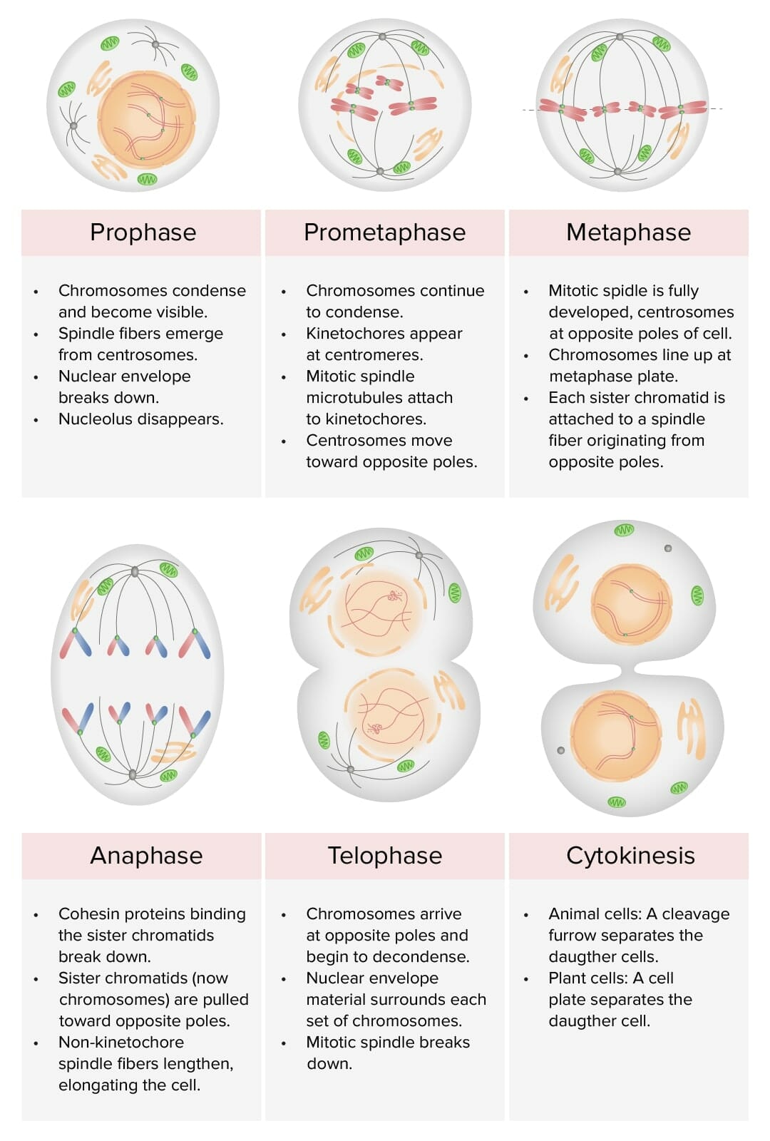
Review of the stages of mitosis: prophase, prometaphase, metaphase, anaphase, and telophase:
During karyotyping, the cell cycle is halted in metaphase, usually by adding an agent like colchicine (inhibits microtubule polymerization to halt mitosis).
Karyotyping is useful for identifying and diagnosing:
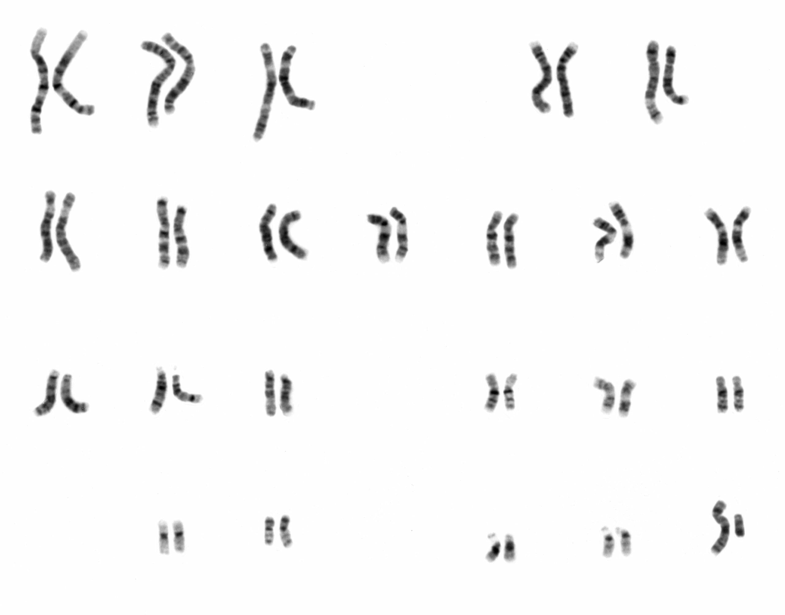
Karyotype of a normal human male: A karyotype is a laboratory technique whereby an individual’s chromosomes are paired and photographed.
Image: “NHGRI human male karyotype” by National Human Genome Research Institute. License: Public Domain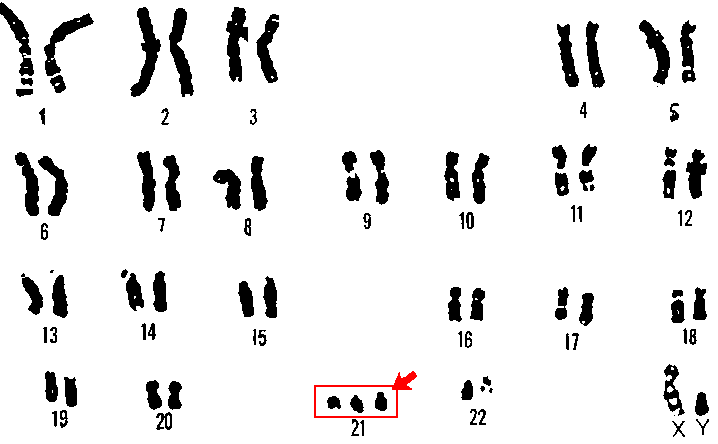
Karyotype of trisomy 21 (Down syndrome): In an individual with Down syndrome, 3 copies of chromosome 21 are usually visible (red box and arrow).
Image: “21 trisomy – Down syndrome” by U.S. Department of Energy Human Genome Program. License: Public Domain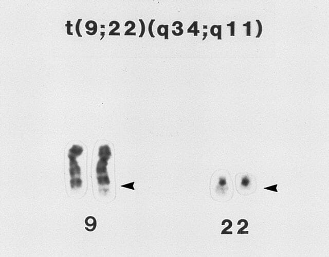
Partial karyotype of a myeloid cell from the bone marrow of an individual with chronic myeloid leukemia: The image demonstrates t(9:22) translocation, also known as the Philadelphia chromosome (bottom portion of chromosome 22 and chromosome 9 are switched (arrows)).
Image: “ BONE MARROW: CHRONIC MYELOID LEUKEMIA: PHILADELPHIA CHROMOSOME” by The Armed Forces Institute of Pathology (AFIP). License: Public DomainChromosomal microarray analysis is a high-resolution technique that utilizes fluorescent tags to identify small chromosomal imbalances.
A technique in which a fluorescent DNA DNA A deoxyribonucleotide polymer that is the primary genetic material of all cells. Eukaryotic and prokaryotic organisms normally contain DNA in a double-stranded state, yet several important biological processes transiently involve single-stranded regions. DNA, which consists of a polysugar-phosphate backbone possessing projections of purines (adenine and guanine) and pyrimidines (thymine and cytosine), forms a double helix that is held together by hydrogen bonds between these purines and pyrimidines (adenine to thymine and guanine to cytosine). DNA Types and Structure (or RNA RNA A polynucleotide consisting essentially of chains with a repeating backbone of phosphate and ribose units to which nitrogenous bases are attached. RNA is unique among biological macromolecules in that it can encode genetic information, serve as an abundant structural component of cells, and also possesses catalytic activity. RNA Types and Structure) probe Probe A device placed on the patient’s body to visualize a target Ultrasound (Sonography) is utilized to identify and locate specific genes Genes A category of nucleic acid sequences that function as units of heredity and which code for the basic instructions for the development, reproduction, and maintenance of organisms. DNA Types and Structure on chromosomes Chromosomes In a prokaryotic cell or in the nucleus of a eukaryotic cell, a structure consisting of or containing DNA which carries the genetic information essential to the cell. DNA Types and Structure
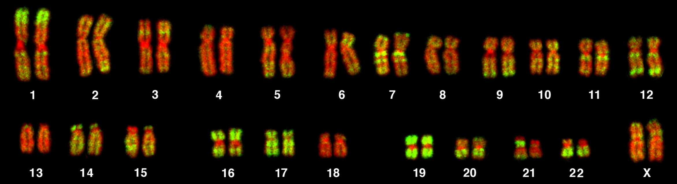
Female human karyotype subjected to FISH with a probe to the Alu sequence (green) and counterstained for DNA (red)
Image: “ChromosomesAluFish” by Dietzel65. License: CC BY 2.5