Advertisement

Advertisement

Advertisement

Advertisement

Testicular Cancer

Testicular cancer is the most common solid malignancy Malignancy Hemothorax affecting men 15–35 years of age. Most of the testicular cancers are of the germ cell tumor Tumor Inflammation type, and they can be classified as seminomas and nonseminomas. The most common presentation of testicular cancer is a painless testicular mass Mass Three-dimensional lesion that occupies a space within the breast Imaging of the Breast. Diagnosis is via physical exam, testicular ultrasonography, and serum tumor Tumor Inflammation markers. Additional imaging helps with staging Staging Methods which attempt to express in replicable terms the extent of the neoplasm in the patient. Grading, Staging, and Metastasis and assessment of metastasis Metastasis The transfer of a neoplasm from one organ or part of the body to another remote from the primary site. Grading, Staging, and Metastasis. Treatment consists of surgical inguinal orchiectomy, and further adjuvant Adjuvant Substances that augment, stimulate, activate, potentiate, or modulate the immune response at either the cellular or humoral level. The classical agents (freund's adjuvant, bcg, corynebacterium parvum, et al.) contain bacterial antigens. Some are endogenous (e.g., histamine, interferon, transfer factor, tuftsin, interleukin-1). Their mode of action is either non-specific, resulting in increased immune responsiveness to a wide variety of antigens, or antigen-specific, i.e., affecting a restricted type of immune response to a narrow group of antigens. The therapeutic efficacy of many biological response modifiers is related to their antigen-specific immunoadjuvanticity. Vaccination therapy is based on disease pathology and stage. Subsequent strategies include disease surveillance Surveillance Developmental Milestones and Normal Growth, radiotherapy, and chemotherapy Chemotherapy Osteosarcoma. Disease prognosis Prognosis A prediction of the probable outcome of a disease based on a individual's condition and the usual course of the disease as seen in similar situations. Non-Hodgkin Lymphomas is excellent, as testicular cancer is one of the most curable solid neoplasms Neoplasms New abnormal growth of tissue. Malignant neoplasms show a greater degree of anaplasia and have the properties of invasion and metastasis, compared to benign neoplasms. Benign Bone Tumors.

Last updated: May 17, 2024

Editorial responsibility: Stanley Oiseth, Lindsay Jones, Evelin Maza

Advertisement

Advertisement

Advertisement

Advertisement

Advertisement

Advertisement

Overview

Epidemiology

  • Testicular neoplasms Neoplasms New abnormal growth of tissue. Malignant neoplasms show a greater degree of anaplasia and have the properties of invasion and metastasis, compared to benign neoplasms. Benign Bone Tumors are the most common tumor Tumor Inflammation in men 15–35 years of age:
    • Germ cell tumors Germ cell tumors A true neoplasm composed of a number of different types of tissue, none of which is native to the area in which it occurs. It is composed of tissues that are derived from three germinal layers, the endoderm, mesoderm, and ectoderm. They are classified histologically as mature (benign) or immature (malignant). Ovarian Cancer (GCTs) comprise 95% of all testicular cancers.
    • Most commonly presents as localized seminoma in about 50% of cases 
    • Only 10%–30% of patients Patients Individuals participating in the health care system for the purpose of receiving therapeutic, diagnostic, or preventive procedures. Clinician–Patient Relationship will have metastatic disease. 
  • Annual incidence Incidence The number of new cases of a given disease during a given period in a specified population. It also is used for the rate at which new events occur in a defined population. It is differentiated from prevalence, which refers to all cases in the population at a given time. Measures of Disease Frequency in the United States: 6 in 100,000 men:
    • Incidence Incidence The number of new cases of a given disease during a given period in a specified population. It also is used for the rate at which new events occur in a defined population. It is differentiated from prevalence, which refers to all cases in the population at a given time. Measures of Disease Frequency is highest among non-Hispanic Whites.
    • Lowest incidence Incidence The number of new cases of a given disease during a given period in a specified population. It also is used for the rate at which new events occur in a defined population. It is differentiated from prevalence, which refers to all cases in the population at a given time. Measures of Disease Frequency among African Americans 

Risk factors

  • History of a GCT or germ cell neoplasia in situ (GCNIS)
  • History of cryptorchidism Cryptorchidism Cryptorchidism is one of the most common congenital anomalies in young boys. Typically, this asymptomatic condition presents during a routine well-child examination where 1 or both testicles are not palpable in the scrotum. Cryptorchidism ( relative risk increase Relative risk increase Measures of Risk of 4–6 times) 
  • Family history Family History Adult Health Maintenance of testicular cancer
  • HIV HIV Anti-HIV Drugs
  • Klinefelter syndrome Klinefelter syndrome Klinefelter syndrome is a chromosomal aneuploidy characterized by the presence of 1 or more extra X chromosomes in a male karyotype, most commonly leading to karyotype 47,XXY. Klinefelter syndrome is associated with decreased levels of testosterone and is the most common cause of congenital hypogonadism. Klinefelter Syndrome, Down syndrome Down syndrome Down syndrome, or trisomy 21, is the most common chromosomal aberration and the most frequent genetic cause of developmental delay. Both boys and girls are affected and have characteristic craniofacial and musculoskeletal features, as well as multiple medical anomalies involving the cardiac, gastrointestinal, ocular, and auditory systems. Down syndrome (Trisomy 21)
  • Peutz-Jeghers syndrome Peutz-Jeghers Syndrome Peutz-Jeghers syndrome (PJS) is an autosomal-dominant inherited disorder characterized by GI polyps and mucocutaneous-pigmented macules. Peutz-Jeghers syndrome is 1 of the polyposis syndromes, a group of inherited or acquired conditions characterized by the growth of polyps in the GI tract and associated with other extracolonic features. Peutz-Jeghers Syndrome
  • Carney complex Carney complex Autosomal dominant syndrome characterized by cardiac and cutaneous myxomas; lentiginosis (spotty pigmentation of the skin), and endocrinopathy and its associated endocrine tumors. The cardiac myxomas may lead to sudden cardiac death and other complications in carney complex patients. The gene coding for the prkar1a protein is one of the causative genetic loci (type 1). A second locus is at chromosome 2p16 (type 2). Cardiac Myxoma
  • Infertility Infertility Infertility is the inability to conceive in the context of regular intercourse. The most common causes of infertility in women are related to ovulatory dysfunction or tubal obstruction, whereas, in men, abnormal sperm is a common cause. Infertility
  • Testicular dysgenesis
  • Hypospadias Hypospadias A birth defect due to malformation of the urethra in which the urethral opening is below its normal location. In the male, the malformed urethra generally opens on the ventral surface of the penis or on the perineum. In the female, the malformed urethral opening is in the vagina. Penile Anomalies and Conditions

Classification

  • GCTs (95%):
    • Seminoma
    • Nonseminoma GCT (NSGCT):
      • Embryonal carcinoma
      • Yolk sac Yolk Sac The first of four extra-embryonic membranes to form during embryogenesis. In reptiles and birds, it arises from endoderm and mesoderm to incorporate the egg yolk into the digestive tract for nourishing the embryo. In placental mammals, its nutritional function is vestigial; however, it is the source of intestinal mucosa; blood cells; and germ cells. It is sometimes called the vitelline sac, which should not be confused with the vitelline membrane of the egg. Embryoblast and Trophoblast Development
      • Choriocarcinoma Choriocarcinoma A malignant metastatic form of trophoblastic tumors. Unlike the hydatidiform mole, choriocarcinoma contains no chorionic villi but rather sheets of undifferentiated cytotrophoblasts and syncytiotrophoblasts (trophoblasts). It is characterized by the large amounts of chorionic gonadotropin produced. Tissue origins can be determined by DNA analyses: placental (fetal) origin or non-placental origin. Gestational Trophoblastic Disease
      • Teratoma Teratoma A true neoplasm composed of a number of different types of tissue, none of which is native to the area in which it occurs. It is composed of tissues that are derived from three germinal layers, the endoderm, mesoderm, and ectoderm. They are classified histologically as mature (benign) or immature (malignant). Imaging of the Mediastinum
      • Mixed germ cell tumors Germ cell tumors A true neoplasm composed of a number of different types of tissue, none of which is native to the area in which it occurs. It is composed of tissues that are derived from three germinal layers, the endoderm, mesoderm, and ectoderm. They are classified histologically as mature (benign) or immature (malignant). Ovarian Cancer
  • Non-GCTs (5%):

Pathophysiology

The mechanisms are not fully known, but different factors appear to play a role in the development of testicular cancer.

Testicular dysgenesis syndrome

  • Environmental factors (i.e., in utero exposures) and genetic mutations Genetic Mutations Carcinogenesis affect the germ cell during early development.
  • Disturbed function leads to different phenotypes, ranging from cryptorchidism Cryptorchidism Cryptorchidism is one of the most common congenital anomalies in young boys. Typically, this asymptomatic condition presents during a routine well-child examination where 1 or both testicles are not palpable in the scrotum. Cryptorchidism, hypospadias Hypospadias A birth defect due to malformation of the urethra in which the urethral opening is below its normal location. In the male, the malformed urethra generally opens on the ventral surface of the penis or on the perineum. In the female, the malformed urethral opening is in the vagina. Penile Anomalies and Conditions, and other genital malformations to GCTs/testicular cancer. 

Genetic factors

  • 4× higher risk in sons of affected individuals
  • 8–10× higher risk in brothers of affected individuals
  •  KITLG (KIT ligand) locus Locus Specific regions that are mapped within a genome. Genetic loci are usually identified with a shorthand notation that indicates the chromosome number and the position of a specific band along the P or Q arm of the chromosome where they are found. For example the locus 6p21 is found within band 21 of the P-arm of chromosome 6. Many well known genetic loci are also known by common names that are associated with a genetic function or hereditary disease. Basic Terms of Genetics on chromosome Chromosome In a prokaryotic cell or in the nucleus of a eukaryotic cell, a structure consisting of or containing DNA which carries the genetic information essential to the cell. Basic Terms of Genetics 12 has a strong association with GCT risk in the testicles Testicles The testicles, also known as the testes or the male gonads, are a pair of egg-shaped glands suspended within the scrotum. The testicles have multiple layers: an outer tunica vaginalis, an intermediate tunica albuginea, and an innermost tunica vasculosa. The testicles are composed of testicular lobules and seminiferous tubules. Testicles: Anatomy.

Clinical Presentation

Classic findings

  • Genital:
  • Metastatic disease can be associated with abdominal pain Abdominal Pain Acute Abdomen, back pain Pain An unpleasant sensation induced by noxious stimuli which are detected by nerve endings of nociceptive neurons. Pain: Types and Pathways, and shortness of breath Shortness of breath Dyspnea is the subjective sensation of breathing discomfort. Dyspnea is a normal manifestation of heavy physical or psychological exertion, but also may be caused by underlying conditions (both pulmonary and extrapulmonary). Dyspnea.
  • Seminomas: commonly metastasize through the lymphatic system Lymphatic system A system of organs and tissues that process and transport immune cells and lymph. Primary Lymphatic Organs → present with signs of disseminated disease
  • On exam: negative transillumination Transillumination Passage of light through body tissues or cavities for examination of internal structures. Male Genitourinary Examination test

Histologic subtypes

Table: Germ cell tumors Germ cell tumors A true neoplasm composed of a number of different types of tissue, none of which is native to the area in which it occurs. It is composed of tissues that are derived from three germinal layers, the endoderm, mesoderm, and ectoderm. They are classified histologically as mature (benign) or immature (malignant). Ovarian Cancer (GCTs)
Type Subtype Characteristics
Seminoma
  • Most common testicular tumor Tumor Inflammation
  • Average age at presentation: 40 years
  • Causes homogeneous Homogeneous Imaging of the Spleen testicular enlargement
  • Large cells in lobules Lobules Breasts: Anatomy with a fried-egg appearance
  • Serum markers: ↑ β-hCG
  • ↑ Placental ALP ALP An enzyme that catalyzes the conversion of an orthophosphoric monoester and water to an alcohol and orthophosphate. Osteosarcoma
  • Prognosis Prognosis A prediction of the probable outcome of a disease based on a individual’s condition and the usual course of the disease as seen in similar situations. Non-Hodgkin Lymphomas:
Nonseminoma Yolk sac Yolk Sac The first of four extra-embryonic membranes to form during embryogenesis. In reptiles and birds, it arises from endoderm and mesoderm to incorporate the egg yolk into the digestive tract for nourishing the embryo. In placental mammals, its nutritional function is vestigial; however, it is the source of intestinal mucosa; blood cells; and germ cells. It is sometimes called the vitelline sac, which should not be confused with the vitelline membrane of the egg. Embryoblast and Trophoblast Development or testicular endodermal sinus tumor Tumor Inflammation
  • Common in boys < 3 years old
  • Morphology: Schiller-Duval bodies are present.
  • Serum markers: ↑ AFP AFP The first alpha-globulins to appear in mammalian sera during fetal development and the dominant serum proteins in early embryonic life. Hepatocellular Carcinoma (HCC) and Liver Metastases (highly characteristic)
  • Aggressive
Choriocarcinoma Choriocarcinoma A malignant metastatic form of trophoblastic tumors. Unlike the hydatidiform mole, choriocarcinoma contains no chorionic villi but rather sheets of undifferentiated cytotrophoblasts and syncytiotrophoblasts (trophoblasts). It is characterized by the large amounts of chorionic gonadotropin produced. Tissue origins can be determined by DNA analyses: placental (fetal) origin or non-placental origin. Gestational Trophoblastic Disease
  • Early hematogenous Hematogenous Hepatocellular Carcinoma (HCC) and Liver Metastases spread; can include the brain Brain The part of central nervous system that is contained within the skull (cranium). Arising from the neural tube, the embryonic brain is comprised of three major parts including prosencephalon (the forebrain); mesencephalon (the midbrain); and rhombencephalon (the hindbrain). The developed brain consists of cerebrum; cerebellum; and other structures in the brain stem. Nervous System: Anatomy, Structure, and Classification
  • Alteration of the trophoblastic cells
  • Serum markers: ↑ β-hCG, no AFP AFP The first alpha-globulins to appear in mammalian sera during fetal development and the dominant serum proteins in early embryonic life. Hepatocellular Carcinoma (HCC) and Liver Metastases production
  • Most aggressive; poor prognosis Prognosis A prediction of the probable outcome of a disease based on a individual’s condition and the usual course of the disease as seen in similar situations. Non-Hodgkin Lymphomas
  • Might cause gynecomastia Gynecomastia Gynecomastia is a benign proliferation of male breast glandular ductal tissue, usually bilateral, caused by increased estrogen activity, decreased testosterone activity, or medications. The condition is common and physiological in neonates, adolescent boys, and elderly men. Gynecomastia or symptoms of hyperthyroidism Hyperthyroidism Hypersecretion of thyroid hormones from the thyroid gland. Elevated levels of thyroid hormones increase basal metabolic rate. Thyrotoxicosis and Hyperthyroidism
Embryonal carcinoma
  • Average age at presentation: 30 years
  • Clusters and sheets of cells with atypia Atypia Fibrocystic Change
  • Locally aggressive
  • Serum marker: may have ↑ β-hCG
Teratoma Teratoma A true neoplasm composed of a number of different types of tissue, none of which is native to the area in which it occurs. It is composed of tissues that are derived from three germinal layers, the endoderm, mesoderm, and ectoderm. They are classified histologically as mature (benign) or immature (malignant). Imaging of the Mediastinum
  • Involves tissues from ≥ 1 of the 3 germinal layers
  • Those with malignant transformation Transformation Change brought about to an organism’s genetic composition by unidirectional transfer (transfection; transduction, genetic; conjugation, genetic, etc.) and incorporation of foreign DNA into prokaryotic or eukaryotic cells by recombination of part or all of that DNA into the cell’s genome. Bacteriology are chemotherapy-resistant.
  • Benign Benign Fibroadenoma in children
Mixed GCT
  • Composed of > 1 type
  • About 30% of GCTs
AFP: alpha-fetoprotein
ALP: alkaline phosphatase
Table: Non– germ cells Germ Cells The reproductive cells in multicellular organisms at various stages during gametogenesis. Gametogenesis tumors
Type Characteristics
Leydig cell tumor Tumor Inflammation
  • Golden brown color in gross appearance
  • Contains Reinke crystals
  • Produces androgens Androgens Androgens are naturally occurring steroid hormones responsible for development and maintenance of the male sex characteristics, including penile, scrotal, and clitoral growth, development of sexual hair, deepening of the voice, and musculoskeletal growth. Androgens and Antiandrogens or estrogens → gynecomastia Gynecomastia Gynecomastia is a benign proliferation of male breast glandular ductal tissue, usually bilateral, caused by increased estrogen activity, decreased testosterone activity, or medications. The condition is common and physiological in neonates, adolescent boys, and elderly men. Gynecomastia in men and precocious puberty Precocious puberty Precocious puberty (PP) is the appearance of secondary sexual characteristics due to elevated sex hormones before the age of 6-8 in girls and 9 in boys. Excess hormone secretion may occur only at the level of the sex hormone or may involve the whole hypothalamic-pituitary-gonadal axis. Precocious Puberty in boys
  • In some, can produce corticosteroids Corticosteroids Chorioretinitis
Sertoli cell tumor Tumor Inflammation
  • Testicular mass Mass Three-dimensional lesion that occupies a space within the breast Imaging of the Breast (hormonally silent)
  • Cell arrangement: form cords and tubules
Testicular lymphoma Lymphoma A general term for various neoplastic diseases of the lymphoid tissue. Imaging of the Mediastinum
Retroperitoneal mass biopsy consistent with classic seminoma

H&E stain of biopsy sample of retroperitoneal mass consistent with classic seminoma

Image: “Testicular seminoma presenting with duodenal perforation: a case report” by Miocinovic R, Abaza R. License: CC BY 2.0, cropped by Lecturio.

Diagnosis

Physical examination

Focused genitourinary exam:

  • Testicular exam palpating for masses:
    • Gently palpate testicle, epididymis Epididymis The convoluted cordlike structure attached to the posterior of the testis. Epididymis consists of the head (caput), the body (corpus), and the tail (cauda). A network of ducts leaving the testis joins into a common epididymal tubule proper which provides the transport, storage, and maturation of spermatozoa. Testicles: Anatomy, and scrotum Scrotum A cutaneous pouch of skin containing the testicles and spermatic cords. Testicles: Anatomy.
    • Assess for masses, lesions, and nodules.
  • General abdominal exam to palpate for masses 
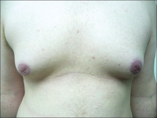
  • Chest exam to assess for gynecomastia Gynecomastia Gynecomastia is a benign proliferation of male breast glandular ductal tissue, usually bilateral, caused by increased estrogen activity, decreased testosterone activity, or medications. The condition is common and physiological in neonates, adolescent boys, and elderly men. Gynecomastia 
  • Inguinal exam to palpate for enlarged inguinal lymph nodes Lymph Nodes They are oval or bean shaped bodies (1 – 30 mm in diameter) located along the lymphatic system. Lymphatic Drainage System: Anatomy 
Bilateral gynecomastia

Bilateral gynecomastia in a patient

Image: “Finasteride induced Gynecomastia: Case report and Review of the Literature” by Ramot Y, Czarnowicki T, Zlotogorski A. License: CC BY 2.0

Laboratory tests

Obtain tumor Tumor Inflammation markers: 

  • Alpha-fetoprotein Alpha-fetoprotein The first alpha-globulins to appear in mammalian sera during fetal development and the dominant serum proteins in early embryonic life. Hepatocellular Carcinoma (HCC) and Liver Metastases ( AFP AFP The first alpha-globulins to appear in mammalian sera during fetal development and the dominant serum proteins in early embryonic life. Hepatocellular Carcinoma (HCC) and Liver Metastases): 
    • Half-life Half-Life The time it takes for a substance (drug, radioactive nuclide, or other) to lose half of its pharmacologic, physiologic, or radiologic activity. Pharmacokinetics and Pharmacodynamics: 5–7 days 
    • Elevated in about 50%–80% of NSGCT 
    • AFP AFP The first alpha-globulins to appear in mammalian sera during fetal development and the dominant serum proteins in early embryonic life. Hepatocellular Carcinoma (HCC) and Liver Metastases may also be elevated in liver Liver The liver is the largest gland in the human body. The liver is found in the superior right quadrant of the abdomen and weighs approximately 1.5 kilograms. Its main functions are detoxification, metabolism, nutrient storage (e.g., iron and vitamins), synthesis of coagulation factors, formation of bile, filtration, and storage of blood. Liver: Anatomy disease and abdominal cancers such as hepatocellular carcinoma Hepatocellular carcinoma Hepatocellular carcinoma (HCC) typically arises in a chronically diseased or cirrhotic liver and is the most common primary liver cancer. Diagnosis may include ultrasound, CT, MRI, biopsy (if inconclusive imaging), and/or biomarkers. Hepatocellular Carcinoma (HCC) and Liver Metastases
    • Infants will have temporary elevation of AFP AFP The first alpha-globulins to appear in mammalian sera during fetal development and the dominant serum proteins in early embryonic life. Hepatocellular Carcinoma (HCC) and Liver Metastases that will normalize. 
  • β-hCG: 
    • Half-life Half-Life The time it takes for a substance (drug, radioactive nuclide, or other) to lose half of its pharmacologic, physiologic, or radiologic activity. Pharmacokinetics and Pharmacodynamics: 24–36 hours 
    • Elevated in 20%–60% NSGCT and 15% of seminomas 
    • May see false elevations with marijuana use 
    • Hypogonadism Hypogonadism Hypogonadism is a condition characterized by reduced or no sex hormone production by the testes or ovaries. Hypogonadism can result from primary (hypergonadotropic) or secondary (hypogonadotropic) failure. Symptoms include infertility, increased risk of osteoporosis, erectile dysfunction, decreased libido, and regression (or absence) of secondary sexual characteristics. Hypogonadism may cause low levels of β-hCG.
  • LDH LDH Osteosarcoma
    • Half-life Half-Life The time it takes for a substance (drug, radioactive nuclide, or other) to lose half of its pharmacologic, physiologic, or radiologic activity. Pharmacokinetics and Pharmacodynamics: 24 hours 
    • Most common isoenzyme elevated in GCTs 
    • Used as a surrogate for disease burden, bulky tumor Tumor Inflammation

Imaging

  • Ultrasonography:
  • Chest X-ray X-ray Penetrating electromagnetic radiation emitted when the inner orbital electrons of an atom are excited and release radiant energy. X-ray wavelengths range from 1 pm to 10 nm. Hard x-rays are the higher energy, shorter wavelength x-rays. Soft x-rays or grenz rays are less energetic and longer in wavelength. The short wavelength end of the x-ray spectrum overlaps the gamma rays wavelength range. The distinction between gamma rays and x-rays is based on their radiation source. Pulmonary Function Tests to rule out pulmonary metastasis Metastasis The transfer of a neoplasm from one organ or part of the body to another remote from the primary site. Grading, Staging, and Metastasis 
  • Abdominal and pelvic CT scans: helpful in staging Staging Methods which attempt to express in replicable terms the extent of the neoplasm in the patient. Grading, Staging, and Metastasis the disease
  • Other imaging (e.g., brain Brain The part of central nervous system that is contained within the skull (cranium). Arising from the neural tube, the embryonic brain is comprised of three major parts including prosencephalon (the forebrain); mesencephalon (the midbrain); and rhombencephalon (the hindbrain). The developed brain consists of cerebrum; cerebellum; and other structures in the brain stem. Nervous System: Anatomy, Structure, and Classification CT): assess metastasis Metastasis The transfer of a neoplasm from one organ or part of the body to another remote from the primary site. Grading, Staging, and Metastasis

Staging Staging Methods which attempt to express in replicable terms the extent of the neoplasm in the patient. Grading, Staging, and Metastasis

Testicular cancer is staged according to the TNM Staging System TNM staging system Grading, Staging, and Metastasis of the American Joint Committee on Cancer and the Union for International Cancer Control (UICC).

Table: Staging Staging Methods which attempt to express in replicable terms the extent of the neoplasm in the patient. Grading, Staging, and Metastasis of testicular cancer
Stage Description
I Limited to testis, no lymph nodes Lymph Nodes They are oval or bean shaped bodies (1 – 30 mm in diameter) located along the lymphatic system. Lymphatic Drainage System: Anatomy, no metastasis Metastasis The transfer of a neoplasm from one organ or part of the body to another remote from the primary site. Grading, Staging, and Metastasis
II Testis + lymph nodes Lymph Nodes They are oval or bean shaped bodies (1 – 30 mm in diameter) located along the lymphatic system. Lymphatic Drainage System: Anatomy, no metastasis Metastasis The transfer of a neoplasm from one organ or part of the body to another remote from the primary site. Grading, Staging, and Metastasis:
III Distant metastasis Metastasis The transfer of a neoplasm from one organ or part of the body to another remote from the primary site. Grading, Staging, and Metastasis, significantly elevated tumor Tumor Inflammation markers
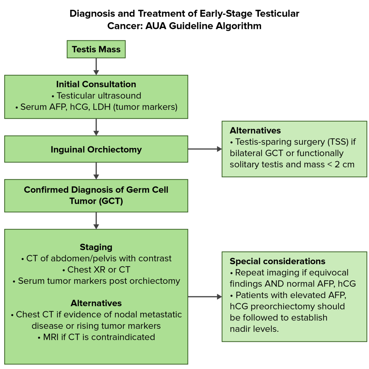
Aua guideline algorithm

Overview of the diagnostic and initial management of testicular cancer:
AFP: alpha-fetoprotein
AUA: American Urological Association

Image by Lecturio. License: CC BY-NC-SA 4.0

Management

Treatment of seminomas

Fertility preservation (i.e., sperm banking) should be discussed extensively with men of reproductive age before any treatment. Primary treatment is radical inguinal orchiectomy, followed by therapy, depending on the stage:

  • Stage I seminomas (high cure rate): primary treatment is inguinal orchiectomy, followed by: 
  • Stage II seminomas:
    • IIA: 
    • IIB or IIC: 
      • Chemotherapy Chemotherapy Osteosarcoma with bleomycin Bleomycin A complex of related glycopeptide antibiotics from streptomyces verticillus consisting of bleomycin a2 and b2. It inhibits DNA metabolism and is used as an antineoplastic, especially for solid tumors. Antitumor Antibiotics, etoposide Etoposide A semisynthetic derivative of podophyllotoxin that exhibits antitumor activity. Etoposide inhibits DNA synthesis by forming a complex with topoisomerase II and DNA. This complex induces breaks in double stranded DNA and prevents repair by topoisomerase II binding. Accumulated breaks in DNA prevent entry into the mitotic phase of cell division, and lead to cell death. Etoposide acts primarily in the g2 and s phases of the cell cycle. Microtubule and Topoisomerase Inhibitors, and cisplatin Cisplatin An inorganic and water-soluble platinum complex. After undergoing hydrolysis, it reacts with DNA to produce both intra and interstrand crosslinks. These cross links appear to impair replication and transcription of DNA. The cytotoxicity of cisplatin correlates with cellular arrest in the g2 phase of the cell cycle. Alkylating Agents and Platinum (BEP), or 
      • Etoposide Etoposide A semisynthetic derivative of podophyllotoxin that exhibits antitumor activity. Etoposide inhibits DNA synthesis by forming a complex with topoisomerase II and DNA. This complex induces breaks in double stranded DNA and prevents repair by topoisomerase II binding. Accumulated breaks in DNA prevent entry into the mitotic phase of cell division, and lead to cell death. Etoposide acts primarily in the g2 and s phases of the cell cycle. Microtubule and Topoisomerase Inhibitors and cisplatin Cisplatin An inorganic and water-soluble platinum complex. After undergoing hydrolysis, it reacts with DNA to produce both intra and interstrand crosslinks. These cross links appear to impair replication and transcription of DNA. The cytotoxicity of cisplatin correlates with cellular arrest in the g2 phase of the cell cycle. Alkylating Agents and Platinum alone ( EP EP Ectopic pregnancy refers to the implantation of a fertilized egg (embryo) outside the uterine cavity. The main cause is disruption of the normal anatomy of the fallopian tube. Ectopic Pregnancy)
      • Bleomycin Bleomycin A complex of related glycopeptide antibiotics from streptomyces verticillus consisting of bleomycin a2 and b2. It inhibits DNA metabolism and is used as an antineoplastic, especially for solid tumors. Antitumor Antibiotics: associated with dose-related pulmonary toxicity Toxicity Dosage Calculation 
      • Chemotherapy Chemotherapy Osteosarcoma also associated with short-term thrombocytopenia Thrombocytopenia Thrombocytopenia occurs when the platelet count is < 150,000 per microliter. The normal range for platelets is usually 150,000-450,000/µL of whole blood. Thrombocytopenia can be a result of decreased production, increased destruction, or splenic sequestration of platelets. Patients are often asymptomatic until platelet counts are < 50,000/µL. Thrombocytopenia and GI complications
  • Stage III (pulmonary or nonpulmonary metastatic seminomas): chemotherapy Chemotherapy Osteosarcoma with BEP or EP EP Ectopic pregnancy refers to the implantation of a fertilized egg (embryo) outside the uterine cavity. The main cause is disruption of the normal anatomy of the fallopian tube. Ectopic Pregnancy

Treatment of nonseminomas

Fertility preservation (i.e., sperm banking) should be discussed extensively with men of reproductive age before any treatment. Primary treatment is inguinal orchiectomy, followed by therapy depending on the stage:

  • Stage I nonseminomas
    • Postorchiectomy surveillance Surveillance Developmental Milestones and Normal Growth with serial physical exams, serum tumor Tumor Inflammation markers, abdominopelvic imaging as indicated 
    • Retroperitoneal Retroperitoneal Peritoneum: Anatomy lymph Lymph The interstitial fluid that is in the lymphatic system. Secondary Lymphatic Organs node dissection: for men with high-risk pathologic features and high risk of disease recurrence 
    • Adjuvant Adjuvant Substances that augment, stimulate, activate, potentiate, or modulate the immune response at either the cellular or humoral level. The classical agents (freund’s adjuvant, bcg, corynebacterium parvum, et al.) contain bacterial antigens. Some are endogenous (e.g., histamine, interferon, transfer factor, tuftsin, interleukin-1). Their mode of action is either non-specific, resulting in increased immune responsiveness to a wide variety of antigens, or antigen-specific, i.e., affecting a restricted type of immune response to a narrow group of antigens. The therapeutic efficacy of many biological response modifiers is related to their antigen-specific immunoadjuvanticity. Vaccination chemotherapy Chemotherapy Osteosarcoma: BEP 
  • Stage IIA and IIB nonseminomas:
  • Stage IIC and III nonseminomas: standard regimens for advanced tumors (includes BEP or EP EP Ectopic pregnancy refers to the implantation of a fertilized egg (embryo) outside the uterine cavity. The main cause is disruption of the normal anatomy of the fallopian tube. Ectopic Pregnancy)

Prognosis Prognosis A prediction of the probable outcome of a disease based on a individual’s condition and the usual course of the disease as seen in similar situations. Non-Hodgkin Lymphomas

  • Testicular GCTs: 5-year survival rates of approximately 95%
  • About 1%–2% present with brain Brain The part of central nervous system that is contained within the skull (cranium). Arising from the neural tube, the embryonic brain is comprised of three major parts including prosencephalon (the forebrain); mesencephalon (the midbrain); and rhombencephalon (the hindbrain). The developed brain consists of cerebrum; cerebellum; and other structures in the brain stem. Nervous System: Anatomy, Structure, and Classification metastasis Metastasis The transfer of a neoplasm from one organ or part of the body to another remote from the primary site. Grading, Staging, and Metastasis.
  • Relapse Relapse Relapsing Fever is rare after 2 years.
  • Hypogonadism Hypogonadism Hypogonadism is a condition characterized by reduced or no sex hormone production by the testes or ovaries. Hypogonadism can result from primary (hypergonadotropic) or secondary (hypogonadotropic) failure. Symptoms include infertility, increased risk of osteoporosis, erectile dysfunction, decreased libido, and regression (or absence) of secondary sexual characteristics. Hypogonadism is a potential complication (can be associated with sexual dysfunction Sexual dysfunction Physiological disturbances in normal sexual performance in either the male or the female. Sexual Physiology and infertility Infertility Infertility is the inability to conceive in the context of regular intercourse. The most common causes of infertility in women are related to ovulatory dysfunction or tubal obstruction, whereas, in men, abnormal sperm is a common cause. Infertility).
  • There is risk of a 2nd malignancy Malignancy Hemothorax in those treated with RT and chemotherapy Chemotherapy Osteosarcoma.

Differential Diagnosis

  • Varicocele Varicocele A condition characterized by the dilated tortuous veins of the spermatic cord with a marked left-sided predominance. Adverse effect on male fertility occurs when varicocele leads to an increased scrotal (and testicular) temperature and reduced testicular volume. Varicocele, Hydrocele, and Spermatocele: dilatation of the pampiniform venous plexus. Varicocele Varicocele A condition characterized by the dilated tortuous veins of the spermatic cord with a marked left-sided predominance. Adverse effect on male fertility occurs when varicocele leads to an increased scrotal (and testicular) temperature and reduced testicular volume. Varicocele, Hydrocele, and Spermatocele presents without pain Pain An unpleasant sensation induced by noxious stimuli which are detected by nerve endings of nociceptive neurons. Pain: Types and Pathways, like testicular cancer. Varicoceles have the characteristic “bag of worms” appearance on exam. Ultrasound shows negative transillumination Transillumination Passage of light through body tissues or cavities for examination of internal structures. Male Genitourinary Examination and dilated venous complex with retrograde blood flow Blood flow Blood flow refers to the movement of a certain volume of blood through the vasculature over a given unit of time (e.g., mL per minute). Vascular Resistance, Flow, and Mean Arterial Pressure
  • Spermatocele Spermatocele A cystic dilation of the epididymis, usually in the head portion (caput epididymis). The cyst fluid contains dead spermatozoa and can be easily differentiated from testicular hydrocele and other testicular lesions. Varicocele, Hydrocele, and Spermatocele: presents as an epididymal cyst, commonly arising from the head of the epididymis Epididymis The convoluted cordlike structure attached to the posterior of the testis. Epididymis consists of the head (caput), the body (corpus), and the tail (cauda). A network of ducts leaving the testis joins into a common epididymal tubule proper which provides the transport, storage, and maturation of spermatozoa. Testicles: Anatomy. Like testicular cancer, spermatocele Spermatocele A cystic dilation of the epididymis, usually in the head portion (caput epididymis). The cyst fluid contains dead spermatozoa and can be easily differentiated from testicular hydrocele and other testicular lesions. Varicocele, Hydrocele, and Spermatocele is also a painless mass Mass Three-dimensional lesion that occupies a space within the breast Imaging of the Breast often found incidentally. Furthermore, on imaging, cystic Cystic Fibrocystic Change lesions are noted, and on color Doppler Doppler Ultrasonography applying the doppler effect, with frequency-shifted ultrasound reflections produced by moving targets (usually red blood cells) in the bloodstream along the ultrasound axis in direct proportion to the velocity of movement of the targets, to determine both direction and velocity of blood flow. Ultrasound (Sonography), there is a characteristic “falling snow” appearance/sign (movement of internal echoes away from transducer Transducer A device placed on the patient’s body to visualize a target Ultrasound (Sonography), indicating solid particles). Spermatocele Spermatocele A cystic dilation of the epididymis, usually in the head portion (caput epididymis). The cyst fluid contains dead spermatozoa and can be easily differentiated from testicular hydrocele and other testicular lesions. Varicocele, Hydrocele, and Spermatocele requires conservative management.
  • Hydrocele Hydrocele Accumulation of serous fluid between the layers of membrane (tunica vaginalis) covering the testis in the scrotum. Varicocele, Hydrocele, and Spermatocele: represents extra fluid in the tunica vaginalis Tunica vaginalis Testicles: Anatomy leading to a swollen scrotum Scrotum A cutaneous pouch of skin containing the testicles and spermatic cords. Testicles: Anatomy. The condition arises from different fluid and anatomic factors. Findings include a painless scrotal mass Mass Three-dimensional lesion that occupies a space within the breast Imaging of the Breast that can be increasing in size or progressive heaviness or aching in the scrotum Scrotum A cutaneous pouch of skin containing the testicles and spermatic cords. Testicles: Anatomy. Compared to testicular cancer, a hydrocele Hydrocele Accumulation of serous fluid between the layers of membrane (tunica vaginalis) covering the testis in the scrotum. Varicocele, Hydrocele, and Spermatocele will have a positive transillumination Transillumination Passage of light through body tissues or cavities for examination of internal structures. Male Genitourinary Examination test on ultrasonography.
  • Indirect inguinal hernia Hernia Protrusion of tissue, structure, or part of an organ through the bone, muscular tissue, or the membrane by which it is normally contained. Hernia may involve tissues such as the abdominal wall or the respiratory diaphragm. Hernias may be internal, external, congenital, or acquired. Abdominal Hernias: congenital Congenital Chorioretinitis inguinal hernia Hernia Protrusion of tissue, structure, or part of an organ through the bone, muscular tissue, or the membrane by which it is normally contained. Hernia may involve tissues such as the abdominal wall or the respiratory diaphragm. Hernias may be internal, external, congenital, or acquired. Abdominal Hernias occurs when the processus vaginalis fails to close after the testis has descended. Indirect inguinal hernia Hernia Protrusion of tissue, structure, or part of an organ through the bone, muscular tissue, or the membrane by which it is normally contained. Hernia may involve tissues such as the abdominal wall or the respiratory diaphragm. Hernias may be internal, external, congenital, or acquired. Abdominal Hernias allows abdominal contents to pass through. In contrast with testicular cancer, the hernia Hernia Protrusion of tissue, structure, or part of an organ through the bone, muscular tissue, or the membrane by which it is normally contained. Hernia may involve tissues such as the abdominal wall or the respiratory diaphragm. Hernias may be internal, external, congenital, or acquired. Abdominal Hernias presents as a bulge in the groin Groin The external junctural region between the lower part of the abdomen and the thigh. Male Genitourinary Examination that can reach the scrotum Scrotum A cutaneous pouch of skin containing the testicles and spermatic cords. Testicles: Anatomy, which becomes more prominent with increased intraabdominal pressure (straining, coughing). Management is surgical repair.

References

  1. Gaddam, S., Chesnut, G. (2021). Testicle cancer. StatPearls. Retrieved May 21, 2021, from https://www.ncbi.nlm.nih.gov/books/NBK563159/
  2. Oh, W. (2021). Overview of the treatment of testicular germ cell tumors. UpToDate. Retrieved May 21, 2021, from https://www.uptodate.com/contents/overview-of-the-treatment-of-testicular-germ-cell-tumors
  3. Rose, T. (2021). Testis neoplasms. AUA Core Curriculum. Retrieved May 22, 2021, from https://auau.auanet.org/core 
  4. Steele, G. (2021). Clinical manifestations, diagnosis, and staging of testicular germ cell tumors. UpToDate. Retrieved May 21, 2021, from https://www.uptodate.com/contents/clinical-manifestations-diagnosis-and-staging-of-testicular-germ-cell-tumors
  5. Rajpert-De Meyts, E., Skakkebaek, N.E., Toppari, J. (2018). Testicular Cancer Pathogenesis, Diagnosis and Endocrine Aspects. Feingold, K.R., et al. (Ed.). Endotext. South Dartmouth (MA): MDText.com, Inc. https://www.ncbi.nlm.nih.gov/books/NBK278992/

Back to School, Back to Success

📚
Get 50% off
all Premium plans!

Create your free account or log in to continue reading!

Sign up now and get free access to Lecturio with concept pages, medical videos, and questions for your medical education.

User Reviews

Details