Cardiac myxoma is the most common of the primary tumors of the adult heart, all of which are very rare. Cardiac myxoma is a benign Benign Fibroadenoma neoplasm that arises from primitive multipotent mesenchymal cells. Most occur sporadically, but some are a part of some familial syndromes. All 4 chambers may give rise to myxoma, but 90% originate and grow in the atria, with a left-to-right ratio of approximately 4:1. Diagnosis is made by echocardiography Echocardiography Ultrasonic recording of the size, motion, and composition of the heart and surrounding tissues. The standard approach is transthoracic. Tricuspid Valve Atresia (TVA), cardiac magnetic resonance Cardiac magnetic resonance Aortic Regurgitation imaging (MRI), or cardiac computed tomography (CT). Complete surgical excision is required because of the substantial risk of embolization Embolization A method of hemostasis utilizing various agents such as gelfoam, silastic, metal, glass, or plastic pellets, autologous clot, fat, and muscle as emboli. It has been used in the treatment of spinal cord and intracranial arteriovenous malformations, renal arteriovenous fistulas, gastrointestinal bleeding, epistaxis, hypersplenism, certain highly vascular tumors, traumatic rupture of blood vessels, and control of operative hemorrhage. Gastrointestinal Bleeding and cardiovascular complications, including sudden death.
Last updated: Dec 15, 2025
Cardiac myxoma is a benign Benign Fibroadenoma primary tumor Tumor Inflammation of the heart.
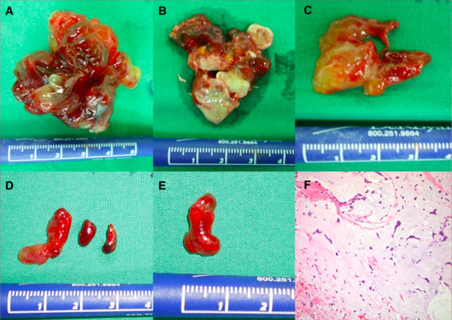
Surgical gross pathology specimens (A, B, C, D, E) and histologic (F) findings of bilateral atrial myxomas
A: myxoma from left atrium
B: myxoma from right atrium
C: embolic myxoma from aortic bifurcation
D: embolic myxoma from left popliteal artery
E: embolic myxoma from right popliteal artery
F: histologic findings of myxoma (H&E (hematoxylin and eosin) x400): characteristic stellate tumor cells within an abundant myxoid stroma
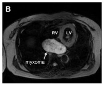
Magnetic resonance image of large biatrial myxoma
Image: “The large tumor” by Yuki Saito et al. License: CC BY 2.0, edited by Lecturio.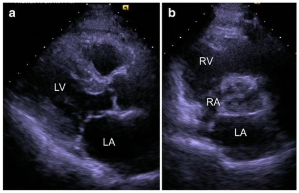
Transthoracic echocardiogram showing a: no mass; post myxoma resection view; b: cardiac myxoma (mass) arising from left atrium (pre-resection)
Image: “Fig. 2” by Yuki Saito et al. License: CC BY 4.0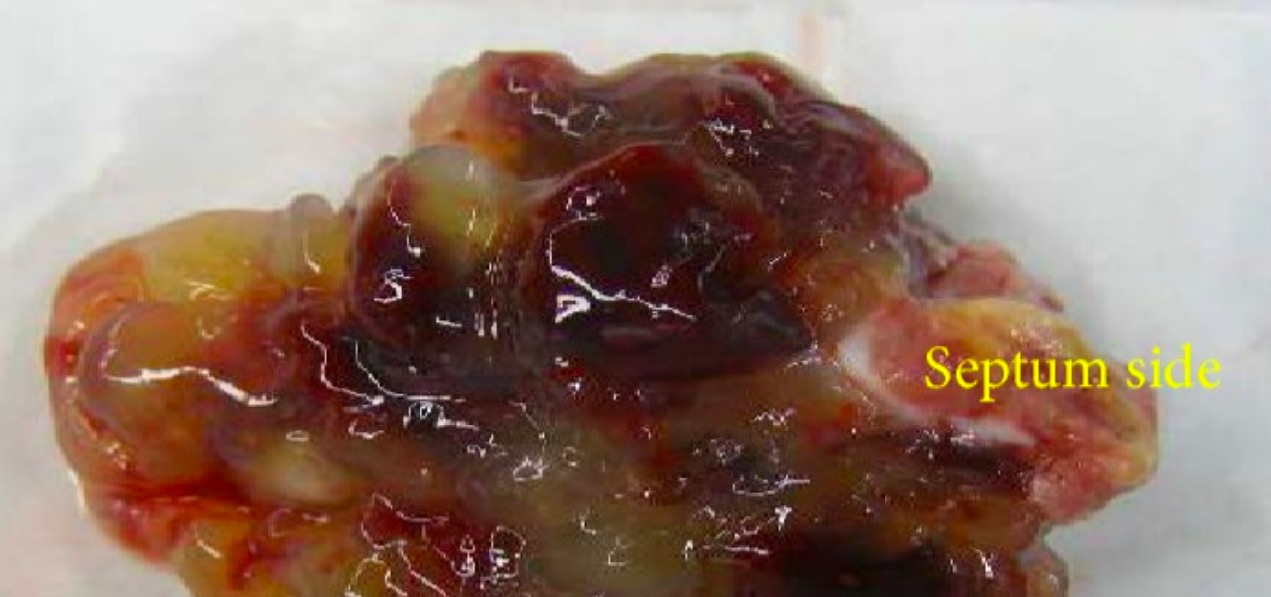
Gross pathology of excised left atrial myxoma, measuring 7 × 6 × 4 cm: Note the translucency of the tumor, due to its abundant content of acid mucopolysaccharide ground substance.
Image: “Gross pathology” by Department of Neurology, Hospital of International University of Health and Welfare, 537-3 Iguchi, Nasushiobara, Tochigi 329-2763, Japan. License: CC BY 3.0