The prenatal period begins with the formation of the embryo Embryo The entity of a developing mammal, generally from the cleavage of a zygote to the end of embryonic differentiation of basic structures. For the human embryo, this represents the first two months of intrauterine development preceding the stages of the fetus. Fertilization and First Week and continues through the development of the fetus, terminating with birth. Neonatal physiology during prenatal life differs significantly from that during postnatal life. Before birth, nutrient, gas exchange Gas exchange Human cells are primarily reliant on aerobic metabolism. The respiratory system is involved in pulmonary ventilation and external respiration, while the circulatory system is responsible for transport and internal respiration. Pulmonary ventilation (breathing) represents movement of air into and out of the lungs. External respiration, or gas exchange, is represented by the O2 and CO2 exchange between the lungs and the blood. Gas Exchange, and elimination Elimination The initial damage and destruction of tumor cells by innate and adaptive immunity. Completion of the phase means no cancer growth. Cancer Immunotherapy of waste products occur via the placenta Placenta A highly vascularized mammalian fetal-maternal organ and major site of transport of oxygen, nutrients, and fetal waste products. It includes a fetal portion (chorionic villi) derived from trophoblasts and a maternal portion (decidua) derived from the uterine endometrium. The placenta produces an array of steroid, protein and peptide hormones (placental hormones). Placenta, Umbilical Cord, and Amniotic Cavity. The fetus receives oxygenated, nutrient-rich blood via the umbilical vein, and deoxygenated blood is returned to the placenta Placenta A highly vascularized mammalian fetal-maternal organ and major site of transport of oxygen, nutrients, and fetal waste products. It includes a fetal portion (chorionic villi) derived from trophoblasts and a maternal portion (decidua) derived from the uterine endometrium. The placenta produces an array of steroid, protein and peptide hormones (placental hormones). Placenta, Umbilical Cord, and Amniotic Cavity for removal of waste via the umbilical arteries Arteries Arteries are tubular collections of cells that transport oxygenated blood and nutrients from the heart to the tissues of the body. The blood passes through the arteries in order of decreasing luminal diameter, starting in the largest artery (the aorta) and ending in the small arterioles. Arteries are classified into 3 types: large elastic arteries, medium muscular arteries, and small arteries and arterioles. Arteries: Histology. The 3 shunts that help redirect the fetal circulation Circulation The movement of the blood as it is pumped through the cardiovascular system. ABCDE Assessment are the ductus venosus Ductus venosus Development of the Heart, foramen ovale Foramen ovale An opening in the wall between the right and the left upper chambers (heart atria) of a fetal heart. Oval foramen normally closes soon after birth; when it fails to close the condition is called patent oval foramen. Patent Foramen Ovale, and ductus arteriosus Ductus arteriosus A fetal blood vessel connecting the pulmonary artery with the descending aorta. Patent Ductus Arteriosus (PDA). These shunts close after birth, leaving behind vestigial remnants. Postnatally, the fetal circulatory system and organ systems adapt to the extrauterine environment. Placental blood supply is cut off, causing the neonate Neonate An infant during the first 28 days after birth. Physical Examination of the Newborn to make adaptive changes.
Last updated: Jun 30, 2026
| Gestational age Gestational age The age of the conceptus, beginning from the time of fertilization. In clinical obstetrics, the gestational age is often estimated as the time from the last day of the last menstruation which is about 2 weeks before ovulation and fertilization. Pregnancy: Diagnosis, Physiology, and Care in weeks | Characteristics |
|---|---|
| Weeks 3–4 |
|
| Weeks 5–8 |
|
| Weeks 9–12 |
|
| Weeks 13–16 |
|
| Weeks 17–20 |
|
| Weeks 21–24 |
|
| Weeks 25–28 |
|
| Weeks 29–32 |
|
| Weeks 33–36 |
|
| Weeks 37–40 |
|

Embryological milestones during the prenatal period
Image by Lecturio.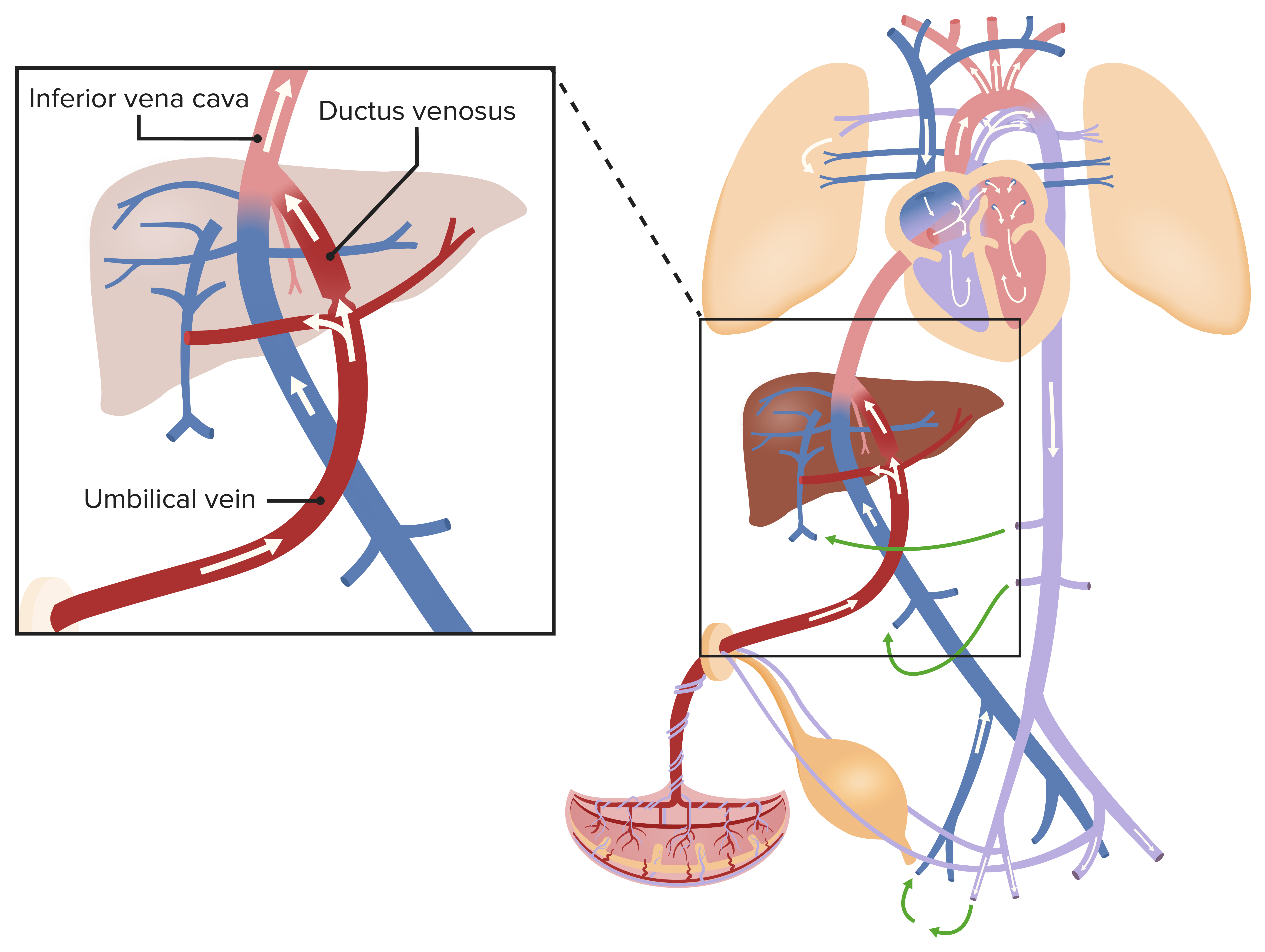
Fetal circulation:
Oxygenated blood from the umbilical vein drains into the inferior vena cava (IVC), which also collects the blood from the organs and lower portion of the body, causing the blood to become slightly deoxygenated.

Fetal circulation in the heart:
Blood from the inferior vena cava (IVC) is preferentially directed through the foramen ovale and flows into the left side of the heart, where it mixes in the left atrium (LA) with a tiny amount of deoxygenated blood from the pulmonary veins. The blood leaves the left side of the heart through the aorta and flows to the iliac arteries, which are connected to the placenta by the umbilical arteries. Blood from the superior vena cava (SVC) is preferentially directed downwards into the right ventricle (RV) and then to the pulmonary trunk, where most of it passes directly into the descending aorta through the ductus arteriosus. A small amount of blood from the pulmonary trunk passes through the lungs, is deoxygenated, and returns to the LA where it mixes with blood coming across the foramen ovale.
| Structure | Remnants |
|---|---|
| Umbilical arteries Arteries Arteries are tubular collections of cells that transport oxygenated blood and nutrients from the heart to the tissues of the body. The blood passes through the arteries in order of decreasing luminal diameter, starting in the largest artery (the aorta) and ending in the small arterioles. Arteries are classified into 3 types: large elastic arteries, medium muscular arteries, and small arteries and arterioles. Arteries: Histology |
|
| Umbilical vein |
|
| Ductus venosus Ductus venosus Development of the Heart |
|
| Ductus arteriosus Ductus arteriosus A fetal blood vessel connecting the pulmonary artery with the descending aorta. Patent Ductus Arteriosus (PDA) |
|
| Foramen ovale Foramen ovale An opening in the wall between the right and the left upper chambers (heart atria) of a fetal heart. Oval foramen normally closes soon after birth; when it fails to close the condition is called patent oval foramen. Patent Foramen Ovale |
|
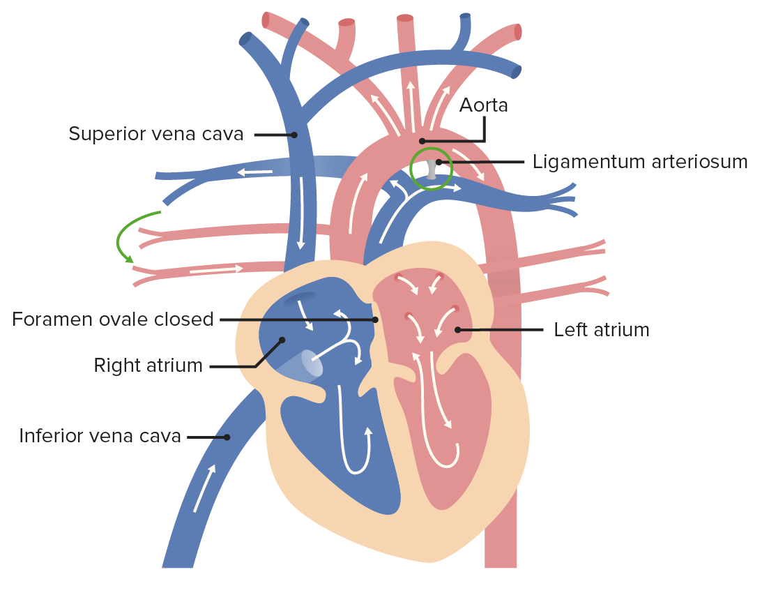
After birth, the ductus arteriosus becomes the ligamentum arteriosum, as blood is able to enter the lungs to be oxygenated. The foramen ovale closes and becomes the fossa ovalis due to increased pressure in the left atrium (LA).
Image by Lecturio.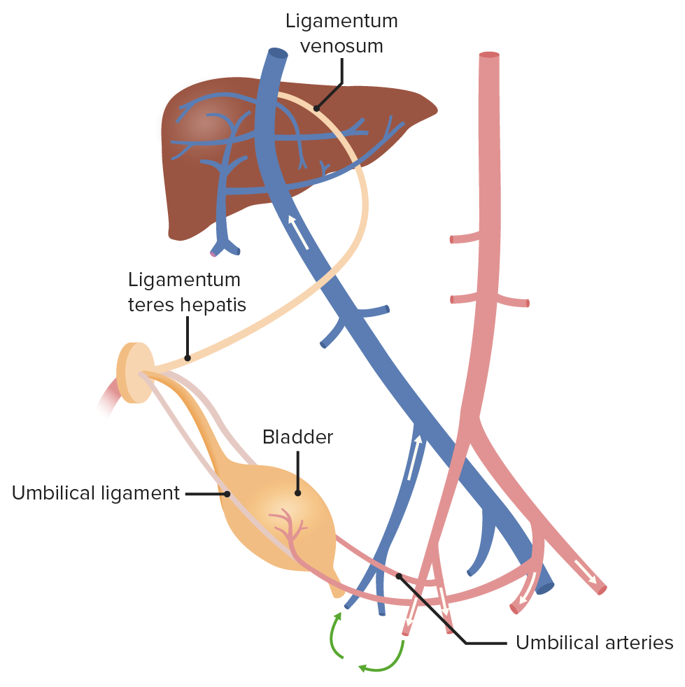
After birth, the umbilical arteries become the medial umbilical ligaments, while the umbilical vein becomes the ligamentum teres hepatis. The ductus venosus becomes the ligamentum venosum.
Image by Lecturio.Fetal metabolic rates are relatively low and must significantly increase to meet the metabolic needs of a growing neonate Neonate An infant during the first 28 days after birth. Physical Examination of the Newborn after birth.
| Organ system | Before birth | After birth |
|---|---|---|
| Respiratory system |
|
|
| Liver Liver The liver is the largest gland in the human body. The liver is found in the superior right quadrant of the abdomen and weighs approximately 1.5 kilograms. Its main functions are detoxification, metabolism, nutrient storage (e.g., iron and vitamins), synthesis of coagulation factors, formation of bile, filtration, and storage of blood. Liver: Anatomy |
|
|
| Blood and immune system Immune system The body’s defense mechanism against foreign organisms or substances and deviant native cells. It includes the humoral immune response and the cell-mediated response and consists of a complex of interrelated cellular, molecular, and genetic components. Primary Lymphatic Organs |
|
|
| GI tract |
|
|
| Urinary system |
|
|
| Thermoregulation Thermoregulation Body temperature can be divided into external temperature, which involves the skin, and core temperature, which involves the CNS and viscera. While external temperature can be variable, the core temperature is maintained within a narrow range of 36.5-37.5ºC (97.7-99.5ºF). Body Temperature Regulation system |
|
|