Achieve Mastery of Medical Concepts

Study for medical school and boards with Lecturio

Patent Foramen Ovale

A patent foramen ovale (PFO) is an abnormal communication Communication The exchange or transmission of ideas, attitudes, or beliefs between individuals or groups. Decision-making Capacity and Legal Competence between the atria that persists after birth. The condition results from incomplete closure of the foramen ovale. Small, isolated, and asymptomatic PFOs are a common incidental finding on echocardiography Echocardiography Ultrasonic recording of the size, motion, and composition of the heart and surrounding tissues. The standard approach is transthoracic. Tricuspid Valve Atresia (TVA) and require no treatment. Larger PFOs and PFOs associated with paradoxical thromboembolic stroke or other cardiac anomalies may require treatment with anticoagulation Anticoagulation Pulmonary Hypertension Drugs. Surgical or percutaneous closure may be indicated in select cases.

Last updated: Sep 26, 2022

Editorial responsibility: Stanley Oiseth, Lindsay Jones, Evelin Maza

Overview

Definition

Patent foramen ovale (PFO) is a persistent, post-natal communication Communication The exchange or transmission of ideas, attitudes, or beliefs between individuals or groups. Decision-making Capacity and Legal Competence between the atria due to the failure of the closure of the foramen ovale during the transition from in-utero to ex-utero neonatal circulation Circulation The movement of the blood as it is pumped through the cardiovascular system. ABCDE Assessment.

Epidemiology

  • Autopsy studies show a prevalence Prevalence The total number of cases of a given disease in a specified population at a designated time. It is differentiated from incidence, which refers to the number of new cases in the population at a given time. Measures of Disease Frequency rate of approximately 27% in the general population.
  • Often associated with other cardiac anomalies:
    • Atrial septal aneurysm Aneurysm An aneurysm is a bulging, weakened area of a blood vessel that causes an abnormal widening of its diameter > 1.5 times the size of the native vessel. Aneurysms occur more often in arteries than in veins and are at risk of dissection and rupture, which can be life-threatening. Thoracic Aortic Aneurysms
    • Atrial septal defect Atrial Septal Defect Atrial septal defects (ASDs) are benign acyanotic congenital heart defects characterized by an opening in the interatrial septum that causes blood to flow from the left atrium (LA) to the right atrium (RA) (left-to-right shunt). Atrial Septal Defect (ASD) ( ASD ASD Autism spectrum disorder (ASD) is a neurodevelopmental disorder marked by poor social skills, restricted interests/social interactions, and repetitive/stereotyped behaviors. The condition is termed a “spectrum” because of the wide variability in the severity of symptoms exhibited. Autism Spectrum Disorder)
    • Eustachian valve: located at the junction of the inferior vena cava Inferior vena cava The venous trunk which receives blood from the lower extremities and from the pelvic and abdominal organs. Mediastinum and Great Vessels: Anatomy and right atrium
    • Ebstein’s anomaly (EA): congenital Congenital Chorioretinitis apical displacement Displacement The process by which an emotional or behavioral response that is appropriate for one situation appears in another situation for which it is inappropriate. Defense Mechanisms of the septal and posterior tricuspid valve Tricuspid valve The valve consisting of three cusps situated between the right atrium and right ventricle of the heart. Heart: Anatomy leaflets and an abnormal anterior leaflet

Embryology

Normal closure of foramen ovale and patent foramen ovale

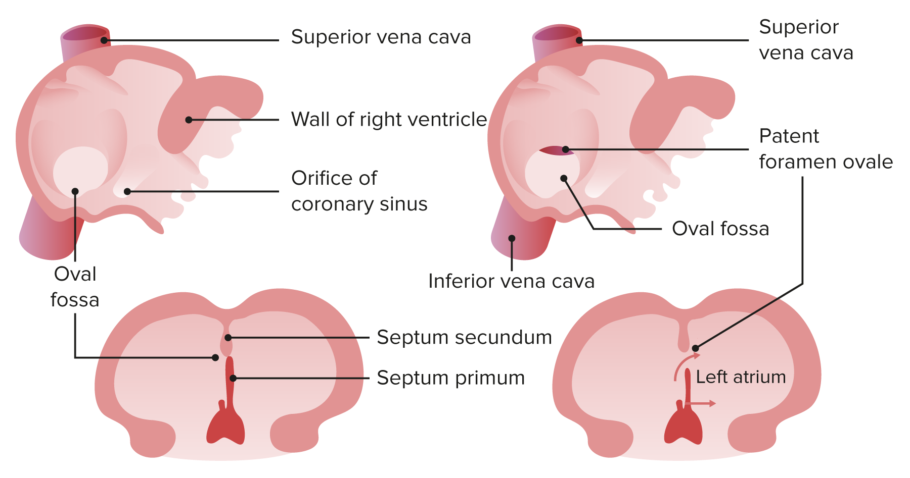
Normal closure of the foramen ovale, and a patent foramen ovale

Image by Lecturio.
Heart before and after birth

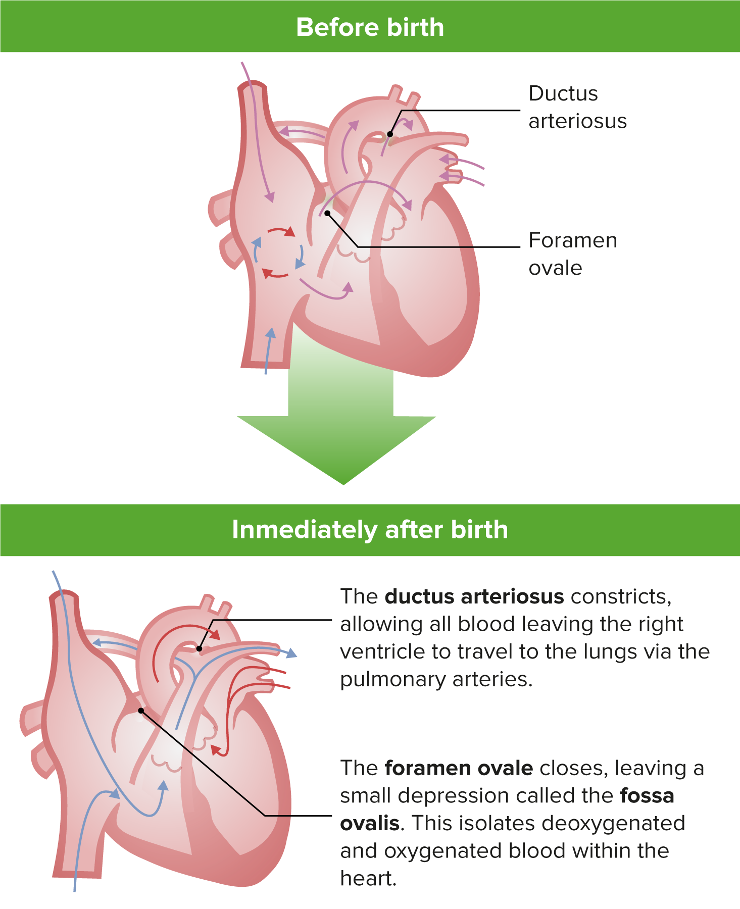
Normal transition from in-utero cardiac circulation to ex-utero cardiac circulation

Image by Lecturio.

Clinical Presentation and Imaging

Isolated PFO

  • Frequently asymptomatic
  • Cryptogenic strokes:
    • Occurring in the absence of blood clots in the heart
    • Caused by an embolus that passes from the right to the left side of the heart without going through the natural filter of the lung vasculature (paradoxical embolus)
    • Present with signs of transient ischemic attack Transient ischemic attack Transient ischemic attack (TIA) is a temporary episode of neurologic dysfunction caused by ischemia without infarction that resolves completely when blood supply is restored. Transient ischemic attack is a neurologic emergency that warrants urgent medical attention. Transient Ischemic Attack (TIA) or ischemic stroke Ischemic Stroke An ischemic stroke (also known as cerebrovascular accident) is an acute neurologic injury that occurs as a result of brain ischemia; this condition may be due to cerebral blood vessel occlusion by thrombosis or embolism, or rarely due to systemic hypoperfusion. Ischemic Stroke including headache Headache The symptom of pain in the cranial region. It may be an isolated benign occurrence or manifestation of a wide variety of headache disorders. Brain Abscess, hemisensory deficits, paresis Paresis A general term referring to a mild to moderate degree of muscular weakness, occasionally used as a synonym for paralysis (severe or complete loss of motor function). In the older literature, paresis often referred specifically to paretic neurosyphilis. ‘general paresis’ and ‘general paralysis’ may still carry that connotation. Bilateral lower extremity paresis is referred to as paraparesis. Spinal Disk Herniation, and ataxia Ataxia Impairment of the ability to perform smoothly coordinated voluntary movements. This condition may affect the limbs, trunk, eyes, pharynx, larynx, and other structures. Ataxia may result from impaired sensory or motor function. Sensory ataxia may result from posterior column injury or peripheral nerve diseases. Motor ataxia may be associated with cerebellar diseases; cerebral cortex diseases; thalamic diseases; basal ganglia diseases; injury to the red nucleus; and other conditions. Ataxia-telangiectasia
  • Vascular headaches:
    • Caused by hypo-oxygenation of the brain Brain The part of central nervous system that is contained within the skull (cranium). Arising from the neural tube, the embryonic brain is comprised of three major parts including prosencephalon (the forebrain); mesencephalon (the midbrain); and rhombencephalon (the hindbrain). The developed brain consists of cerebrum; cerebellum; and other structures in the brain stem. Nervous System: Anatomy, Structure, and Classification 
    • Due to right-to-left shunting of oxygenated blood through the PFO
  • Less common manifestations:
    • Decompression sickness with nitrogen Nitrogen An element with the atomic symbol n, atomic number 7, and atomic weight [14. 00643; 14. 00728]. Nitrogen exists as a diatomic gas and makes up about 78% of the earth’s atmosphere by volume. It is a constituent of proteins and nucleic acids and found in all living cells. Urea Cycle gas embolism when emerging from deep-sea scuba diving 
    • Myocardial or other systemic infarctions

Patent foramen ovale associated with other cardiac anomalies

  • Often associated with pathological processes that cause elevated pulmonary vascular pressure:
    • Pulmonary stenosis Stenosis Hypoplastic Left Heart Syndrome (HLHS)
    • Tricuspid valve Tricuspid valve The valve consisting of three cusps situated between the right atrium and right ventricle of the heart. Heart: Anatomy abnormalities
    • Pulmonary hypertension Pulmonary Hypertension Pulmonary hypertension (PH) or pulmonary arterial hypertension (PAH) is characterized by elevated pulmonary arterial pressure, which can lead to chronic progressive right heart failure. Pulmonary hypertension is grouped into 5 categories based on etiology, which include primary PAH, and PH due to cardiac disease, lung or hypoxic disease, chronic thromboembolic disease, and multifactorial or unclear etiologies. Pulmonary Hypertension 
  • Atrial septum aneurysm Aneurysm An aneurysm is a bulging, weakened area of a blood vessel that causes an abnormal widening of its diameter > 1.5 times the size of the native vessel. Aneurysms occur more often in arteries than in veins and are at risk of dissection and rupture, which can be life-threatening. Thoracic Aortic Aneurysms: aneurysm Aneurysm An aneurysm is a bulging, weakened area of a blood vessel that causes an abnormal widening of its diameter > 1.5 times the size of the native vessel. Aneurysms occur more often in arteries than in veins and are at risk of dissection and rupture, which can be life-threatening. Thoracic Aortic Aneurysms of the membrane of the fossa ovalis Fossa ovalis Prenatal and Postnatal Physiology of the Neonate
  • EA:

Diagnostic imaging

  • Color-flow Doppler Doppler Ultrasonography applying the doppler effect, with frequency-shifted ultrasound reflections produced by moving targets (usually red blood cells) in the bloodstream along the ultrasound axis in direct proportion to the velocity of movement of the targets, to determine both direction and velocity of blood flow. Ultrasound (Sonography) echocardiography Echocardiography Ultrasonic recording of the size, motion, and composition of the heart and surrounding tissues. The standard approach is transthoracic. Tricuspid Valve Atresia (TVA) is commonly used to identify PFOs.
  • Contrast-enhanced 2-dimensional 2-Dimensional Imaging of the Heart and Great Vessels or 3-dimensional transesophageal echocardiography Echocardiography Ultrasonic recording of the size, motion, and composition of the heart and surrounding tissues. The standard approach is transthoracic. Tricuspid Valve Atresia (TVA) may identify small PFOs.
Pfo ultrasound

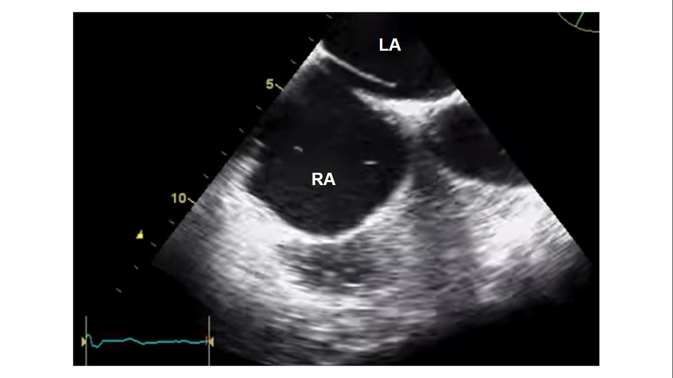
Transesophageal echocardiogram showing a patent foramen ovale in a patient who presented with cryptogenic ischemic stroke. The patent foramen is observed in the septum between the right atrium (RA) and left atrium (LA).

Image: “TEE showing a PFO in 62°” by Knebel, F. at al. License: CC BY 2.0, screenshot edited by Lecturio.

Management

  • Isolated asymptomatic PFO: no treatment indicated
  • Isolated PFO associated with an otherwise unexplained stroke:
    • All patients Patients Individuals participating in the health care system for the purpose of receiving therapeutic, diagnostic, or preventive procedures. Clinician–Patient Relationship: lifestyle modifications aimed at reducing heart disease and risk of strokes including diet, exercise, and smoking Smoking Willful or deliberate act of inhaling and exhaling smoke from burning substances or agents held by hand. Interstitial Lung Diseases cessation
    • Low risk for recurrent stroke: antiplatelet agents Antiplatelet agents Antiplatelet agents are medications that inhibit platelet aggregation, a critical step in the formation of the initial platelet plug. Abnormal, or inappropriate, platelet aggregation is a key step in the pathophysiology of arterial ischemic events. The primary categories of antiplatelet agents include aspirin, ADP inhibitors, phosphodiesterase/adenosine uptake inhibitors, and glycoprotein IIb/IIIa inhibitors. Antiplatelet Drugs (e.g., aspirin Aspirin The prototypical analgesic used in the treatment of mild to moderate pain. It has anti-inflammatory and antipyretic properties and acts as an inhibitor of cyclooxygenase which results in the inhibition of the biosynthesis of prostaglandins. Aspirin also inhibits platelet aggregation and is used in the prevention of arterial and venous thrombosis. Nonsteroidal Antiinflammatory Drugs (NSAIDs), clopidogrel Clopidogrel A ticlopidine analog and platelet purinergic p2y receptor antagonist that inhibits adenosine diphosphate-mediated platelet aggregation. It is used to prevent thromboembolism in patients with arterial occlusive diseases; myocardial infarction; stroke; or atrial fibrillation. Antiplatelet Drugs)
    • High risk for recurrent stroke: aspirin Aspirin The prototypical analgesic used in the treatment of mild to moderate pain. It has anti-inflammatory and antipyretic properties and acts as an inhibitor of cyclooxygenase which results in the inhibition of the biosynthesis of prostaglandins. Aspirin also inhibits platelet aggregation and is used in the prevention of arterial and venous thrombosis. Nonsteroidal Antiinflammatory Drugs (NSAIDs) + warfarin Warfarin An anticoagulant that acts by inhibiting the synthesis of vitamin K-dependent coagulation factors. Warfarin is indicated for the prophylaxis and/or treatment of venous thrombosis and its extension, pulmonary embolism, and atrial fibrillation with embolization. It is also used as an adjunct in the prophylaxis of systemic embolism after myocardial infarction. Warfarin is also used as a rodenticide. Anticoagulants
  • Open surgical closure or percutaneous closure: indicated in patients Patients Individuals participating in the health care system for the purpose of receiving therapeutic, diagnostic, or preventive procedures. Clinician–Patient Relationship with recurrent stroke, large PFOs, contraindications Contraindications A condition or factor associated with a recipient that makes the use of a drug, procedure, or physical agent improper or inadvisable. Contraindications may be absolute (life threatening) or relative (higher risk of complications in which benefits may outweigh risks). Noninvasive Ventilation to anticoagulant therapy, etc ETC The electron transport chain (ETC) sends electrons through a series of proteins, which generate an electrochemical proton gradient that produces energy in the form of adenosine triphosphate (ATP). Electron Transport Chain (ETC).

References

  1. Snyder, B., & Neuman, T. (2016). Diving disorders. In J. E. Tintinalli et al. Tintinalli’s emergency medicine: A comprehensive study guide, 8e. New York, NY: McGraw-Hill Education. accessmedicine.mhmedical.com/content.aspx?aid=1121493927
  2. Therrien, J., & Marelli, A. J. (2020). Congenital heart disease in adults. In L. Goldman MD, & A. I. Schafer MD (Eds.), Goldman-Cecil medicine (pp. 35-365.e4) https://www.clinicalkey.es/#!/content/3-s2.0-B9780323532662000618
  3. Suradi, Hussam S., MD, et al. (2016). Patent foramen ovale. Cardiology Clinics, 34(2), 231-240. doi:http://dx.doi.org/10.1016/j.ccl.2015.12.001
  4. Geva, T., Prof, Martins, J. D., M.D., & Wald, R. M., M.D. (2014). Atrial septal defects. Lancet, The, 383(9932), 1921-1932. doi:http://dx.doi.org/10.1016/S0140-6736(13)62145-5
  5. Dattilo, P. B., M.D., Kim, M. S., M.D., & Carroll, J. D., M.D. (2013). Patent foramen ovale. Cardiology Clinics, 31(3), 401-415. doi:http://dx.doi.org/10.1016/j.ccl.2013.05.002

Create your free account or log in to continue reading!

Sign up now and get free access to Lecturio with concept pages, medical videos, and questions for your medical education.

User Reviews

Details