The primary lymphoid organs, also referred to as central lymphoid/lymphatic organs, are the tissues responsible for the production of lymphoid cells from progenitor cells, incuding the bone marrow Bone marrow The soft tissue filling the cavities of bones. Bone marrow exists in two types, yellow and red. Yellow marrow is found in the large cavities of large bones and consists mostly of fat cells and a few primitive blood cells. Red marrow is a hematopoietic tissue and is the site of production of erythrocytes and granular leukocytes. Bone marrow is made up of a framework of connective tissue containing branching fibers with the frame being filled with marrow cells. Bone Marrow: Composition and Hematopoiesis and the thymus Thymus A single, unpaired primary lymphoid organ situated in the mediastinum, extending superiorly into the neck to the lower edge of the thyroid gland and inferiorly to the fourth costal cartilage. It is necessary for normal development of immunologic function early in life. By puberty, it begins to involute and much of the tissue is replaced by fat. Lymphatic Drainage System: Anatomy. In the bone marrow Bone marrow The soft tissue filling the cavities of bones. Bone marrow exists in two types, yellow and red. Yellow marrow is found in the large cavities of large bones and consists mostly of fat cells and a few primitive blood cells. Red marrow is a hematopoietic tissue and is the site of production of erythrocytes and granular leukocytes. Bone marrow is made up of a framework of connective tissue containing branching fibers with the frame being filled with marrow cells. Bone Marrow: Composition and Hematopoiesis, hematopoietic stem cells Hematopoietic stem cells Progenitor cells from which all blood cells derived. They are found primarily in the bone marrow and also in small numbers in the peripheral blood. Bone Marrow: Composition and Hematopoiesis progress to become oligopotent progenitors Oligopotent progenitors Bone Marrow: Composition and Hematopoiesis (in the case of lymphocytes Lymphocytes Lymphocytes are heterogeneous WBCs involved in immune response. Lymphocytes develop from the bone marrow, starting from hematopoietic stem cells (HSCs) and progressing to common lymphoid progenitors (CLPs). B and T lymphocytes and natural killer (NK) cells arise from the lineage. Lymphocytes: Histology, the common lymphoid progenitor). B lymphocytes B lymphocytes Lymphoid cells concerned with humoral immunity. They are short-lived cells resembling bursa-derived lymphocytes of birds in their production of immunoglobulin upon appropriate stimulation. B cells: Types and Functions stay and undergo processes for differentiation before migrating to the secondary lymphoid organs (such as lymph nodes Lymph Nodes They are oval or bean shaped bodies (1 - 30 mm in diameter) located along the lymphatic system. Lymphatic Drainage System: Anatomy). The progenitor cells that are to become T lymphocytes T lymphocytes Lymphocytes responsible for cell-mediated immunity. Two types have been identified - cytotoxic (t-lymphocytes, cytotoxic) and helper T-lymphocytes (t-lymphocytes, helper-inducer). They are formed when lymphocytes circulate through the thymus gland and differentiate to thymocytes. When exposed to an antigen, they divide rapidly and produce large numbers of new T cells sensitized to that antigen. T cells: Types and Functions proceed to the thymus Thymus A single, unpaired primary lymphoid organ situated in the mediastinum, extending superiorly into the neck to the lower edge of the thyroid gland and inferiorly to the fourth costal cartilage. It is necessary for normal development of immunologic function early in life. By puberty, it begins to involute and much of the tissue is replaced by fat. Lymphatic Drainage System: Anatomy for further maturation.
Last updated: Dec 15, 2025
The lymphatic system ( lymph Lymph The interstitial fluid that is in the lymphatic system. Secondary Lymphatic Organs vessels, lymph Lymph The interstitial fluid that is in the lymphatic system. Secondary Lymphatic Organs fluid, and lymphoid organs) is part of the body’s immune system.
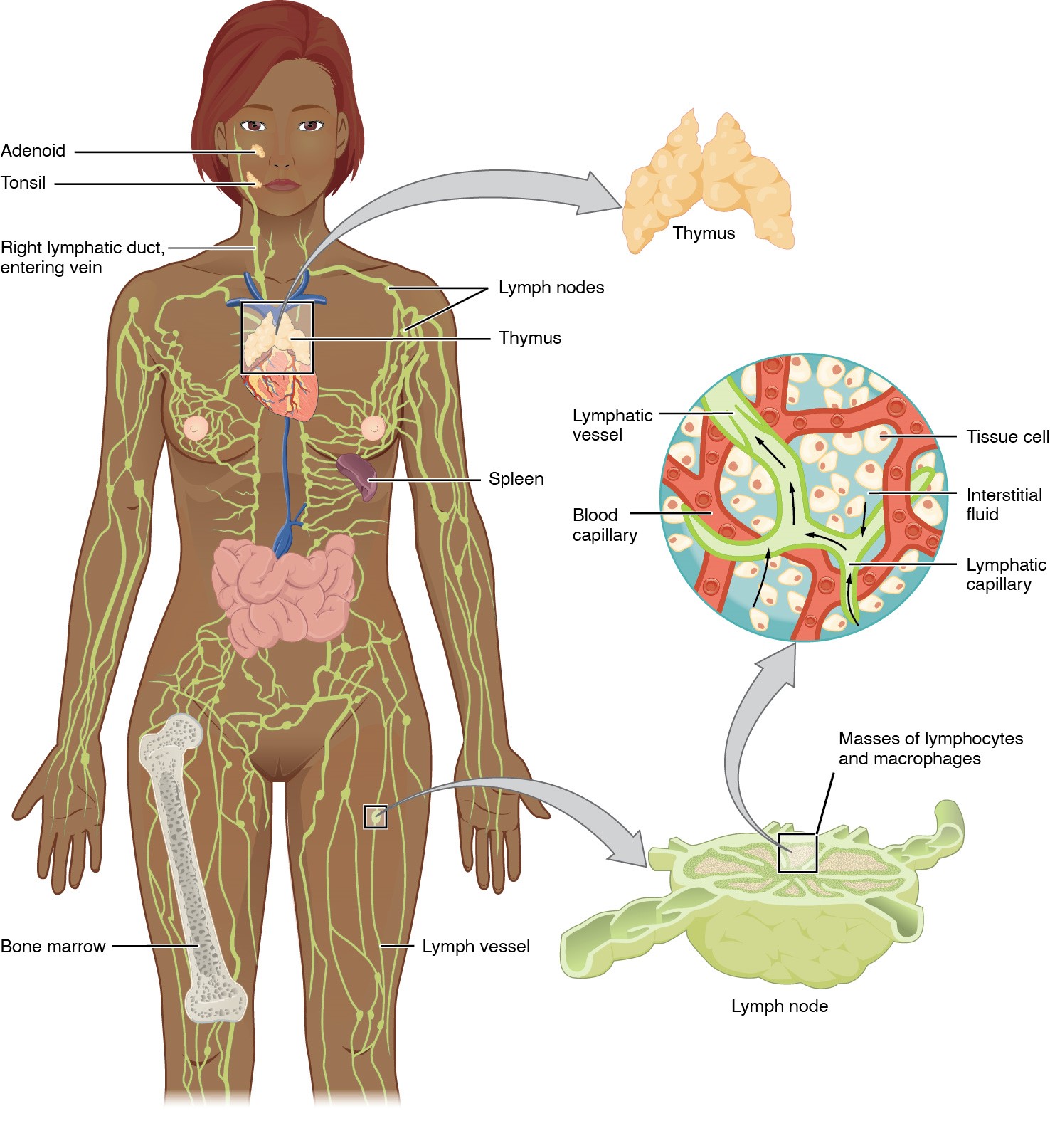
Anatomy of the lymphatic system:
Includes the primary (bone marrow, thymus) and secondary (spleen, lymph nodes, and MALT) lymphoid organs
Lymphatic vessels convey lymph to the larger lymphatic vessels in the torso, transporting fluid back to the venous circulation.
Bone marrow Bone marrow The soft tissue filling the cavities of bones. Bone marrow exists in two types, yellow and red. Yellow marrow is found in the large cavities of large bones and consists mostly of fat cells and a few primitive blood cells. Red marrow is a hematopoietic tissue and is the site of production of erythrocytes and granular leukocytes. Bone marrow is made up of a framework of connective tissue containing branching fibers with the frame being filled with marrow cells. Bone Marrow: Composition and Hematopoiesis is the spongy tissue found in the medullary canals Medullary canals Bone Marrow: Composition and Hematopoiesis of long bones Long bones Length greater than width. Bones: Structure and Types and cavities of the cancellous bones.
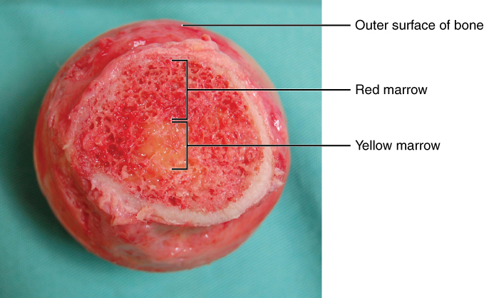
Bone marrow:
Cross section showing the red and yellow marrow of the bone
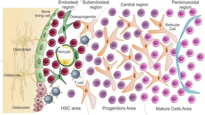
Schematic representation of the bone marrow microenvironment:
The hematopoietic stem cell (HSC) area contains the HSCs and uncommitted progenitors. These cells are in close association with endosteal osteoblasts and bone-lining cells. As HSCs change from quiescence to proliferative states, they migrate and colonize the subendosteal region and then the central region of the marrow (progenitors).
Differentiated cells reach the mature area, closer to the sinusoids. To be released into circulation, these mature cells pass through the endothelium (lining the sinusoids) by transcellular migration.
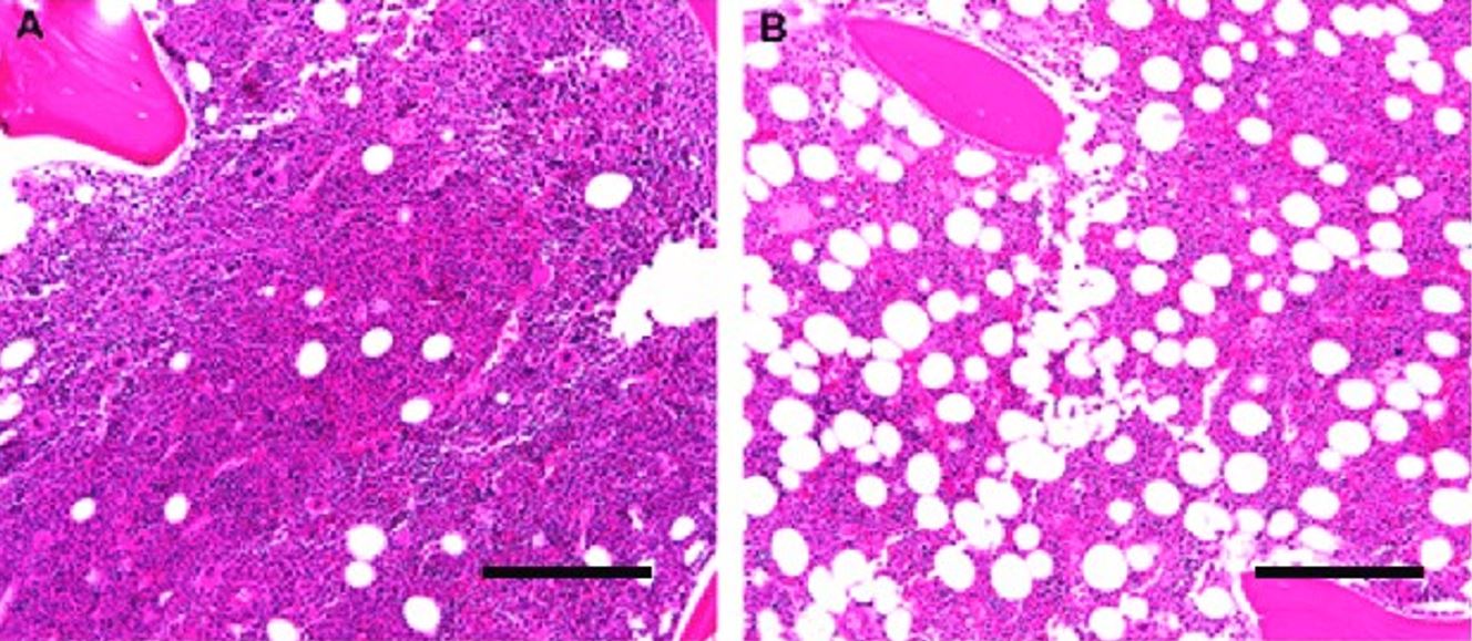
Human bone marrow histologic architecture:
Images show the different cells and structures that are in the marrow.
A: A bone marrow biopsy from a 5-year-old child is > 90% cellular, with a predominance of trilineage hematopoiesis and little admixed adipose tissue.
B: A bone marrow biopsy from a 60-year-old adult is composed of 50% hematopoietic elements and 50% admixed mature adipose tissue.
A and B: Original magnification 10×; scale bar = 100 μm
The main function of the thymus Thymus A single, unpaired primary lymphoid organ situated in the mediastinum, extending superiorly into the neck to the lower edge of the thyroid gland and inferiorly to the fourth costal cartilage. It is necessary for normal development of immunologic function early in life. By puberty, it begins to involute and much of the tissue is replaced by fat. Lymphatic Drainage System: Anatomy is to act as the site of T-cell differentiation and maturation (from the bone marrow Bone marrow The soft tissue filling the cavities of bones. Bone marrow exists in two types, yellow and red. Yellow marrow is found in the large cavities of large bones and consists mostly of fat cells and a few primitive blood cells. Red marrow is a hematopoietic tissue and is the site of production of erythrocytes and granular leukocytes. Bone marrow is made up of a framework of connective tissue containing branching fibers with the frame being filled with marrow cells. Bone Marrow: Composition and Hematopoiesis, progenitor cells go to the thymus Thymus A single, unpaired primary lymphoid organ situated in the mediastinum, extending superiorly into the neck to the lower edge of the thyroid gland and inferiorly to the fourth costal cartilage. It is necessary for normal development of immunologic function early in life. By puberty, it begins to involute and much of the tissue is replaced by fat. Lymphatic Drainage System: Anatomy).
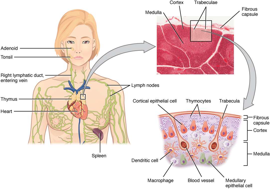
The thymus:
A primary lymphoid organ located in the anterosuperior mediastinum.
The architecture consists of an outer capsule with lobules separated by trabeculae. Each lobule has an outer dark-staining cortex and a central, pale-staining medulla. The cortex contains immature T lymphocytes, which eventually migrate to the medulla as they mature. Antigen-presenting cells (dendritic cells and macrophages) are in the corticomedullary junction.
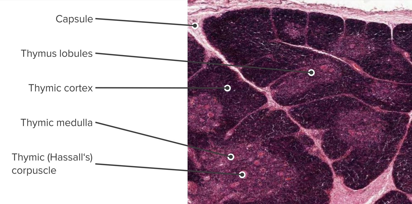
Histologic section showing the structure of a thymus
Image by Geoffrey Meyer, edited by Lecturio.