Advertisement

Advertisement

Advertisement

Advertisement

Diaphragm: Anatomy

The diaphragm is a large, dome-shaped muscle that separates the thoracic cavity from the abdominal cavity. The diaphragm consists of muscle fibers and a large central tendon, which is divided into right and left parts. As the primary muscle of inspiration Inspiration Ventilation: Mechanics of Breathing, the diaphragm contributes 75% of the total inspiratory muscle force, and its contraction leads to flattening of the dome of the diaphragm. This flattening increases the volume of the thoracic cavity and allows the lungs Lungs Lungs are the main organs of the respiratory system. Lungs are paired viscera located in the thoracic cavity and are composed of spongy tissue. The primary function of the lungs is to oxygenate blood and eliminate CO2. Lungs: Anatomy to expand during inspiration Inspiration Ventilation: Mechanics of Breathing.

Last updated: Jan 2, 2024

Editorial responsibility: Stanley Oiseth, Lindsay Jones, Evelin Maza

Advertisement

Advertisement

Advertisement

Advertisement

Advertisement

Advertisement

Advertisement

Advertisement

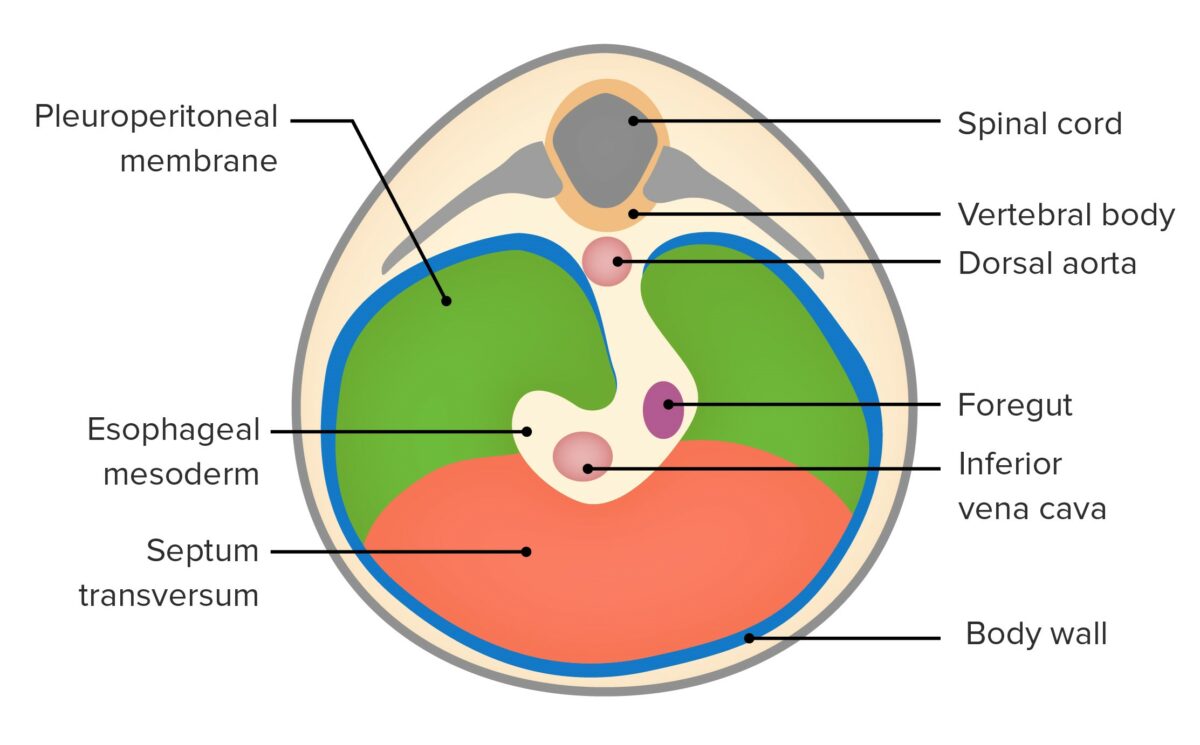
Embryology

  • Develops between the 4th and 7th weeks of gestation
  • Develops from fusion of the following parts:
    • Septum transversum: located in the central portion; becomes the central tendon
    • Pleuroperitoneal membranes: becomes the muscular portion
    • Mesentery Mesentery A layer of the peritoneum which attaches the abdominal viscera to the abdominal wall and conveys their blood vessels and nerves. Peritoneum: Anatomy of the esophagus Esophagus The esophagus is a muscular tube-shaped organ of around 25 centimeters in length that connects the pharynx to the stomach. The organ extends from approximately the 6th cervical vertebra to the 11th thoracic vertebra and can be divided grossly into 3 parts: the cervical part, the thoracic part, and the abdominal part. Esophagus: Anatomy
    • Mesoderm Mesoderm The middle germ layer of an embryo derived from three paired mesenchymal aggregates along the neural tube. Gastrulation and Neurulation of the body wall
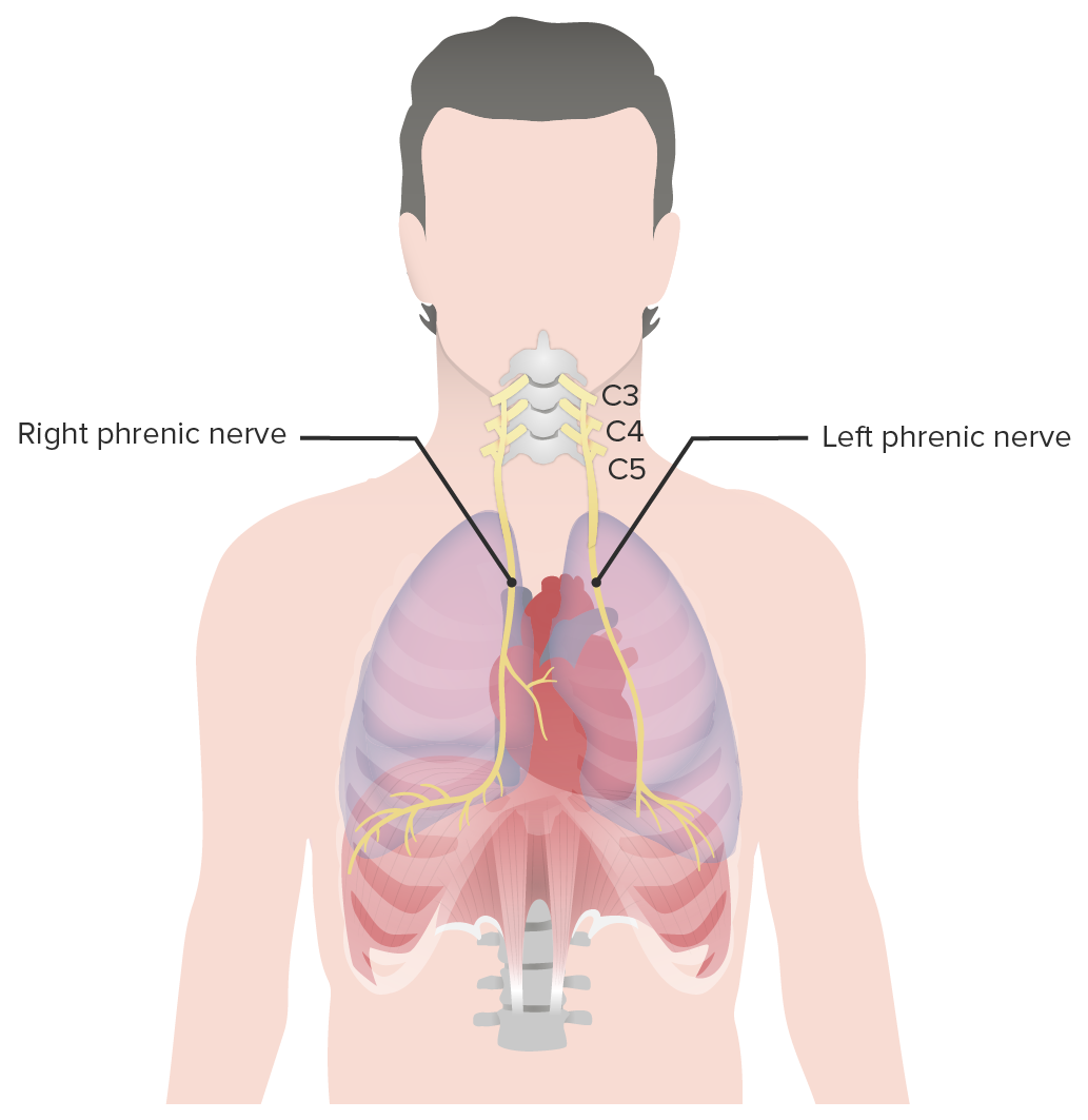
  • Septum transversum originates in the cervical region Cervical region Spinal Cord: Anatomy → picks up myoblasts Myoblasts Embryonic (precursor) cells of the myogenic lineage that develop from the mesoderm. They undergo proliferation, migrate to their various sites, and then differentiate into the appropriate form of myocytes (skeletal, cardiac, smooth muscle). Muscle Tissue: Histology, which are innervated by C3, C4, and C5
  • As the diaphragm moves caudally → drags innervation → becomes phrenic nerve
  • Myoblasts Myoblasts Embryonic (precursor) cells of the myogenic lineage that develop from the mesoderm. They undergo proliferation, migrate to their various sites, and then differentiate into the appropriate form of myocytes (skeletal, cardiac, smooth muscle). Muscle Tissue: Histology invade other components of the diaphragm after fusion → form diaphragm muscle
Developing diaphragm

Developing diaphragm:
Embryonic development of the diaphragm from the pleuroperitoneal membranes and the septum transversum.

Image by Lecturio.

Anatomy

Parts

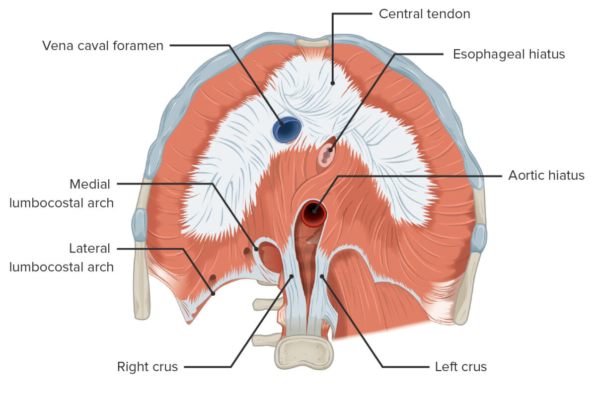
The diaphragm consists of 3 parts, all of which insert into the central tendon.

  1. Sternal: originates from the back of the xiphoid process Xiphoid process Chest Wall: Anatomy
  2. Costal: originates from cartilage Cartilage Cartilage is a type of connective tissue derived from embryonic mesenchyme that is responsible for structural support, resilience, and the smoothness of physical actions. Perichondrium (connective tissue membrane surrounding cartilage) compensates for the absence of vasculature in cartilage by providing nutrition and support. Cartilage: Histology and adjacent inner surfaces of the lower 6 ribs Ribs A set of twelve curved bones which connect to the vertebral column posteriorly, and terminate anteriorly as costal cartilage. Together, they form a protective cage around the internal thoracic organs. Chest Wall: Anatomy
  3. Lumbar: connects to the spine Spine The human spine, or vertebral column, is the most important anatomical and functional axis of the human body. It consists of 7 cervical vertebrae, 12 thoracic vertebrae, and 5 lumbar vertebrae and is limited cranially by the skull and caudally by the sacrum. Vertebral Column: Anatomy through:
    • Medial lumbocostal arch: connects diaphragm to L1
    • Lateral lumbocostal arch: connects diaphragm to L1 and 12th rib
    • Right crus: attaches to the L1, L2, and L3 vertebrae
    • Left crus: attaches to the L1 and L2 vertebrae

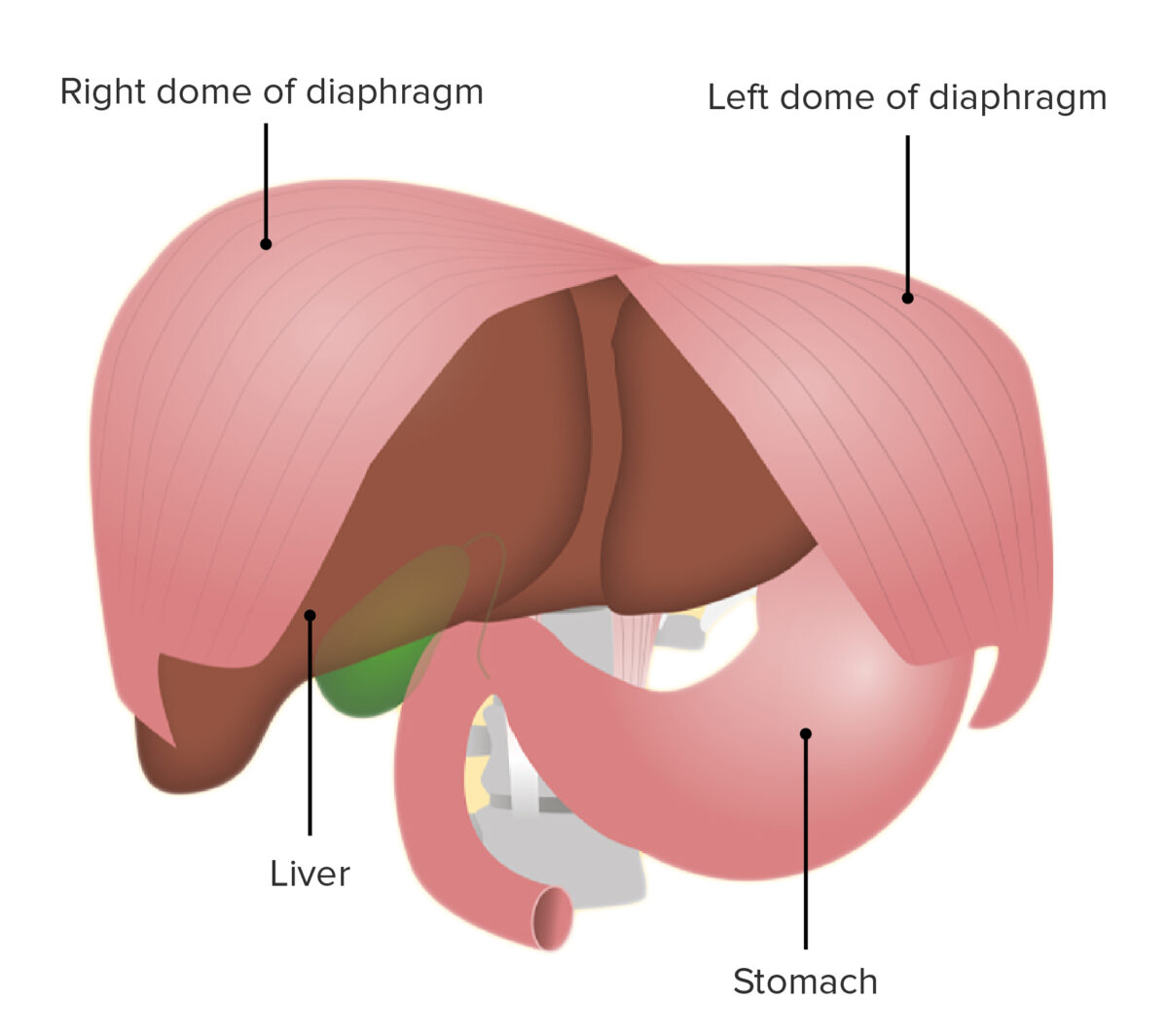
Domes

  • Right dome: positioned higher than the left because of the liver Liver The liver is the largest gland in the human body. The liver is found in the superior right quadrant of the abdomen and weighs approximately 1.5 kilograms. Its main functions are detoxification, metabolism, nutrient storage (e.g., iron and vitamins), synthesis of coagulation factors, formation of bile, filtration, and storage of blood. Liver: Anatomy underneath it
  • Left dome: positioned above the stomach Stomach The stomach is a muscular sac in the upper left portion of the abdomen that plays a critical role in digestion. The stomach develops from the foregut and connects the esophagus with the duodenum. Structurally, the stomach is C-shaped and forms a greater and lesser curvature and is divided grossly into regions: the cardia, fundus, body, and pylorus. Stomach: Anatomy

Surfaces

  • Thoracic surface: in contact with the pleura Pleura The pleura is a serous membrane that lines the walls of the thoracic cavity and the surface of the lungs. This structure of mesodermal origin covers both lungs, the mediastinum, the thoracic surface of the diaphragm, and the inner part of the thoracic cage. The pleura is divided into a visceral pleura and parietal pleura. Pleura: Anatomy and the pericardium Pericardium A conical fibroserous sac surrounding the heart and the roots of the great vessels (aorta; venae cavae; pulmonary artery). Pericardium consists of two sacs: the outer fibrous pericardium and the inner serous pericardium. The latter consists of an outer parietal layer facing the fibrous pericardium, and an inner visceral layer (epicardium) resting next to the heart, and a pericardial cavity between these two layers. Heart: Anatomy
  • Abdominal surface: in contact with the liver Liver The liver is the largest gland in the human body. The liver is found in the superior right quadrant of the abdomen and weighs approximately 1.5 kilograms. Its main functions are detoxification, metabolism, nutrient storage (e.g., iron and vitamins), synthesis of coagulation factors, formation of bile, filtration, and storage of blood. Liver: Anatomy on the right and the stomach Stomach The stomach is a muscular sac in the upper left portion of the abdomen that plays a critical role in digestion. The stomach develops from the foregut and connects the esophagus with the duodenum. Structurally, the stomach is C-shaped and forms a greater and lesser curvature and is divided grossly into regions: the cardia, fundus, body, and pylorus. Stomach: Anatomy and spleen Spleen The spleen is the largest lymphoid organ in the body, located in the LUQ of the abdomen, superior to the left kidney and posterior to the stomach at the level of the 9th-11th ribs just below the diaphragm. The spleen is highly vascular and acts as an important blood filter, cleansing the blood of pathogens and damaged erythrocytes. Spleen: Anatomy on the left

Openings

Table: Openings in the diaphragm through which the chest communicates with the abdomen
Opening Location Passage for
Vena caval foramen At level of T8–T9
Esophageal hiatus At level of T10
  • Esophagus Esophagus The esophagus is a muscular tube-shaped organ of around 25 centimeters in length that connects the pharynx to the stomach. The organ extends from approximately the 6th cervical vertebra to the 11th thoracic vertebra and can be divided grossly into 3 parts: the cervical part, the thoracic part, and the abdominal part. Esophagus: Anatomy
  • Vagus nerve Vagus nerve The 10th cranial nerve. The vagus is a mixed nerve which contains somatic afferents (from skin in back of the ear and the external auditory meatus), visceral afferents (from the pharynx, larynx, thorax, and abdomen), parasympathetic efferents (to the thorax and abdomen), and efferents to striated muscle (of the larynx and pharynx). Pharynx: Anatomy
  • Sympathetic nerves
  • Esophageal branches of the left gastric arteries and veins Arteries And Veins ACES and RUSH: Resuscitation Ultrasound Protocols
Aortic hiatus At level of T12

Minor diaphragmatic foramina (openings): 
  • Lesser aperture of left crus: opening for the hemiazygos vein and lesser and greater splanchnic nerves 
  • Lesser aperture of right crus: opening for lesser and greater splanchnic nerves
  • Foramen of Morgagni
    • Between sternal and costal aspects of the diaphragm 
    • Contains lymphatics of the abdominal wall Abdominal wall The outer margins of the abdomen, extending from the osteocartilaginous thoracic cage to the pelvis. Though its major part is muscular, the abdominal wall consists of at least seven layers: the skin, subcutaneous fat, deep fascia; abdominal muscles, transversalis fascia, extraperitoneal fat, and the parietal peritoneum. Surgical Anatomy of the Abdomen and the superior epigastric artery
  • Medial and lateral lumbocostal arches
    • Separate the pleura Pleura The pleura is a serous membrane that lines the walls of the thoracic cavity and the surface of the lungs. This structure of mesodermal origin covers both lungs, the mediastinum, the thoracic surface of the diaphragm, and the inner part of the thoracic cage. The pleura is divided into a visceral pleura and parietal pleura. Pleura: Anatomy from the superior and posterior surface of the kidneys Kidneys The kidneys are a pair of bean-shaped organs located retroperitoneally against the posterior wall of the abdomen on either side of the spine. As part of the urinary tract, the kidneys are responsible for blood filtration and excretion of water-soluble waste in the urine. Kidneys: Anatomy
    • Sympathetic trunk: runs posteriorly below the medial lumbocostal arches

Function

Respiration Respiration The act of breathing with the lungs, consisting of inhalation, or the taking into the lungs of the ambient air, and of exhalation, or the expelling of the modified air which contains more carbon dioxide than the air taken in. Nose Anatomy (External & Internal)

Inspiration Inspiration Ventilation: Mechanics of Breathing:

Expiration Expiration Ventilation: Mechanics of Breathing:

  • During expiration Expiration Ventilation: Mechanics of Breathing, the diaphragm returns passively to its domed configuration.
  • Thoracic cavity becomes smaller.
  • Negative pressure inside the thoracic cavity decreases.
  • Air is passively expelled from the lung. 
Respiration

Respiration:
Image displays the changes of the thorax with the contracting and relaxing of the diaphragm muscle.
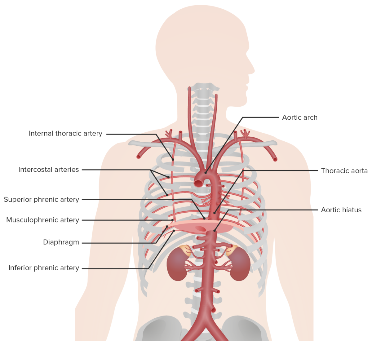
Image by Lecturio.

Other functions

Neurovasculature

Vasculature

  • Arterial supply:
    • Superior phrenic artery: branch of the aorta Aorta The main trunk of the systemic arteries. Mediastinum and Great Vessels: Anatomy
    • Inferior phrenic artery
    • Musculophrenic artery: branch of the internal thoracic artery
    • Lower 5 intercostal arteries Arteries Arteries are tubular collections of cells that transport oxygenated blood and nutrients from the heart to the tissues of the body. The blood passes through the arteries in order of decreasing luminal diameter, starting in the largest artery (the aorta) and ending in the small arterioles. Arteries are classified into 3 types: large elastic arteries, medium muscular arteries, and small arteries and arterioles. Arteries: Histology
  • Venous drainage:
    • Left suprarenal vein
    • Azygos system

Innervation

  • Motor Motor Neurons which send impulses peripherally to activate muscles or secretory cells. Nervous System: Histology nerve supply: right and left phrenic nerves (originate from cervical spinal cord Spinal cord The spinal cord is the major conduction pathway connecting the brain to the body; it is part of the CNS. In cross section, the spinal cord is divided into an H-shaped area of gray matter (consisting of synapsing neuronal cell bodies) and a surrounding area of white matter (consisting of ascending and descending tracts of myelinated axons). Spinal Cord: Anatomy: C3–C5)
  • Sensory Sensory Neurons which conduct nerve impulses to the central nervous system. Nervous System: Histology nerve supply:
    • Phrenic nerve: for central part of diaphragm
    • 6th–11th intercostal nerves for periphery of diaphragm
      • Activation of sensory Sensory Neurons which conduct nerve impulses to the central nervous system. Nervous System: Histology phrenic nerves occurs throughout the respiratory cycle.
      • Input from sensory Sensory Neurons which conduct nerve impulses to the central nervous system. Nervous System: Histology nerves to the CNS is important for regulation of the breathing cycle.

Clinical Relevance

  • Congenital Congenital Chorioretinitis diaphragmatic hernias: embryologically derived defects in the diaphragm through which abdominal structures can pass into the chest cavity. Hernias most often occur on the left posterolateral portion of the diaphragm. Prenatal diagnosis is commonly made by ultrasonography during pregnancy Pregnancy The status during which female mammals carry their developing young (embryos or fetuses) in utero before birth, beginning from fertilization to birth. Pregnancy: Diagnosis, Physiology, and Care, followed by confirmation by chest x-ray X-ray Penetrating electromagnetic radiation emitted when the inner orbital electrons of an atom are excited and release radiant energy. X-ray wavelengths range from 1 pm to 10 nm. Hard x-rays are the higher energy, shorter wavelength x-rays. Soft x-rays or grenz rays are less energetic and longer in wavelength. The short wavelength end of the x-ray spectrum overlaps the gamma rays wavelength range. The distinction between gamma rays and x-rays is based on their radiation source. Pulmonary Function Tests after birth. Immediate respiratory resuscitation Resuscitation The restoration to life or consciousness of one apparently dead. . Neonatal Respiratory Distress Syndrome at birth with endotracheal intubation Intubation Peritonsillar Abscess and mechanical ventilation Ventilation The total volume of gas inspired or expired per unit of time, usually measured in liters per minute. Ventilation: Mechanics of Breathing are required. Surgical repair is the only curative option. 
  • Diaphragmatic paralysis: can be unilateral or bilateral. Diaphragmatic paralysis is due to disruption of the integrity or function of 1 or both phrenic nerves or, in more rare cases, paralysis of the diaphragmatic muscle. Paralysis can be due to mass Mass Three-dimensional lesion that occupies a space within the breast Imaging of the Breast effect from a tumor Tumor Inflammation, trauma, infection, or autoimmune inflammation Inflammation Inflammation is a complex set of responses to infection and injury involving leukocytes as the principal cellular mediators in the body’s defense against pathogenic organisms. Inflammation is also seen as a response to tissue injury in the process of wound healing. The 5 cardinal signs of inflammation are pain, heat, redness, swelling, and loss of function. Inflammation. Presentation is with dyspnea Dyspnea Dyspnea is the subjective sensation of breathing discomfort. Dyspnea is a normal manifestation of heavy physical or psychological exertion, but also may be caused by underlying conditions (both pulmonary and extrapulmonary). Dyspnea and orthopnea Orthopnea Pulmonary Edema ( shortness of breath Shortness of breath Dyspnea is the subjective sensation of breathing discomfort. Dyspnea is a normal manifestation of heavy physical or psychological exertion, but also may be caused by underlying conditions (both pulmonary and extrapulmonary). Dyspnea when lying flat). Hypoxemia Hypoxemia Neonatal Respiratory Distress Syndrome and hypercapnia Hypercapnia A clinical manifestation of abnormal increase in the amount of carbon dioxide in arterial blood. Neonatal Respiratory Distress Syndrome may be noted. Treatment depends on the underlying cause. Occasionally, invasive or noninvasive mechanical ventilatory support is needed.
  • Hiccups: very common condition that occurs because of involuntary intermittent contractions of the diaphragm. A usual episode of hiccups lasts no more than a few minutes and only 1 hemidiaphragm is affected—the left in most cases. Treatment is often unnecessary because of the self-limited nature of hiccups. Pharyngeal stimulation (directly through a catheter, via drinking iced water, etc ETC The electron transport chain (ETC) sends electrons through a series of proteins, which generate an electrochemical proton gradient that produces energy in the form of adenosine triphosphate (ATP). Electron Transport Chain (ETC).) is a very effective nonpharmacologic intervention, as is Valsalva maneuver Valsalva maneuver Forced expiratory effort against a closed glottis. Rectal Prolapse.

References

  1. Bains, K. N. S, Kashyap, S., & Lappin, S. L. (2021). Anatomy, thorax, diaphragm. StatPearls. Retrieved August 17, 2021, from https://www.ncbi.nlm.nih.gov/books/NBK519558/
  2. Gamache, J. (2018). Diaphragmatic paralysis. Medscape. Retrieved August 17, 2021, from https://reference.medscape.com/article/298200-overview
  3. McCool, F. D., Manzoor, K., & Minami, T. (2018). Disorders of the diaphragm. Clin Chest Med 39, pp. 345–360.
  4. Sefton E. M., Gallardo M., & Kardon, G. (2018). Developmental origin and morphogenesis of the diaphragm, an essential mammalian muscle. Dev Biol 440, pp. 64–73.
  5. Wilkes, G. (2017). Hiccups. Medscape. Retrieved August 17, 2021, from https://reference.medscape.com/article/775746-overview

Create your free account or log in to continue reading!

Sign up now and get free access to Lecturio with concept pages, medical videos, and questions for your medical education.

User Reviews

Details