The pharynx is a component of the digestive system that lies posterior to the nasal cavity Nasal cavity The proximal portion of the respiratory passages on either side of the nasal septum. Nasal cavities, extending from the nares to the nasopharynx, are lined with ciliated nasal mucosa. Nose Anatomy (External & Internal), oral cavity, and larynx Larynx The larynx, also commonly called the voice box, is a cylindrical space located in the neck at the level of the C3-C6 vertebrae. The major structures forming the framework of the larynx are the thyroid cartilage, cricoid cartilage, and epiglottis. The larynx serves to produce sound (phonation), conducts air to the trachea, and prevents large molecules from reaching the lungs. Larynx: Anatomy. The pharynx can be divided into the oropharynx, nasopharynx, and laryngopharynx. Pharyngeal muscles play an integral role in vital processes such as breathing, swallowing Swallowing The act of taking solids and liquids into the gastrointestinal tract through the mouth and throat. Gastrointestinal Motility, and speaking. The muscles of the pharynx receive innervation from the vagus and glossopharyngeal nerve to propel food from the oral cavity into the esophagus Esophagus The esophagus is a muscular tube-shaped organ of around 25 centimeters in length that connects the pharynx to the stomach. The organ extends from approximately the 6th cervical vertebra to the 11th thoracic vertebra and can be divided grossly into 3 parts: the cervical part, the thoracic part, and the abdominal part. Esophagus: Anatomy.
Last updated: Dec 15, 2025
Formation of the pharyngeal (branchial) apparatus is during the 4th and 5th weeks of development.
The pharyngeal apparatus consists of:
Pharyngeal musculature develops from the 3rd, 4th, and 6th arches:
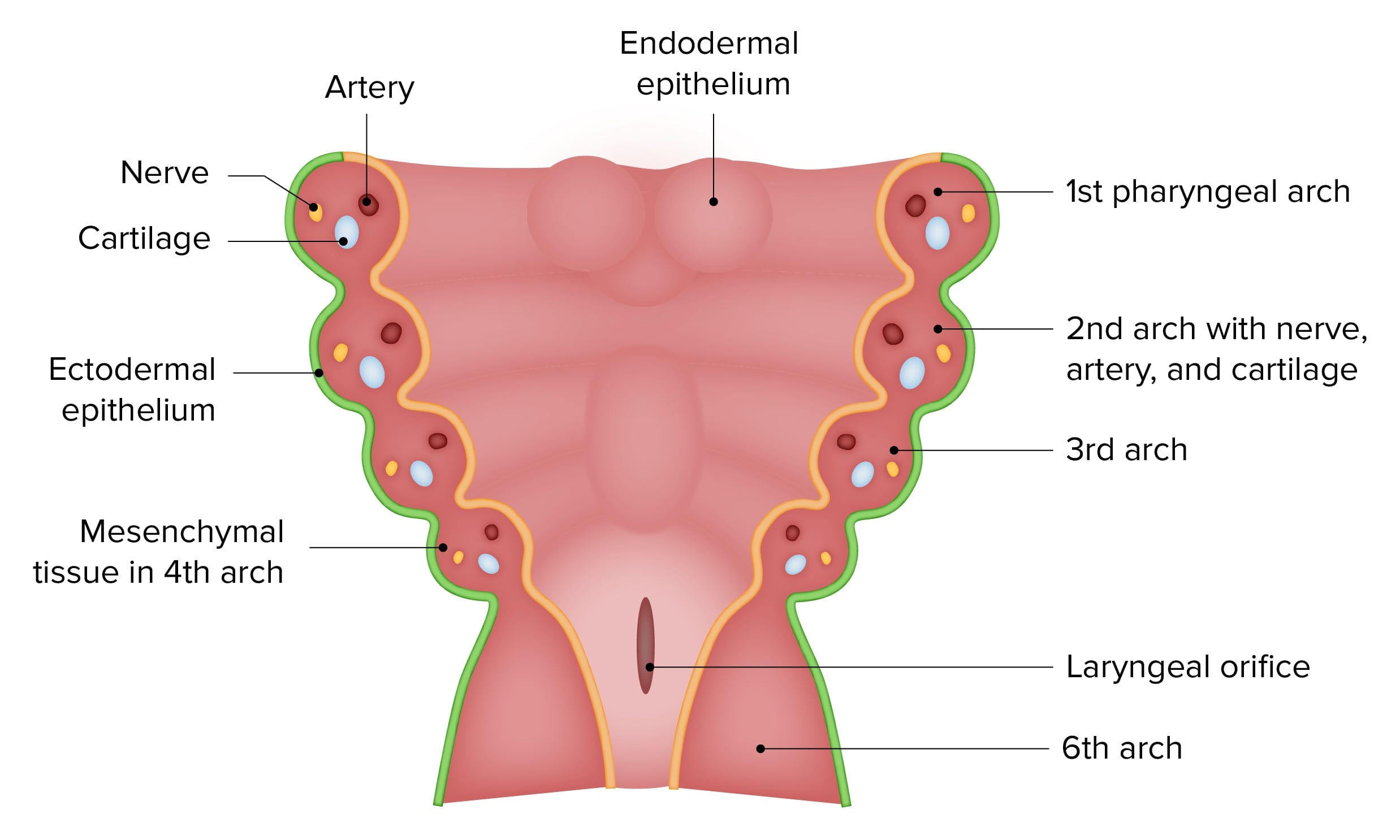
The pharynx arises from the pharyngeal arches:
Pharyngeal pouches are located on the inside of the pharynx (yellow outline), whereas pharyngeal clefts are located on the outside of the pharynx (green outline). The muscles of the pharynx are derived from the 4th and 6th pharyngeal arches.
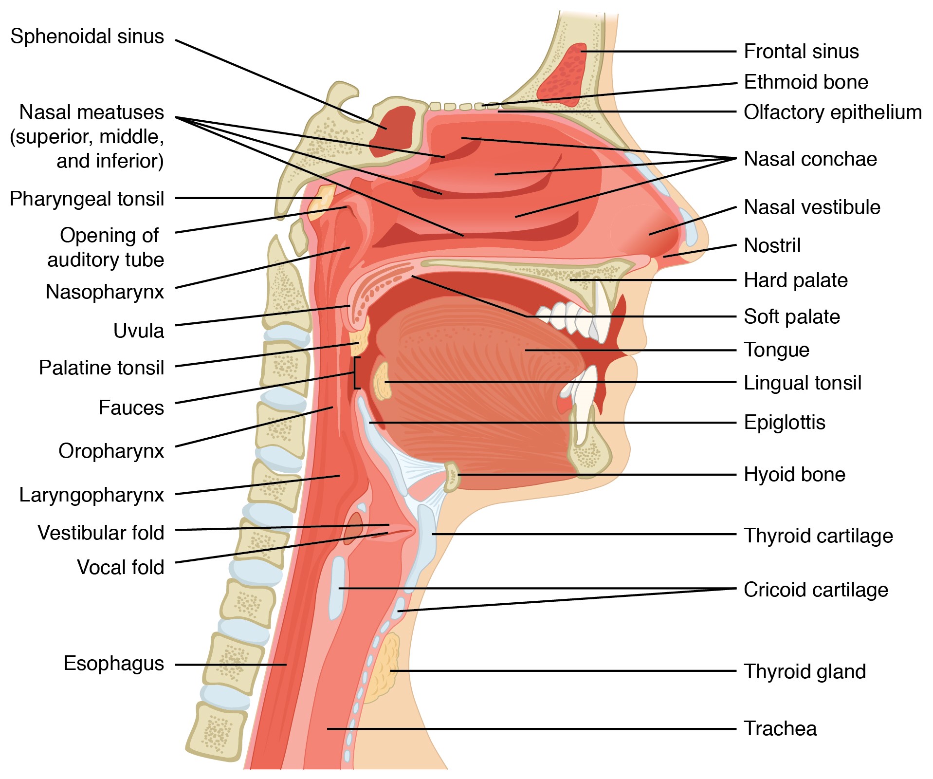
Sagittal view of the head and neck showing the location of the pharynx and its anatomical landmarks
Image: “2303 Anatomy of Nose-Pharynx-Mouth-Larynx” by OpenStax College. License: CC BY 3.0, edited by Lecturio.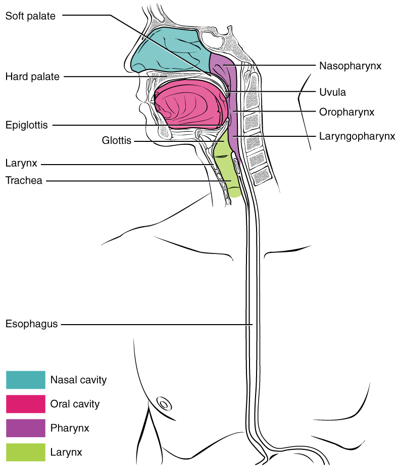
Sagittal view of the head and neck showing the division of the pharynx
Image: “2411_Pharynx” by Phil Schatz. License: CC BY 4.0Constrictor muscles constitute the outer circular layer of muscle. During swallowing Swallowing The act of taking solids and liquids into the gastrointestinal tract through the mouth and throat. Gastrointestinal Motility, constrictor muscles constrict to propel the food bolus downward.
Longitudinal muscles constitute the inner muscular layer and play a role in elevating the pharynx and larynx Larynx The larynx, also commonly called the voice box, is a cylindrical space located in the neck at the level of the C3-C6 vertebrae. The major structures forming the framework of the larynx are the thyroid cartilage, cricoid cartilage, and epiglottis. The larynx serves to produce sound (phonation), conducts air to the trachea, and prevents large molecules from reaching the lungs. Larynx: Anatomy during swallowing Swallowing The act of taking solids and liquids into the gastrointestinal tract through the mouth and throat. Gastrointestinal Motility and speaking.
| Muscle | Origin | Insertion | Neurovasculature |
|---|---|---|---|
| Superior constrictor muscle |
|
|
Blood supply:
Innervation: Pharyngeal plexus of the vagus nerve |
| Middle constrictor muscle |
|
Pharyngeal raphe Raphe Testicles: Anatomy | Blood supply: Ascending pharyngeal artery Innervation: Pharyngeal branch of the vagus nerve (CN X) and pharyngeal plexus |
| Inferior constrictor muscle |
|
Cricopharyngeal part encircles the pharyngoesophageal junction Pharyngoesophageal junction Esophagus: Anatomy without forming a raphe Raphe Testicles: Anatomy. | Blood supply:
Innervation: Pharyngeal branch of the vagus nerve (CN X) and pharyngeal plexus |
| Muscle | Origin | Insertion | Neurovasculature |
|---|---|---|---|
| Palatopharyngeus |
|
Posterior border of the lamina of the thyroid Thyroid The thyroid gland is one of the largest endocrine glands in the human body. The thyroid gland is a highly vascular, brownish-red gland located in the visceral compartment of the anterior region of the neck. Thyroid Gland: Anatomy cartilage Cartilage Cartilage is a type of connective tissue derived from embryonic mesenchyme that is responsible for structural support, resilience, and the smoothness of physical actions. Perichondrium (connective tissue membrane surrounding cartilage) compensates for the absence of vasculature in cartilage by providing nutrition and support. Cartilage: Histology and side of the pharynx and esophagus Esophagus The esophagus is a muscular tube-shaped organ of around 25 centimeters in length that connects the pharynx to the stomach. The organ extends from approximately the 6th cervical vertebra to the 11th thoracic vertebra and can be divided grossly into 3 parts: the cervical part, the thoracic part, and the abdominal part. Esophagus: Anatomy | Blood supply: Facial artery Innervation: Pharyngeal branch of the vagus nerve (CN X) and pharyngeal plexus |
| Stylopharyngeus | Styloid process of the temporal bone Temporal bone Either of a pair of compound bones forming the lateral (left and right) surfaces and base of the skull which contains the organs of hearing. It is a large bone formed by the fusion of parts: the squamous (the flattened anterior-superior part), the tympanic (the curved anterior-inferior part), the mastoid (the irregular posterior portion), and the petrous (the part at the base of the skull). Jaw and Temporomandibular Joint: Anatomy | Posterior border of the thyroid Thyroid The thyroid gland is one of the largest endocrine glands in the human body. The thyroid gland is a highly vascular, brownish-red gland located in the visceral compartment of the anterior region of the neck. Thyroid Gland: Anatomy cartilage Cartilage Cartilage is a type of connective tissue derived from embryonic mesenchyme that is responsible for structural support, resilience, and the smoothness of physical actions. Perichondrium (connective tissue membrane surrounding cartilage) compensates for the absence of vasculature in cartilage by providing nutrition and support. Cartilage: Histology | Blood supply: Pharyngeal branch of the ascending pharyngeal artery Innervation: Glossopharyngeal nerve |
| Salpingopharyngeus | Cartilaginous part of the Eustachian tube Eustachian tube A narrow passageway that connects the upper part of the throat to the tympanic cavity. Ear: Anatomy | Posterior and superior borders of the thyroid Thyroid The thyroid gland is one of the largest endocrine glands in the human body. The thyroid gland is a highly vascular, brownish-red gland located in the visceral compartment of the anterior region of the neck. Thyroid Gland: Anatomy cartilage Cartilage Cartilage is a type of connective tissue derived from embryonic mesenchyme that is responsible for structural support, resilience, and the smoothness of physical actions. Perichondrium (connective tissue membrane surrounding cartilage) compensates for the absence of vasculature in cartilage by providing nutrition and support. Cartilage: Histology with the palatopharyngeus | Blood supply: Ascending pharyngeal artery Innervation: Pharyngeal branch of the vagus nerve (CN X) and pharyngeal plexus |
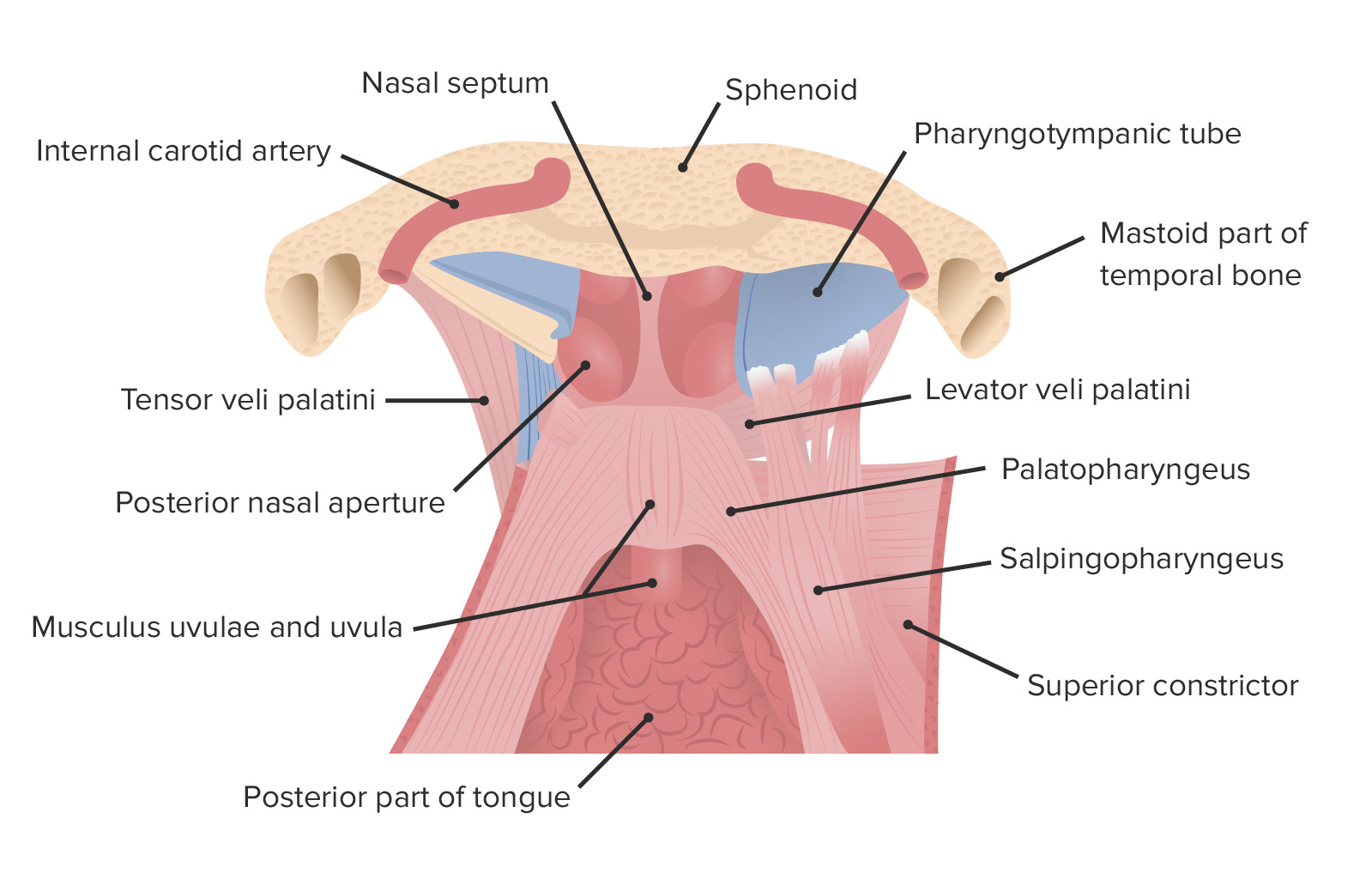
Constrictor and longitudinal muscles of the pharynx
Image by Lecturio.There are 2 layers to the pharyngeal fascia Fascia Layers of connective tissue of variable thickness. The superficial fascia is found immediately below the skin; the deep fascia invests muscles, nerves, and other organs. Cellulitis:
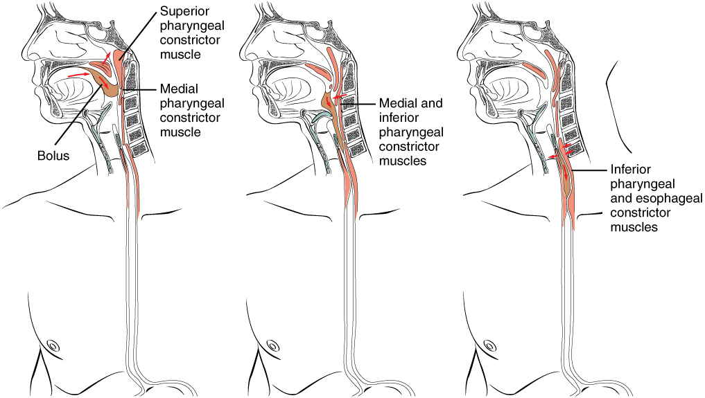
Image showing the role of the pharynx during the deglutition process
Image: “2413 DeglutitionN” by OpenStax College. License: CC BY 3.0