Diphtheria is an infectious disease caused by Corynebacterium diphtheriae that most often results in respiratory disease with membranous inflammation of the pharynx, sore throat, fever, swollen glands, and weakness. The hallmark sign is a sheet of thick, gray material covering the back of the throat. Diphtheria can also manifest as cutaneous disease leading to nonspecific skin lesions. In advanced stages, diphtheria can damage the heart, kidneys, and nervous system. It is diagnosed via a culture of pharyngeal swabs and treated with antibiotic therapy and the diphtheria antitoxin.
Last updated: 26 Jan, 2022
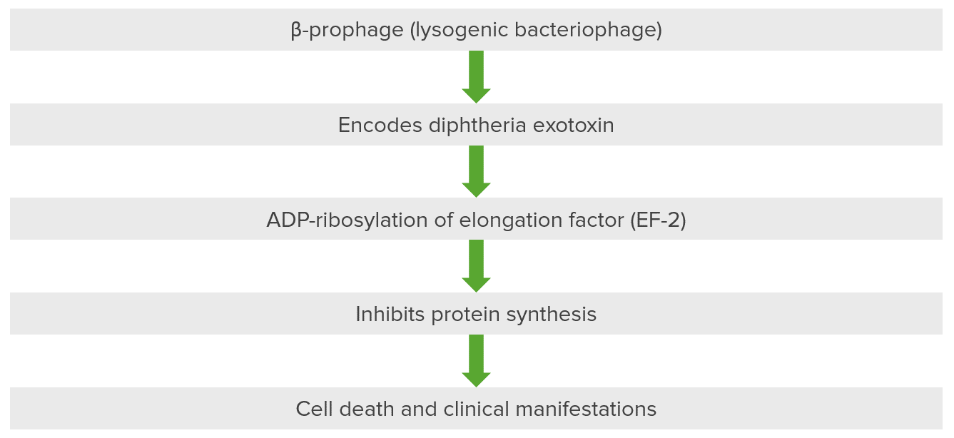
Pathogenesis of diseases associated with
Corynebacterium
Corynebacterium
Corynebacteria are gram-positive, club-shaped bacilli. Corynebacteria are commonly isolated on tellurite or Loeffler’s media and have characteristic metachromatic granules. The major pathogenic species is Corynebacterium diphtheriae, which causes a severe respiratory infection called diphtheria.
Corynebacterium: diphtheria exotoxin inactivates
elongation
Elongation
Polymerase Chain Reaction (PCR) factor (
EF
EF
Cardiac Cycle-2) via ADP-ribosylation.
This bacteriophage, a beta prophage, introduces itself into the cell and
DNA
DNA
A deoxyribonucleotide polymer that is the primary genetic material of all cells. Eukaryotic and prokaryotic organisms normally contain DNA in a double-stranded state, yet several important biological processes transiently involve single-stranded regions. DNA, which consists of a polysugar-phosphate backbone possessing projections of purines (adenine and guanine) and pyrimidines (thymine and cytosine), forms a double helix that is held together by hydrogen bonds between these purines and pyrimidines (adenine to thymine and guanine to cytosine).
DNA Types and Structure of the host organism, resulting in the encoding of diphtheria exotoxin. The exotoxin has 3 domains: 1 is present in fragment A and is catalytic, while 2 are present in fragment B for
receptor
Receptor
Receptors are proteins located either on the surface of or within a cell that can bind to signaling molecules known as ligands (e.g., hormones) and cause some type of response within the cell.
Receptors binding and membrane insertion and translocation. The exotoxin is the major cause of the disease in diphtheria because it ribosylates ADP present in the
elongation
Elongation
Polymerase Chain Reaction (PCR) factor. The
elongation
Elongation
Polymerase Chain Reaction (PCR) factor, specifically
elongation
Elongation
Polymerase Chain Reaction (PCR) factor 2 (
EF
EF
Cardiac Cycle-2), is critical for the
elongation
Elongation
Polymerase Chain Reaction (PCR) of protein chains. Diphtheria exotoxin inhibits
EF
EF
Cardiac Cycle-2 so that protein is not synthesized, resulting in cellular death and secondary clinical manifestations.
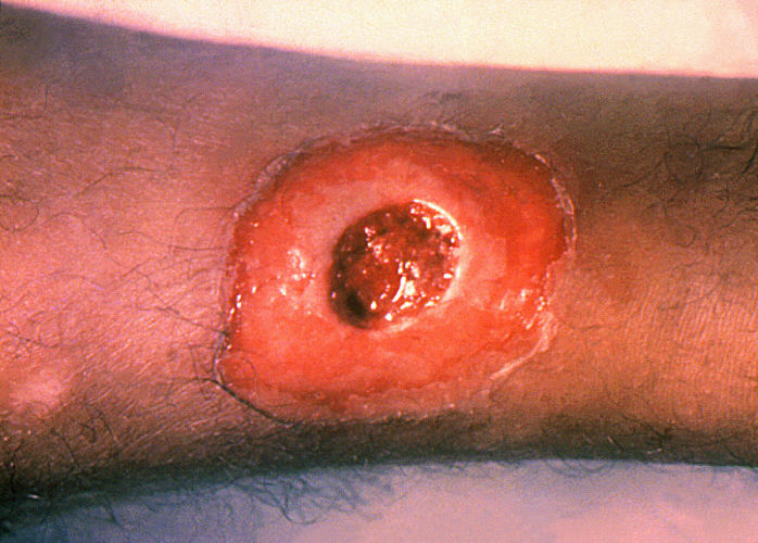
Clinical sign of a patient with cutaneous (wound) diphtheria: chronic non-healing ulcer
Image: “A diphtheria skin Skin The skin, also referred to as the integumentary system, is the largest organ of the body. The skin is primarily composed of the epidermis (outer layer) and dermis (deep layer). The epidermis is primarily composed of keratinocytes that undergo rapid turnover, while the dermis contains dense layers of connective tissue. Skin: Structure and Functions lesion on the leg Leg The lower leg, or just “leg” in anatomical terms, is the part of the lower limb between the knee and the ankle joint. The bony structure is composed of the tibia and fibula bones, and the muscles of the leg are grouped into the anterior, lateral, and posterior compartments by extensions of fascia. Leg: Anatomy” by CDC/Public Health Image Library (PHIL). License: Public Domain
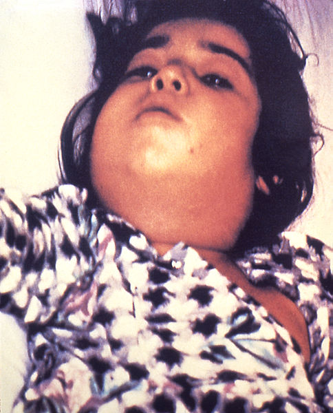
A clinical sign of a patient with respiratory diphtheria: severe lymphadenopathy Lymphadenopathy Lymphadenopathy is lymph node enlargement (> 1 cm) and is benign and self-limited in most patients. Etiologies include malignancy, infection, and autoimmune disorders, as well as iatrogenic causes such as the use of certain medications. Generalized lymphadenopathy often indicates underlying systemic disease. Lymphadenopathy with “bull neck Neck The part of a human or animal body connecting the head to the rest of the body. Peritonsillar Abscess” appearance
Image: “Diphtheria bull neck Neck The part of a human or animal body connecting the head to the rest of the body. Peritonsillar Abscess” by CDC/Public Health Image Library (PHIL). License: Public DomainPediatric patient with diphtheria presenting with characteristic grayish-white membrane covering the posterior pharyngeal wall
Image: “A doctor with the UK’s Emergency Medical Team checks a child for symptoms of Diphtheria in a makeshift clinic in the Kutupalong camp for Rohingya refugees in Bangladesh” by Russell Watkins/Department for International Development. License: CC BY 2.0Key points to remember about diphtheria:
ABCDEFG: ADP-ribosylation, Beta-prophage, Corynebacterium diphtheriae, Elongation factor 2, Granules
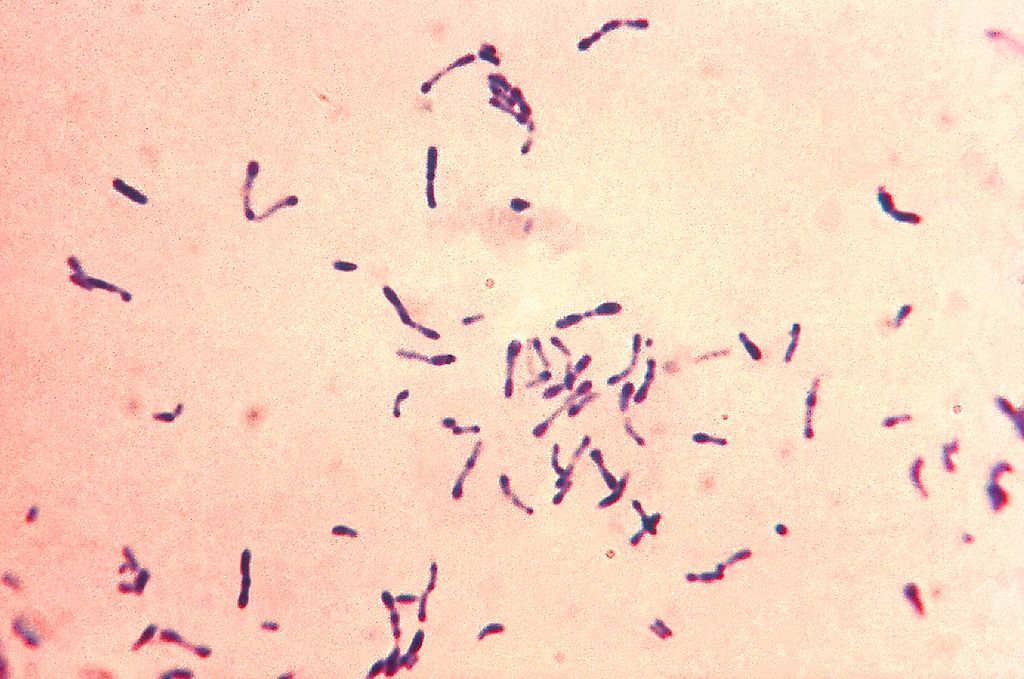
Gram-positive Gram-Positive Penicillins Corynebacterium Corynebacterium Corynebacteria are gram-positive, club-shaped bacilli. Corynebacteria are commonly isolated on tellurite or Loeffler’s media and have characteristic metachromatic granules. The major pathogenic species is Corynebacterium diphtheriae, which causes a severe respiratory infection called diphtheria. Corynebacterium diphtheriae bacteria Bacteria Bacteria are prokaryotic single-celled microorganisms that are metabolically active and divide by binary fission. Some of these organisms play a significant role in the pathogenesis of diseases. Bacteriology with blue metachromatic granules. The bacteria Bacteria Bacteria are prokaryotic single-celled microorganisms that are metabolically active and divide by binary fission. Some of these organisms play a significant role in the pathogenesis of diseases. Bacteriology were grown via Loeffler’s medium to enhance the characteristic metachromatic granules. Notice the club-shaped nature and formation into Y and V.
Image: “ Gram-positive Gram-Positive Penicillins Corynebacterium Corynebacterium Corynebacteria are gram-positive, club-shaped bacilli. Corynebacteria are commonly isolated on tellurite or Loeffler’s media and have characteristic metachromatic granules. The major pathogenic species is Corynebacterium diphtheriae, which causes a severe respiratory infection called diphtheria. Corynebacterium diphtheriae bacteria Bacteria Bacteria are prokaryotic single-celled microorganisms that are metabolically active and divide by binary fission. Some of these organisms play a significant role in the pathogenesis of diseases. Bacteriology” by Centers for Disease Control and Prevention’s Public Health Image Library (PHIL). License: Public DomainA throat Throat The pharynx is a component of the digestive system that lies posterior to the nasal cavity, oral cavity, and larynx. The pharynx can be divided into the oropharynx, nasopharynx, and laryngopharynx. Pharyngeal muscles play an integral role in vital processes such as breathing, swallowing, and speaking. Pharynx: Anatomy swab inoculated with Corynebacterium Corynebacterium Corynebacteria are gram-positive, club-shaped bacilli. Corynebacteria are commonly isolated on tellurite or Loeffler’s media and have characteristic metachromatic granules. The major pathogenic species is Corynebacterium diphtheriae, which causes a severe respiratory infection called diphtheria. Corynebacterium diphtheriae is cultured on a Petri dish plate containing Tinsdale medium supplemented with the agar component Tellurite. The gray-black colonies, each surrounded by a dark brown halo, are characteristic of C. diphtheriae on Tellurite agar.
Image: “ Corynebacterium Corynebacterium Corynebacteria are gram-positive, club-shaped bacilli. Corynebacteria are commonly isolated on tellurite or Loeffler’s media and have characteristic metachromatic granules. The major pathogenic species is Corynebacterium diphtheriae, which causes a severe respiratory infection called diphtheria. Corynebacterium diphtheriae” by Public Health Image Library (PHIL). License: Public DomainPossible complications associated with diphtheria include:
The differential diagnosis of respiratory diphtheria includes: