Long QT syndrome (LQTS) is a disorder of ventricular myocardial repolarization Repolarization Membrane Potential that produces QT prolongation on electrocardiogram Electrocardiogram An electrocardiogram (ECG) is a graphic representation of the electrical activity of the heart plotted against time. Adhesive electrodes are affixed to the skin surface allowing measurement of cardiac impulses from many angles. The ECG provides 3-dimensional information about the conduction system of the heart, the myocardium, and other cardiac structures. Electrocardiogram (ECG) ( ECG ECG An electrocardiogram (ECG) is a graphic representation of the electrical activity of the heart plotted against time. Adhesive electrodes are affixed to the skin surface allowing measurement of cardiac impulses from many angles. The ECG provides 3-dimensional information about the conduction system of the heart, the myocardium, and other cardiac structures. Electrocardiogram (ECG)). Long QT syndrome is associated with an increased risk of developing life-threatening cardiac Cardiac Total Anomalous Pulmonary Venous Return (TAPVR) arrhythmias, specifically torsades de pointes Torsades de pointes A malignant form of polymorphic ventricular tachycardia that is characterized by heart rate between 200 and 250 beats per minute, and QRS complexes with changing amplitude and twisting of the points. The term also describes the syndrome of tachycardia with prolonged ventricular repolarization, long qt intervals exceeding 500 milliseconds or bradycardia. Torsades de pointes may be self-limited or may progress to ventricular fibrillation. Ventricular Tachycardia. The condition may be congenital Congenital Chorioretinitis or acquired. Congenital Congenital Chorioretinitis LQTS is attributed to genetic mutations Genetic Mutations Carcinogenesis affecting cardiac Cardiac Total Anomalous Pulmonary Venous Return (TAPVR) ion channels Channels The Cell: Cell Membrane. Acquired LQTS usually results from drug therapy and/or electrolyte abnormalities. Patients Patients Individuals participating in the health care system for the purpose of receiving therapeutic, diagnostic, or preventive procedures. Clinician–Patient Relationship can be asymptomatic or present with palpitations Palpitations Ebstein’s Anomaly, syncope Syncope Syncope is a short-term loss of consciousness and loss of postural stability followed by spontaneous return of consciousness to the previous neurologic baseline without the need for resuscitation. The condition is caused by transient interruption of cerebral blood flow that may be benign or related to a underlying life-threatening condition. Syncope, seizures Seizures A seizure is abnormal electrical activity of the neurons in the cerebral cortex that can manifest in numerous ways depending on the region of the brain affected. Seizures consist of a sudden imbalance that occurs between the excitatory and inhibitory signals in cortical neurons, creating a net excitation. The 2 major classes of seizures are focal and generalized. Seizures, and even sudden cardiac death Sudden cardiac death Cardiac arrest is the sudden, complete cessation of cardiac output with hemodynamic collapse. Patients present as pulseless, unresponsive, and apneic. Rhythms associated with cardiac arrest are ventricular fibrillation/tachycardia, asystole, or pulseless electrical activity. Cardiac Arrest. Diagnosis is established with ECG ECG An electrocardiogram (ECG) is a graphic representation of the electrical activity of the heart plotted against time. Adhesive electrodes are affixed to the skin surface allowing measurement of cardiac impulses from many angles. The ECG provides 3-dimensional information about the conduction system of the heart, the myocardium, and other cardiac structures. Electrocardiogram (ECG) along with medical and family history Family History Adult Health Maintenance, laboratory workup, and other cardiac Cardiac Total Anomalous Pulmonary Venous Return (TAPVR) tests. Treatment is determined by etiology. Acquired LQTS requires removal of the offending drug and correction of electrolyte abnormalities. Congenital Congenital Chorioretinitis LQTS management involves avoidance of triggers Triggers Hereditary Angioedema (C1 Esterase Inhibitor Deficiency) of arrhythmia, intake of beta-blockers Beta-blockers Drugs that bind to but do not activate beta-adrenergic receptors thereby blocking the actions of beta-adrenergic agonists. Adrenergic beta-antagonists are used for treatment of hypertension, cardiac arrhythmias, angina pectoris, glaucoma, migraine headaches, and anxiety. Class 2 Antiarrhythmic Drugs (Beta Blockers), and placement of an implantable cardioverter- defibrillator Defibrillator Cardiac electrical stimulators that apply brief high-voltage electroshocks to the heart. These stimulators are used to restore normal rhythm and contractile function in hearts of patients who are experiencing ventricular fibrillation or ventricular tachycardia that is not accompanied by a palpable pulse. Some defibrillators may also be used to correct certain noncritical dysrhythmias (called synchronized defibrillation or cardioversion), using relatively low-level discharges synchronized to the patient's ECG waveform. Cardiac Arrest (ICD).
Last updated: 8 Jan, 2021
Pathophysiology:
Types of congenital Congenital Chorioretinitis LQTS:
Variety of associated conditions:
The diagnosis of long QT syndrome can be made via an ECG ECG An electrocardiogram (ECG) is a graphic representation of the electrical activity of the heart plotted against time. Adhesive electrodes are affixed to the skin surface allowing measurement of cardiac impulses from many angles. The ECG provides 3-dimensional information about the conduction system of the heart, the myocardium, and other cardiac structures. Electrocardiogram (ECG) of the patient and/or 1st-degree relatives.
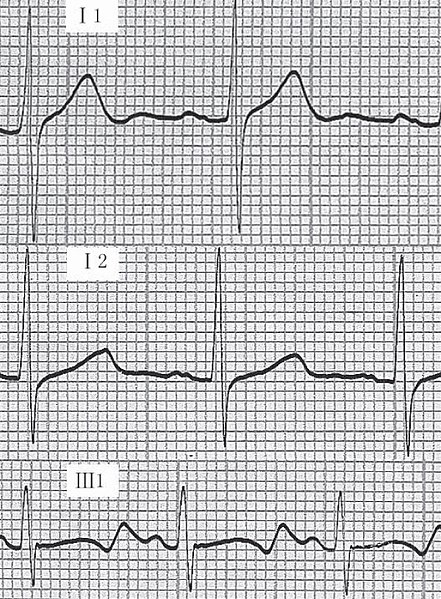
Example of an ECG tracing showing:
i) normal tracing;
ii) Romano-Ward syndrome (prolonged QT);
iii) Jervell-Lange-Nielsen syndrome (prolonged QT).
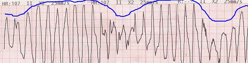
Example of an ECG tracing showing beat-to-beat axis deviation of the QRS complexes around the baseline. This is known as torsades de pointes or polymorphic ventricular tachycardia.
Image: “Tosadesdepointes” by Panthro. License: Public Domain
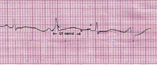
Lead II of ECG showing QT prolongation (QTc = 550 ms). Image: “Lead II of ECG showing QT prolongation” by Department of Pharmacology, Burdwan Medical College, Burdwan, West Bengal 713104, India. License: CC BY 2.0
The following medical conditions may commonly predispose QT prolongation:
The following conditions may be included in a differential diagnosis for prolonged QT interval QT interval Electrocardiogram (ECG):
Untreated QT prolongation may evolve into the following complications: