During the 4th week of gestation, limb buds form on the sides of the developing embryo Embryo The entity of a developing mammal, generally from the cleavage of a zygote to the end of embryonic differentiation of basic structures. For the human embryo, this represents the first two months of intrauterine development preceding the stages of the fetus. Fertilization and First Week. The tips of these buds condense into the apical ectodermal ridge (AER). The AER continues the elongation Elongation Polymerase Chain Reaction (PCR) of the limb buds and maintains its growth by continuously producing fibroblast growth factor Fibroblast growth factor A family of small polypeptide growth factors that share several common features including a strong affinity for heparin, and a central barrel-shaped core region of 140 amino acids that is highly homologous between family members. Although originally studied as proteins that stimulate the growth of fibroblasts this distinction is no longer a requirement for membership in the fibroblast growth factor family. X-linked Hypophosphatemic Rickets 8 (FGF8). As the AER grows away from the body, tissues differentiate. After the cartilage Cartilage Cartilage is a type of connective tissue derived from embryonic mesenchyme that is responsible for structural support, resilience, and the smoothness of physical actions. Perichondrium (connective tissue membrane surrounding cartilage) compensates for the absence of vasculature in cartilage by providing nutrition and support. Cartilage: Histology models are formed in the developing limbs, arteries Arteries Arteries are tubular collections of cells that transport oxygenated blood and nutrients from the heart to the tissues of the body. The blood passes through the arteries in order of decreasing luminal diameter, starting in the largest artery (the aorta) and ending in the small arterioles. Arteries are classified into 3 types: large elastic arteries, medium muscular arteries, and small arteries and arterioles. Arteries: Histology invade central and peripheral areas, giving rise to primary and secondary centers of ossification Ossification The process of bone formation. Histogenesis of bone including ossification. Bones: Development and Ossification. The process of endochondral ossification Ossification The process of bone formation. Histogenesis of bone including ossification. Bones: Development and Ossification is completed when those centers meet and the epiphyseal plate Epiphyseal plate The area between the epiphysis and the diaphysis within which bone growth occurs. Cartilage: Histology is no longer present.
Last updated: Dec 15, 2025
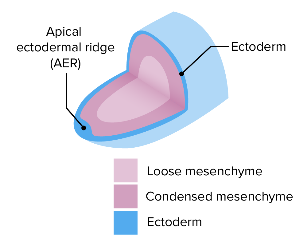
Apical ectodermal ridge:
This ridge is located at the tip of the limb bud. Its function is the production of fibroblast growth factor, which induces proliferation of ectodermal cells to maintain limb elongation.
Mnemonic:
To quickly recall what happens during the 4th week, remember: 4 weeks → 4 limbs, 4 heart chambers
Rotation of the limbs:
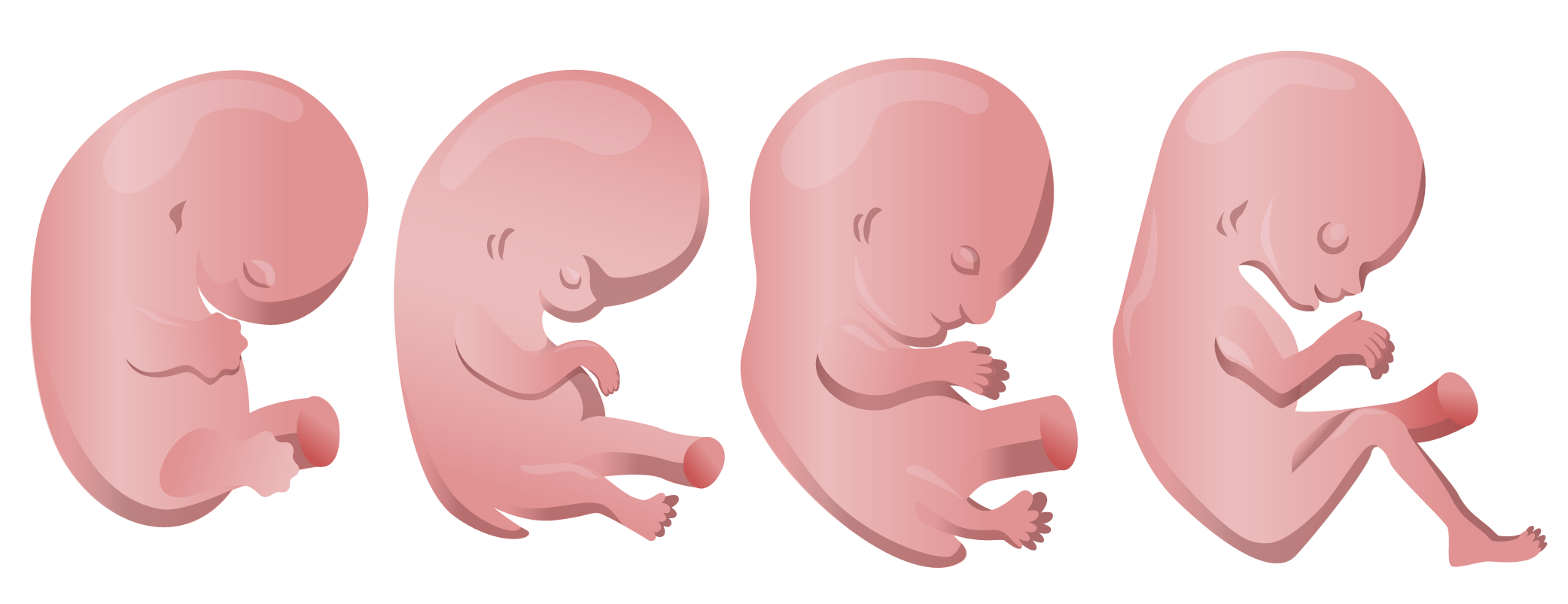
As upper and lower limbs grow, they bend at the elbow and knee with the palms and soles of the feet facing medially. During 90-degree opposite rotation, the elbow points toward the posterior side, whereas the knee points anteriorly.
Muscle comes from myotome: ventrolateral cells originating from somites located on either side of neural tube Neural tube A tube of ectodermal tissue in an embryo that will give rise to the central nervous system, including the spinal cord and the brain. Lumen within the neural tube is called neural canal which gives rise to the central canal of the spinal cord and the ventricles of the brain. Gastrulation and Neurulation that migrate to form muscles. As myotomes migrate, they form:
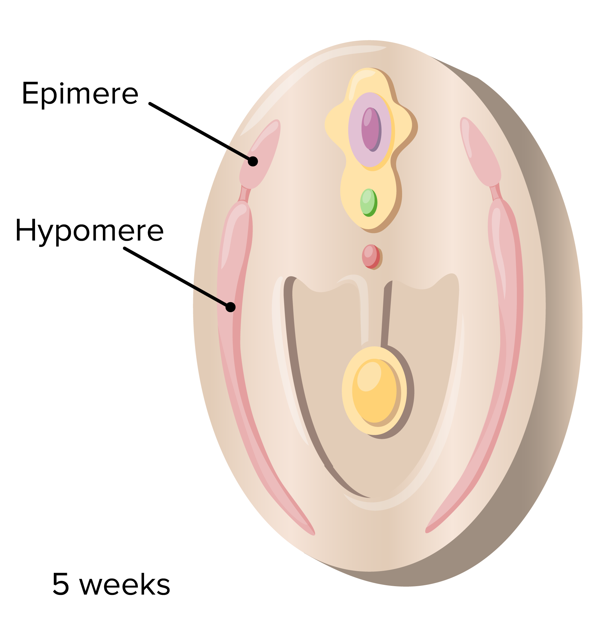
Myotome:
The myotome splits into a dorsal region, the epimere, that becomes the intrinsic muscles of the back, and a ventral region, the hypomere, that will become the muscles of the body wall and limbs.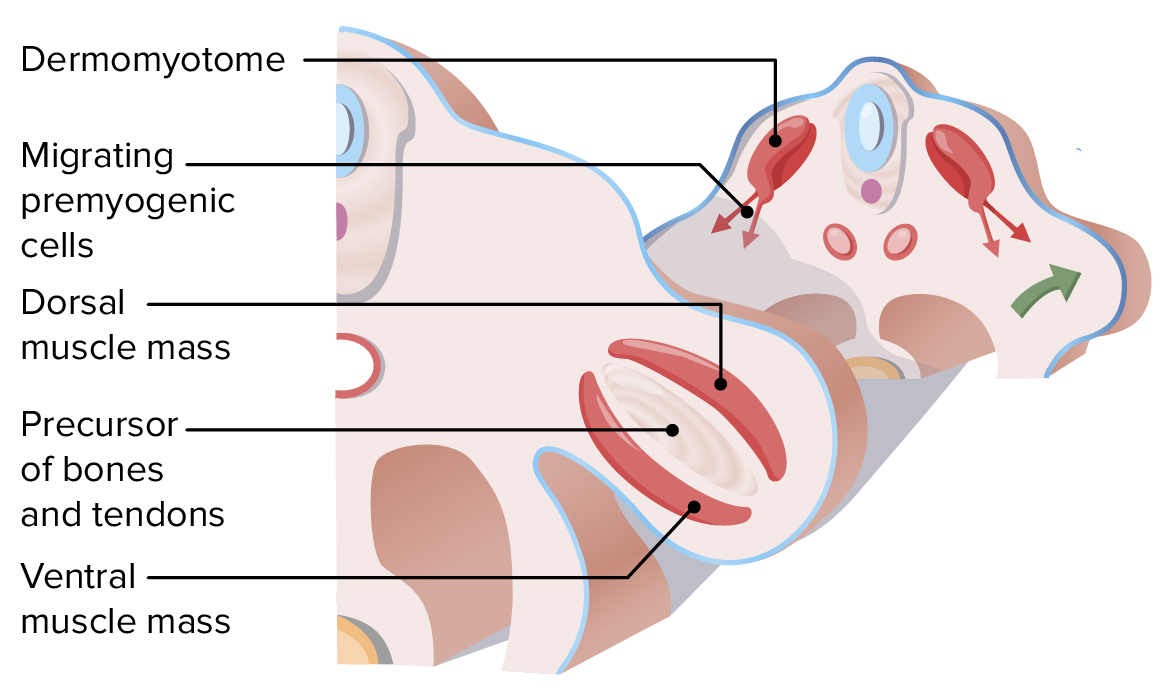
Hypomere:
As the hypomere migrates into the limb buds, it splits into dorsal and ventral muscle masses that will become extensors and flexors, respectively.

Musculature of the upper limbs
Somites corresponding to C4–C8, T1–T2 give rise to posterior and anterior aspects of the musculature of the upper limb. Flexor muscles arise from anterior aspects of these somites, while extensors arise from the dorsal aspects.
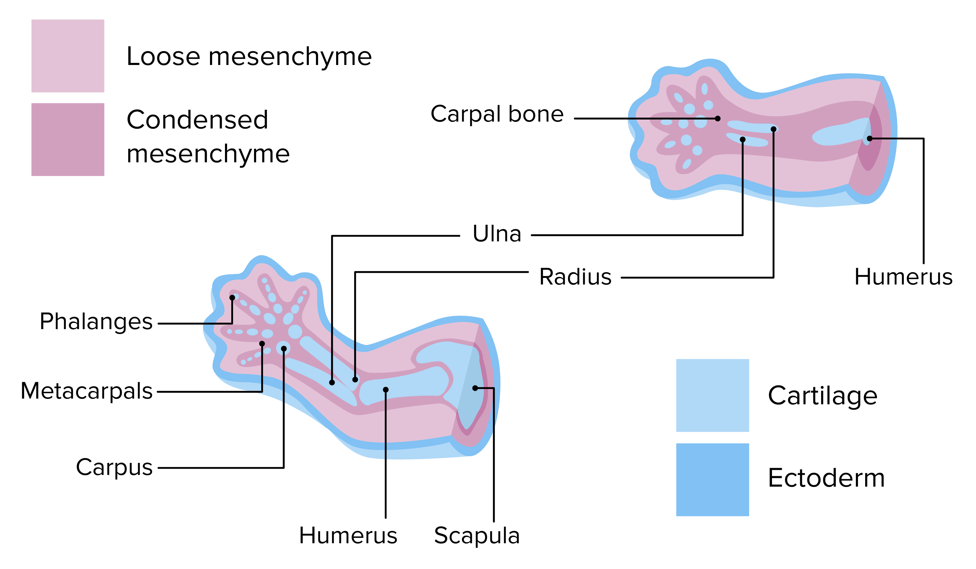
Cartilage is developed from undifferentiated mesenchymal tissue and develops into the bone by a process called endochondral ossification.
Image by Lecturio.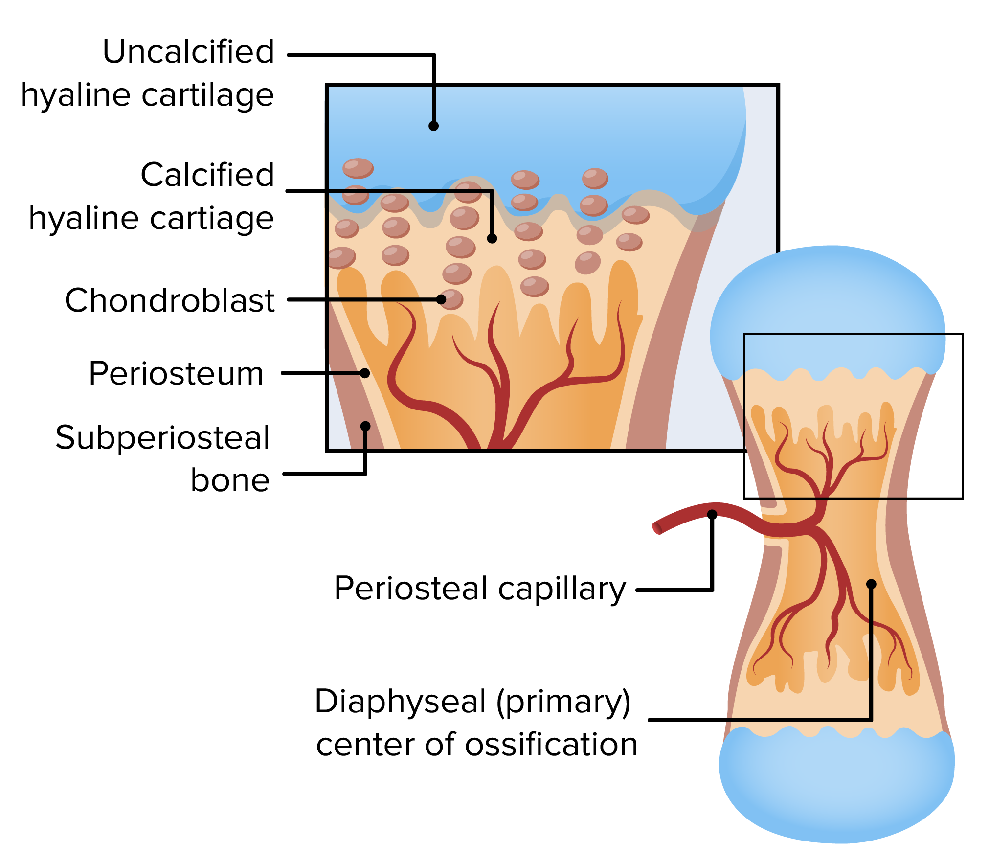
Primary center of ossification is created by arteries invading the middle of the cartilage. Bone formation proceeds outward from it, as the blood also delivers osteoprogenitor cells.
Image by Lecturio.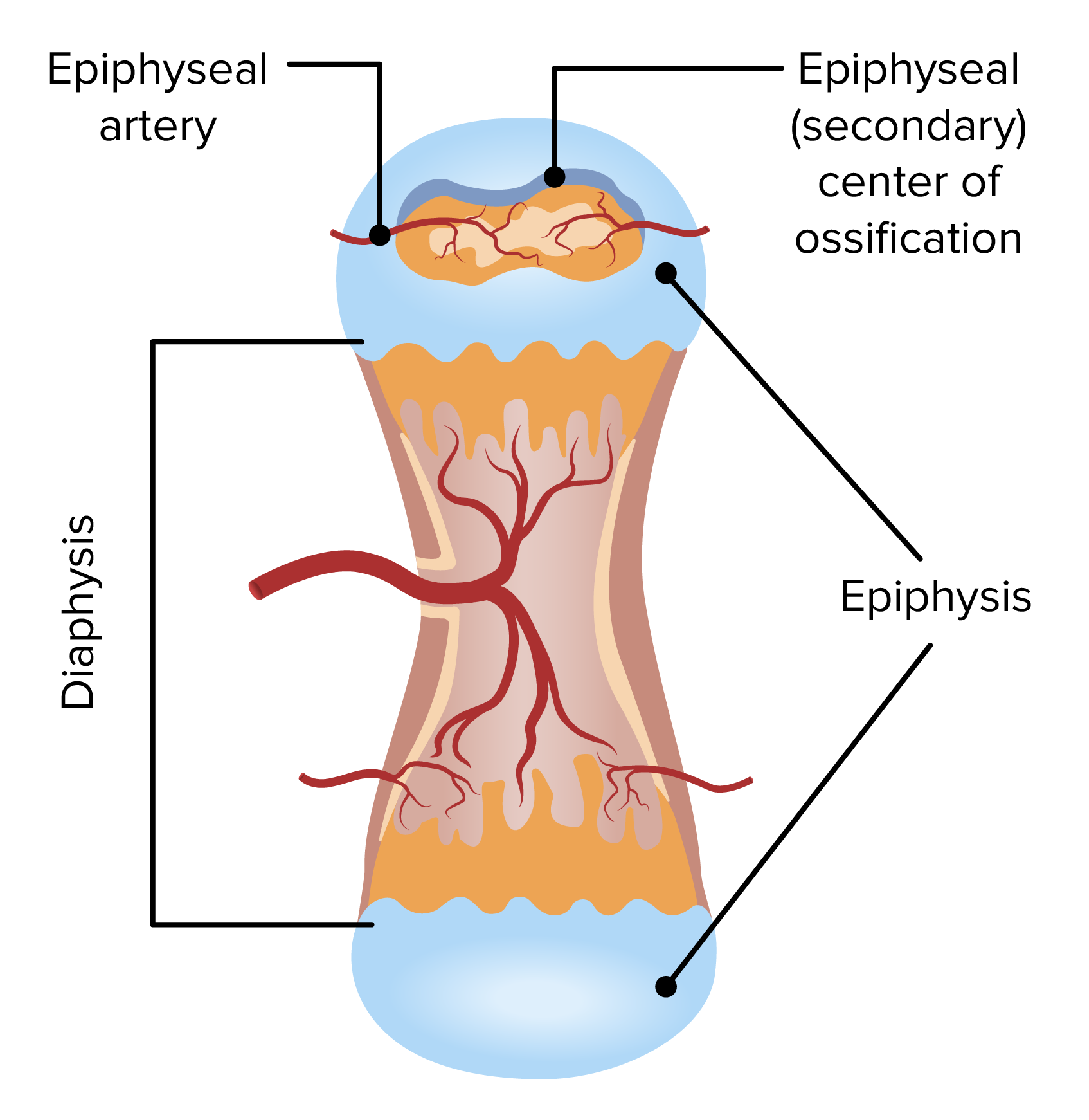
Secondary ossification center is created by arteries invading the bone extremities. The 2 centers of ossification are separated by the epiphyseal plate.
Image by Lecturio.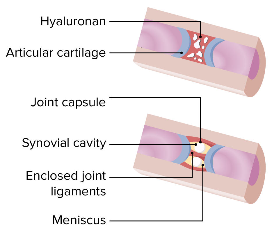
Connective tissue:
As the bones grow, connective tissue remains between them as dense regular connective tissue or fibrocartilage and accumulate synovial fluid.
Positioning, orientation Orientation Awareness of oneself in relation to time, place and person. Psychiatric Assessment, and growth of limbs in the developing embryo Embryo The entity of a developing mammal, generally from the cleavage of a zygote to the end of embryonic differentiation of basic structures. For the human embryo, this represents the first two months of intrauterine development preceding the stages of the fetus. Fertilization and First Week is determined genetically and by the expression of specific timed chemical signals.
Positioning of limbs along the craniocaudal axis is determined by the homeobox (HOX) genes Genes A category of nucleic acid sequences that function as units of heredity and which code for the basic instructions for the development, reproduction, and maintenance of organisms. DNA Types and Structure.