The axilla is a pyramid-shaped space located between the upper thorax and the arm Arm The arm, or "upper arm" in common usage, is the region of the upper limb that extends from the shoulder to the elbow joint and connects inferiorly to the forearm through the cubital fossa. It is divided into 2 fascial compartments (anterior and posterior). Arm: Anatomy. The axilla has a base, an apex, and 4 walls (anterior, medial, lateral, posterior). The base of the pyramid is made up of the axillary skin Skin The skin, also referred to as the integumentary system, is the largest organ of the body. The skin is primarily composed of the epidermis (outer layer) and dermis (deep layer). The epidermis is primarily composed of keratinocytes that undergo rapid turnover, while the dermis contains dense layers of connective tissue. Skin: Structure and Functions. The apex is the axillary inlet, located between the 1st rib, superior border of the scapula, and clavicle Clavicle A bone on the ventral side of the shoulder girdle, which in humans is commonly called the collar bone. Clavicle Fracture. The apex houses various vessels and nerves, including the axillary artery and its branches, the axillary vein and its tributaries, the branches of the brachial plexus Brachial Plexus The large network of nerve fibers which distributes the innervation of the upper extremity. The brachial plexus extends from the neck into the axilla. In humans, the nerves of the plexus usually originate from the lower cervical and the first thoracic spinal cord segments (c5-c8 and T1), but variations are not uncommon. Peripheral Nerve Injuries in the Cervicothoracic Region, and the axillary lymph nodes Lymph Nodes They are oval or bean shaped bodies (1 - 30 mm in diameter) located along the lymphatic system. Lymphatic Drainage System: Anatomy.
Last updated: Dec 15, 2025
The axilla is a pyramid-shaped space below the glenohumeral joint that is the passageway for nerves and vessels to pass into the upper arm Upper Arm The arm, or “upper arm” in common usage, is the region of the upper limb that extends from the shoulder to the elbow joint and connects inferiorly to the forearm through the cubital fossa. It is divided into 2 fascial compartments (anterior and posterior). Arm: Anatomy.
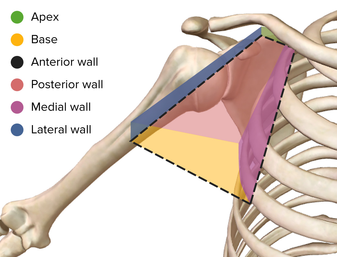
Boundaries of the axilla
Image by BioDigital, edited by Lecturio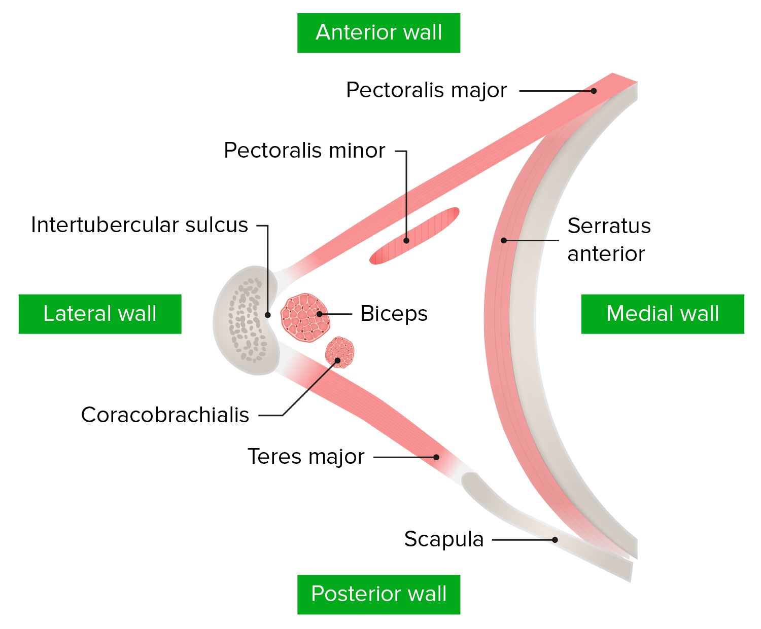
Axillary inlet
Image by Lecturio.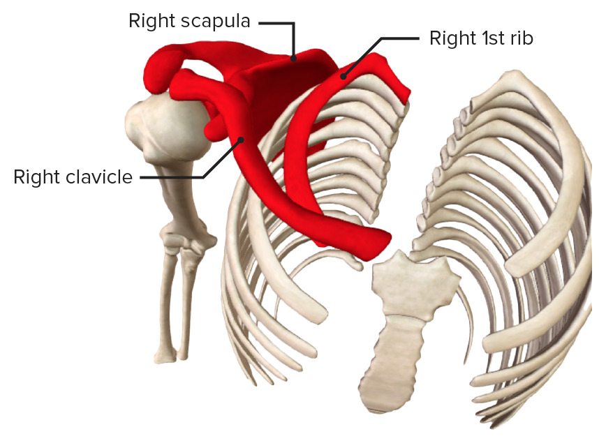
Apex (axillary inlet): between the 1st rib, the scapula, and the clavicle
Image by BioDigital, edited by Lecturio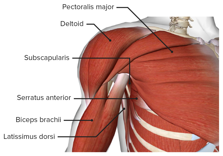
The axilla viewed from the anterior anatomical position
Image by BioDigital, edited by Lecturio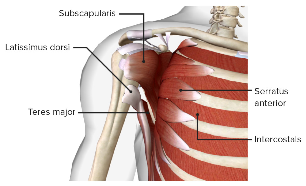
Posterior and medial walls of the axilla: Anterior wall and muscles of the upper limb have been removed.
Image by BioDigital, edited by Lecturio
Anterior view of the axilla, showing the anterior wall and the biceps brachii and coracobrachialis muscles running through the axillary fossa
Image by BioDigital, edited by LecturioThe contents of the axilla are structures enclosed in the axillary sheath.
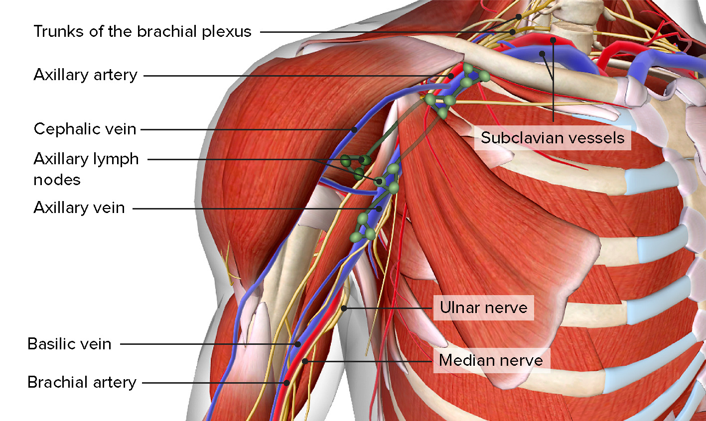
Embedded in the axillary fat, enclosed within the axillary sheath, the contents of the axilla include: the axillary artery and its branches, the axillary vein and its tributaries, the branches of the brachial plexus, and the axillary lymph nodes.
Image by BioDigital, edited by LecturioThe axillary artery is a direct continuation of the subclavian artery and is composed of 3 parts:
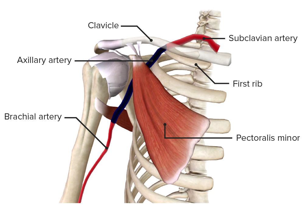
The axillary artery (in blue) originates at the lateral margin of the 1st rib, before which it is called the subclavian artery. After passing the lower margin of teres major, the axillary artery becomes the brachial artery.
Image by BioDigital, edited by Lecturio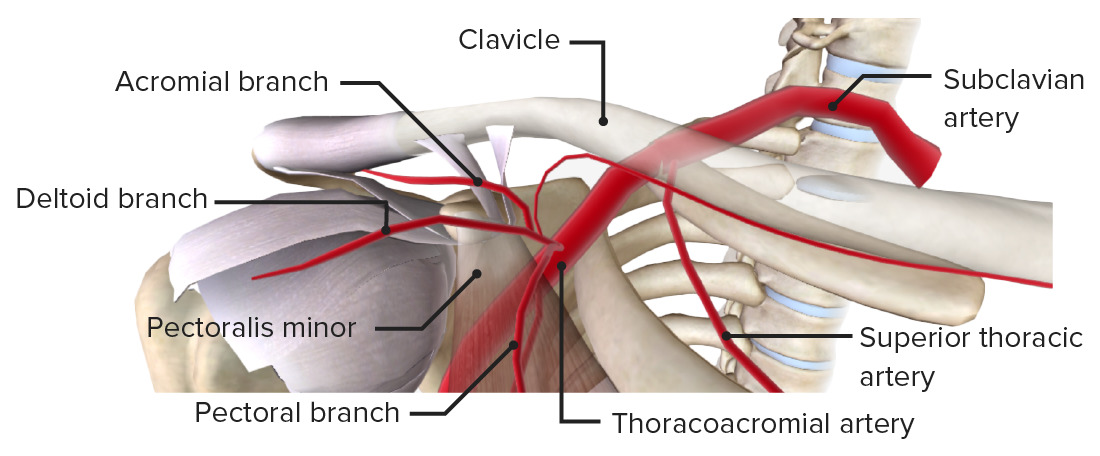
First and 2nd parts of the axillary artery, giving off the superior thoracic and thoracoacromial arteries as branches.
Image by BioDigital, edited by Lecturio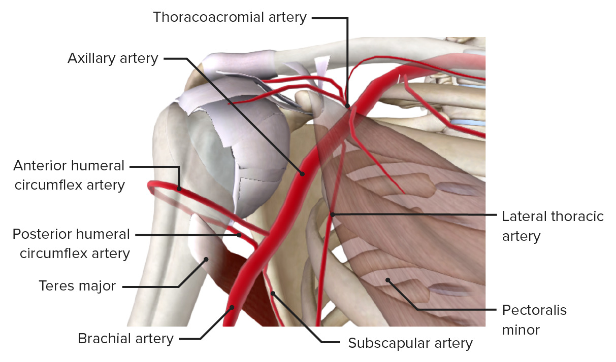
Second and 3rd parts of the axillary artery, giving off the thoracoacromial, lateral thoracic, anterior, and posterior humeral circumflexes, and subscapular arteries as branches.
Image by BioDigital, edited by Lecturio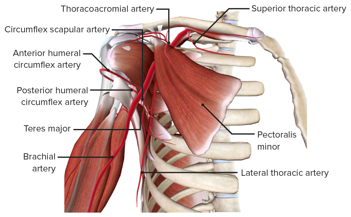
Axillary artery and its branches (humerus faded)
Image by BioDigital, edited by LecturioThe axillary vein is the primary venous drainage of the upper arm Upper Arm The arm, or “upper arm” in common usage, is the region of the upper limb that extends from the shoulder to the elbow joint and connects inferiorly to the forearm through the cubital fossa. It is divided into 2 fascial compartments (anterior and posterior). Arm: Anatomy, formed by the cephalic and basilic veins Veins Veins are tubular collections of cells, which transport deoxygenated blood and waste from the capillary beds back to the heart. Veins are classified into 3 types: small veins/venules, medium veins, and large veins. Each type contains 3 primary layers: tunica intima, tunica media, and tunica adventitia. Veins: Histology.
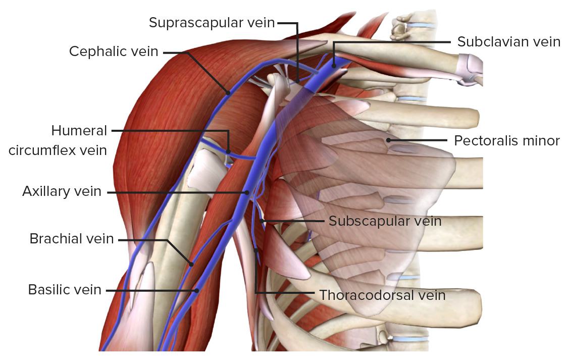
Axillary vein: primary venous drainage of the upper arm, formed by the cephalic and basilic veins
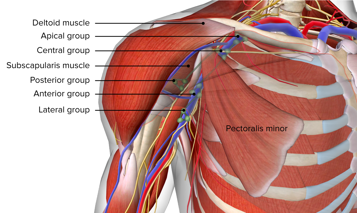
Image by BioDigital, edited by LecturioThere are 5 groups of axillary lymph nodes Lymph Nodes They are oval or bean shaped bodies (1 – 30 mm in diameter) located along the lymphatic system. Lymphatic Drainage System: Anatomy draining to the apical lymph nodes Lymph Nodes They are oval or bean shaped bodies (1 – 30 mm in diameter) located along the lymphatic system. Lymphatic Drainage System: Anatomy:
These lymph nodes Lymph Nodes They are oval or bean shaped bodies (1 – 30 mm in diameter) located along the lymphatic system. Lymphatic Drainage System: Anatomy filter lymph Lymph The interstitial fluid that is in the lymphatic system. Secondary Lymphatic Organs fluid from the arm Arm The arm, or “upper arm” in common usage, is the region of the upper limb that extends from the shoulder to the elbow joint and connects inferiorly to the forearm through the cubital fossa. It is divided into 2 fascial compartments (anterior and posterior). Arm: Anatomy and pectoral region, including the breast (important in the diagnosis and management of breast cancer Breast cancer Breast cancer is a disease characterized by malignant transformation of the epithelial cells of the breast. Breast cancer is the most common form of cancer and 2nd most common cause of cancer-related death among women. Breast Cancer).
Axillary lymph nodes
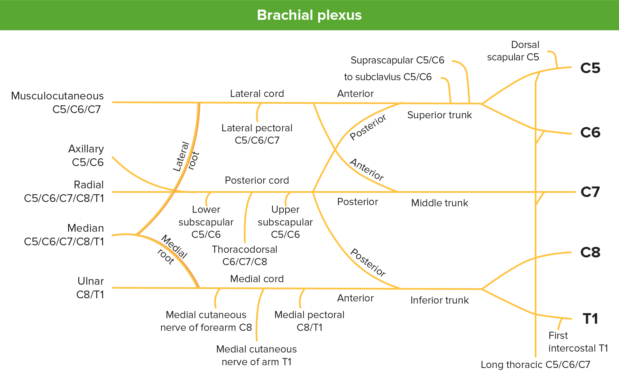
Image by BioDigital, edited by LecturioThe brachial plexus Brachial Plexus The large network of nerve fibers which distributes the innervation of the upper extremity. The brachial plexus extends from the neck into the axilla. In humans, the nerves of the plexus usually originate from the lower cervical and the first thoracic spinal cord segments (c5-c8 and T1), but variations are not uncommon. Peripheral Nerve Injuries in the Cervicothoracic Region is formed from the anterior rami of spinal cord Spinal cord The spinal cord is the major conduction pathway connecting the brain to the body; it is part of the CNS. In cross section, the spinal cord is divided into an H-shaped area of gray matter (consisting of synapsing neuronal cell bodies) and a surrounding area of white matter (consisting of ascending and descending tracts of myelinated axons). Spinal Cord: Anatomy segments C5–T1, which are the roots of the brachial plexus Brachial Plexus The large network of nerve fibers which distributes the innervation of the upper extremity. The brachial plexus extends from the neck into the axilla. In humans, the nerves of the plexus usually originate from the lower cervical and the first thoracic spinal cord segments (c5-c8 and T1), but variations are not uncommon. Peripheral Nerve Injuries in the Cervicothoracic Region and the network of nerves that supply the upper arm Upper Arm The arm, or “upper arm” in common usage, is the region of the upper limb that extends from the shoulder to the elbow joint and connects inferiorly to the forearm through the cubital fossa. It is divided into 2 fascial compartments (anterior and posterior). Arm: Anatomy.
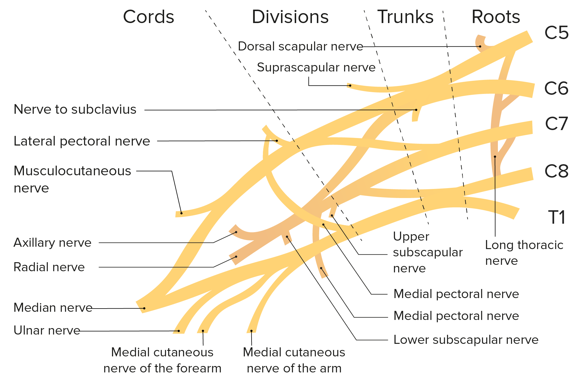
Schematic of the brachial plexus and its branches
Image by LecturioThe brachial plexus Brachial Plexus The large network of nerve fibers which distributes the innervation of the upper extremity. The brachial plexus extends from the neck into the axilla. In humans, the nerves of the plexus usually originate from the lower cervical and the first thoracic spinal cord segments (c5-c8 and T1), but variations are not uncommon. Peripheral Nerve Injuries in the Cervicothoracic Region is a nerve plexus formed by intercommunication of the ventral rami of C5–T1 nerves. The brachial plexus Brachial Plexus The large network of nerve fibers which distributes the innervation of the upper extremity. The brachial plexus extends from the neck into the axilla. In humans, the nerves of the plexus usually originate from the lower cervical and the first thoracic spinal cord segments (c5-c8 and T1), but variations are not uncommon. Peripheral Nerve Injuries in the Cervicothoracic Region serves as the origin of all the peripheral nerves Peripheral Nerves The nerves outside of the brain and spinal cord, including the autonomic, cranial, and spinal nerves. Peripheral nerves contain non-neuronal cells and connective tissue as well as axons. The connective tissue layers include, from the outside to the inside, the epineurium, the perineurium, and the endoneurium. Nervous System: Histology that innervate the upper limb and shoulder.
| Segment | Branch | Function |
|---|---|---|
| Roots | Dorsal scapular nerve (C5) | Supplies the rhomboid major and minor muscles and the levator scapulae muscles |
| Long thoracic nerve (C5–C7) | Supplies the serratus anterior muscles | |
| Upper trunk | Suprascapular nerve (C5–C6) | Supplies the supraspinatus and infraspinatus muscles |
| Nerve to subclavius Subclavius Muscles of the Neck: Anatomy (C5–C6) | Supplies the subclavius muscle Subclavius muscle Chest Wall: Anatomy and the sternoclavicular joint Sternoclavicular Joint Examination of the Upper Limbs | |
| Lateral cord | Lateral pectoral nerve (C5–C7) | Supplies the pectoralis major muscle |
| Musculocutaneous nerve (C5–C7) | Supplies the coracobrachialis Coracobrachialis Arm: Anatomy muscle, biceps Biceps Arm: Anatomy brachii, and brachialis Brachialis Arm: Anatomy muscles, terminates as the lateral cutaneous nerve of forearm Forearm The forearm is the region of the upper limb between the elbow and the wrist. The term “forearm” is used in anatomy to distinguish this area from the arm, a term that is commonly used to describe the entire upper limb. The forearm consists of 2 long bones (the radius and the ulna), the interosseous membrane, and multiple arteries, nerves, and muscles. Forearm: Anatomy | |
| Lateral root of the median nerve Median Nerve A major nerve of the upper extremity. In humans, the fibers of the median nerve originate in the lower cervical and upper thoracic spinal cord (usually C6 to T1), travel via the brachial plexus, and supply sensory and motor innervation to parts of the forearm and hand. Cubital Fossa: Anatomy (C5–C7) | Joins a similar branch from the medial cord to form the median nerve Median Nerve A major nerve of the upper extremity. In humans, the fibers of the median nerve originate in the lower cervical and upper thoracic spinal cord (usually C6 to T1), travel via the brachial plexus, and supply sensory and motor innervation to parts of the forearm and hand. Cubital Fossa: Anatomy (opposition of the thumb and flexion Flexion Examination of the Upper Limbs of the first 3 fingers) | |
| Medial cord | Medial pectoral nerve (C8, T1) | Supplies the pectoralis major and minor muscles |
| Medial cutaneous nerve of the arm Arm The arm, or “upper arm” in common usage, is the region of the upper limb that extends from the shoulder to the elbow joint and connects inferiorly to the forearm through the cubital fossa. It is divided into 2 fascial compartments (anterior and posterior). Arm: Anatomy (medial brachial cutaneous nerve) (C8, T1) | Supplies the skin Skin The skin, also referred to as the integumentary system, is the largest organ of the body. The skin is primarily composed of the epidermis (outer layer) and dermis (deep layer). The epidermis is primarily composed of keratinocytes that undergo rapid turnover, while the dermis contains dense layers of connective tissue. Skin: Structure and Functions on the medial side of the arm Arm The arm, or “upper arm” in common usage, is the region of the upper limb that extends from the shoulder to the elbow joint and connects inferiorly to the forearm through the cubital fossa. It is divided into 2 fascial compartments (anterior and posterior). Arm: Anatomy | |
| Medial cutaneous nerve of the forearm Forearm The forearm is the region of the upper limb between the elbow and the wrist. The term “forearm” is used in anatomy to distinguish this area from the arm, a term that is commonly used to describe the entire upper limb. The forearm consists of 2 long bones (the radius and the ulna), the interosseous membrane, and multiple arteries, nerves, and muscles. Forearm: Anatomy ( medial antebrachial cutaneous nerve Medial antebrachial cutaneous nerve Forearm: Anatomy) (C8, T1) | Supplies the skin Skin The skin, also referred to as the integumentary system, is the largest organ of the body. The skin is primarily composed of the epidermis (outer layer) and dermis (deep layer). The epidermis is primarily composed of keratinocytes that undergo rapid turnover, while the dermis contains dense layers of connective tissue. Skin: Structure and Functions on the medial side of the forearm Forearm The forearm is the region of the upper limb between the elbow and the wrist. The term “forearm” is used in anatomy to distinguish this area from the arm, a term that is commonly used to describe the entire upper limb. The forearm consists of 2 long bones (the radius and the ulna), the interosseous membrane, and multiple arteries, nerves, and muscles. Forearm: Anatomy | |
| Medial root of the median nerve Median Nerve A major nerve of the upper extremity. In humans, the fibers of the median nerve originate in the lower cervical and upper thoracic spinal cord (usually C6 to T1), travel via the brachial plexus, and supply sensory and motor innervation to parts of the forearm and hand. Cubital Fossa: Anatomy (C8, T1) | Joins with the lateral root to form the median nerve Median Nerve A major nerve of the upper extremity. In humans, the fibers of the median nerve originate in the lower cervical and upper thoracic spinal cord (usually C6 to T1), travel via the brachial plexus, and supply sensory and motor innervation to parts of the forearm and hand. Cubital Fossa: Anatomy | |
| Ulnar nerve (C7–T1) | Flexion Flexion Examination of the Upper Limbs of the 4th and 5th fingers | |
| Posterior cord | Upper subscapular nerve (C5–C6) | Supplies the upper part of the subscapularis muscle |
| Thoracodorsal nerve (C6–C8) | Supplies the latissimus dorsi muscle | |
| Lower subscapular nerve (C5–C6) | Supplies the lower part of the subscapularis and teres major muscles | |
| Axillary nerve (C5–C6) | Supplies both the deltoid and teres minor muscles | |
| Radial nerve (C5–T1) | Extension Extension Examination of the Upper Limbs of the wrist and fingers |
Brachial plexus
Image by Lecturio.The axillary spaces are anatomic spaces in the axilla where important nerves and vessels exit the axilla.
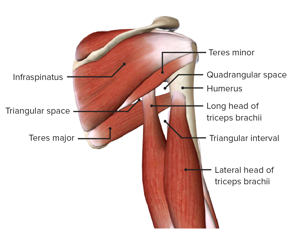
Posterior view of the axilla, featuring the scapulohumeral muscles and the axillary spaces: quadrangular and triangular spaces
Image by BioDigital, edited by Lecturio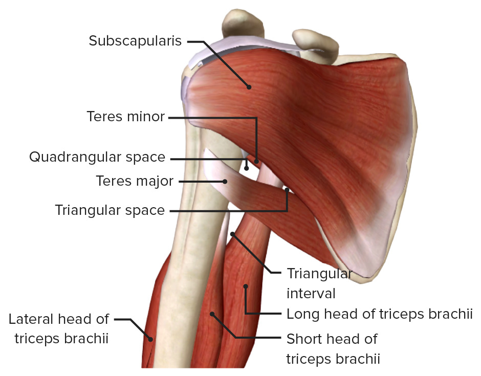
Anterior view of the axilla, featuring the scapulohumeral muscles and the axillary spaces: quadrangular space, triangular space, and triangular interval
Image by BioDigital, edited by Lecturio| Space | Borders | Content |
|---|---|---|
| Quadrangular space |
|
|
| Triangular space |
|
Scapular circumflex artery and vein |
| Triangular interval |
|
|
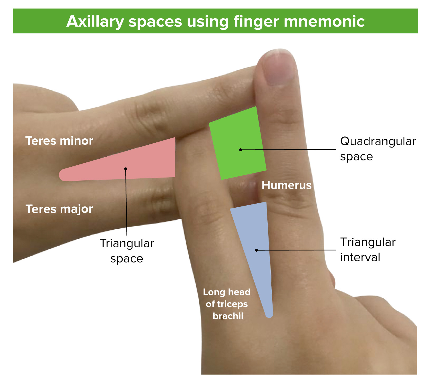
Spaces of arm using a finger arrangement
Image by Lecturio.The following are common conditions and pathologies associated with the axilla: