A clavicular fractureClavicular FractureAcute Shoulder Pain is a common fractureFractureA fracture is a disruption of the cortex of any bone and periosteum and is commonly due to mechanical stress after an injury or accident. Open fractures due to trauma can be a medical emergency. Fractures are frequently associated with automobile accidents, workplace injuries, and trauma.Overview of Bone Fractures usually occurring because of trauma. The trauma may be direct or indirect and is generally of high energy, but it may occur secondary to low-energy traumaLow-Energy TraumaToddler’s Fractures in the elderly. Clinical presentation includes painPainAn unpleasant sensation induced by noxious stimuli which are detected by nerve endings of nociceptive neurons.Pain: Types and Pathways localized to the clavicle, a palpable deformityDeformityExamination of the Upper Limbs over the fractureFractureA fracture is a disruption of the cortex of any bone and periosteum and is commonly due to mechanical stress after an injury or accident. Open fractures due to trauma can be a medical emergency. Fractures are frequently associated with automobile accidents, workplace injuries, and trauma.Overview of Bone Fractures site, and crepitusCrepitusOsteoarthritis. Diagnosis is clinical and confirmed with diagnostic imaging. Management is often conservative, although an increasing number of patientsPatientsIndividuals participating in the health care system for the purpose of receiving therapeutic, diagnostic, or preventive procedures.Clinician–Patient Relationship with clavicular fractureClavicular FractureAcute Shoulder Pain now undergo surgical intervention.
A clavicular fractureClavicular FractureAcute Shoulder Pain is a disruption in the integrity of the bony tissue of the clavicle (also called the collar boneBoneBone is a compact type of hardened connective tissue composed of bone cells, membranes, an extracellular mineralized matrix, and central bone marrow. The 2 primary types of bone are compact and spongy. Bones: Structure and Types). These very common fractures generally heal without incident, although complications are possible.
Epidemiology
Most commonly fractured boneBoneBone is a compact type of hardened connective tissue composed of bone cells, membranes, an extracellular mineralized matrix, and central bone marrow. The 2 primary types of bone are compact and spongy. Bones: Structure and Types in childhood
2%–10% of all fractures
IncidenceIncidenceThe number of new cases of a given disease during a given period in a specified population. It also is used for the rate at which new events occur in a defined population. It is differentiated from prevalence, which refers to all cases in the population at a given time.Measures of Disease Frequency: 1 in 1000 people per year
Most commonly associated with sports injuries in younger patientsPatientsIndividuals participating in the health care system for the purpose of receiving therapeutic, diagnostic, or preventive procedures.Clinician–Patient Relationship
Most commonly associated with falls in older patientsPatientsIndividuals participating in the health care system for the purpose of receiving therapeutic, diagnostic, or preventive procedures.Clinician–Patient Relationship
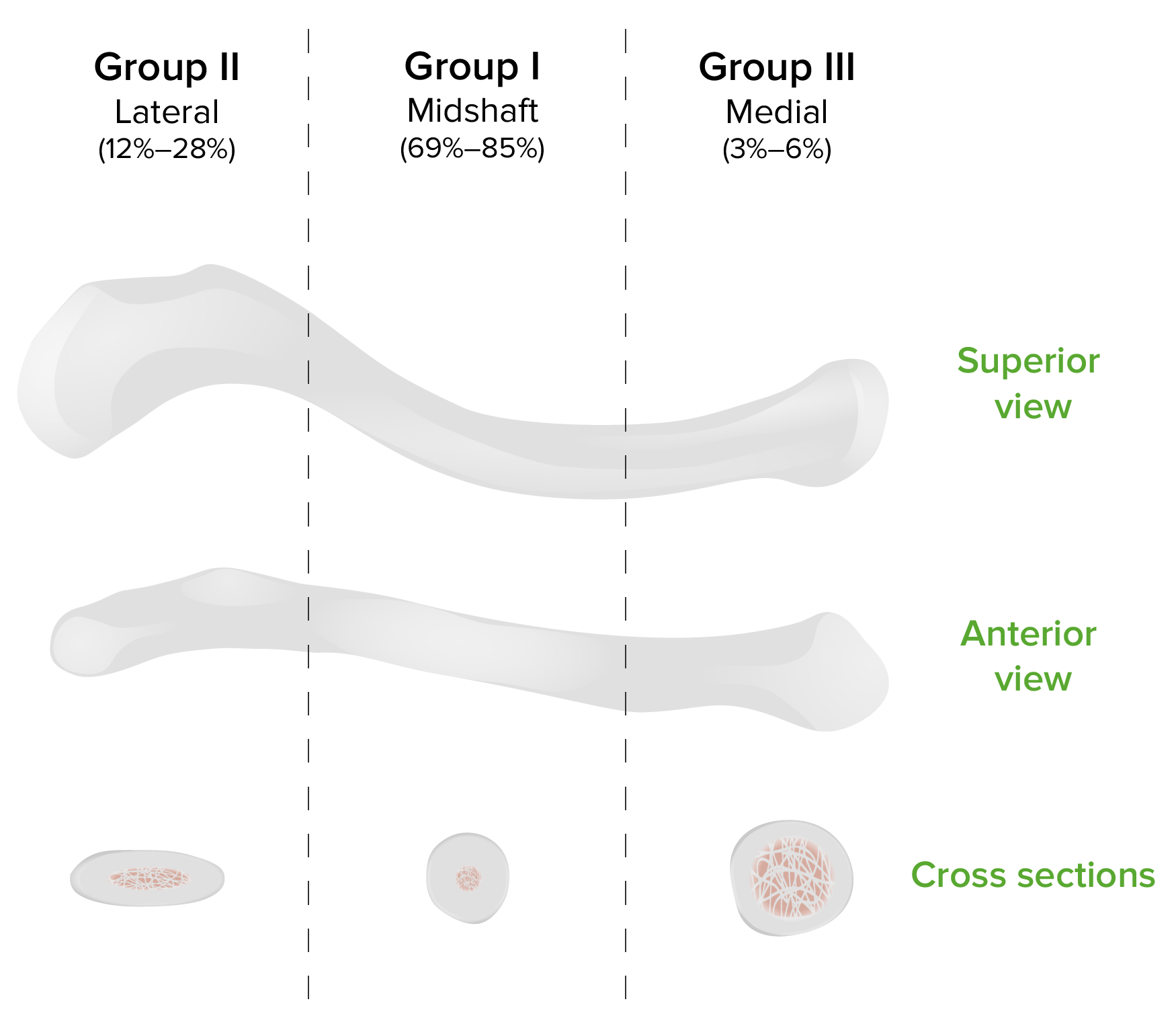
The middle third of the clavicle is the most frequent location, followed by the lateral third and then the medial third.
85% of clavicular fractures occur by a fall onto the lateral shoulder
Most common traumatic birth injury (associated with high birth weight and shoulder dystociaShoulder DystociaObstetric complication during obstetric delivery in which exit of the fetus is delayed due to physical obstruction involving fetal shoulder(s).Complications during Childbirth)
Etiology
High-energy trauma in children, adolescents, and adults
The general principle behind all fractures is that the boneBoneBone is a compact type of hardened connective tissue composed of bone cells, membranes, an extracellular mineralized matrix, and central bone marrow. The 2 primary types of bone are compact and spongy. Bones: Structure and Types is subjected to a load that overcomes its bearing capacity and the boneBoneBone is a compact type of hardened connective tissue composed of bone cells, membranes, an extracellular mineralized matrix, and central bone marrow. The 2 primary types of bone are compact and spongy. Bones: Structure and Types loses its structural integrity. Any traumatic mechanism can induce a fractureFractureA fracture is a disruption of the cortex of any bone and periosteum and is commonly due to mechanical stress after an injury or accident. Open fractures due to trauma can be a medical emergency. Fractures are frequently associated with automobile accidents, workplace injuries, and trauma.Overview of Bone Fractures if the transfer of kinetic energy is great enough.
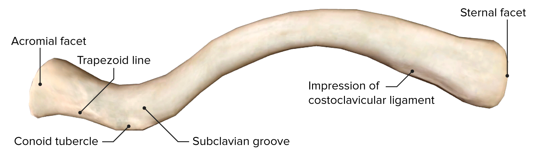
Clinical anatomy
S-shaped boneBoneBone is a compact type of hardened connective tissue composed of bone cells, membranes, an extracellular mineralized matrix, and central bone marrow. The 2 primary types of bone are compact and spongy. Bones: Structure and Types
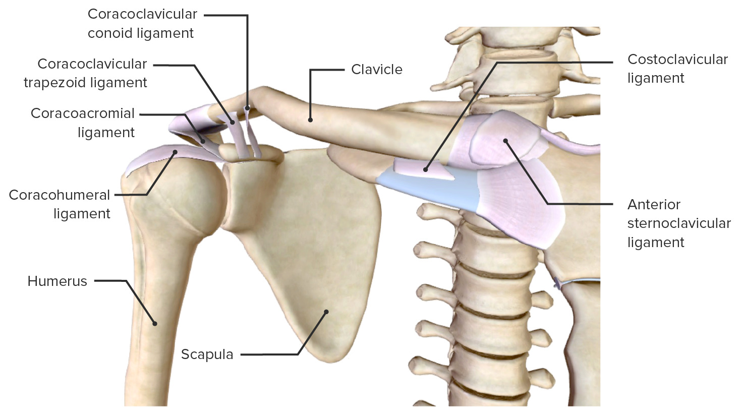
Only bony connection between the trunk and the armArmThe arm, or “upper arm” in common usage, is the region of the upper limb that extends from the shoulder to the elbow joint and connects inferiorly to the forearm through the cubital fossa. It is divided into 2 fascial compartments (anterior and posterior).Arm: Anatomy
Articulates medially with the sternumSternumA long, narrow, and flat bone commonly known as breastbone occurring in the midsection of the anterior thoracic segment or chest region, which stabilizes the rib cage and serves as the point of origin for several muscles that move the arms, head, and neck.Chest Wall: Anatomy and laterally with the acromion
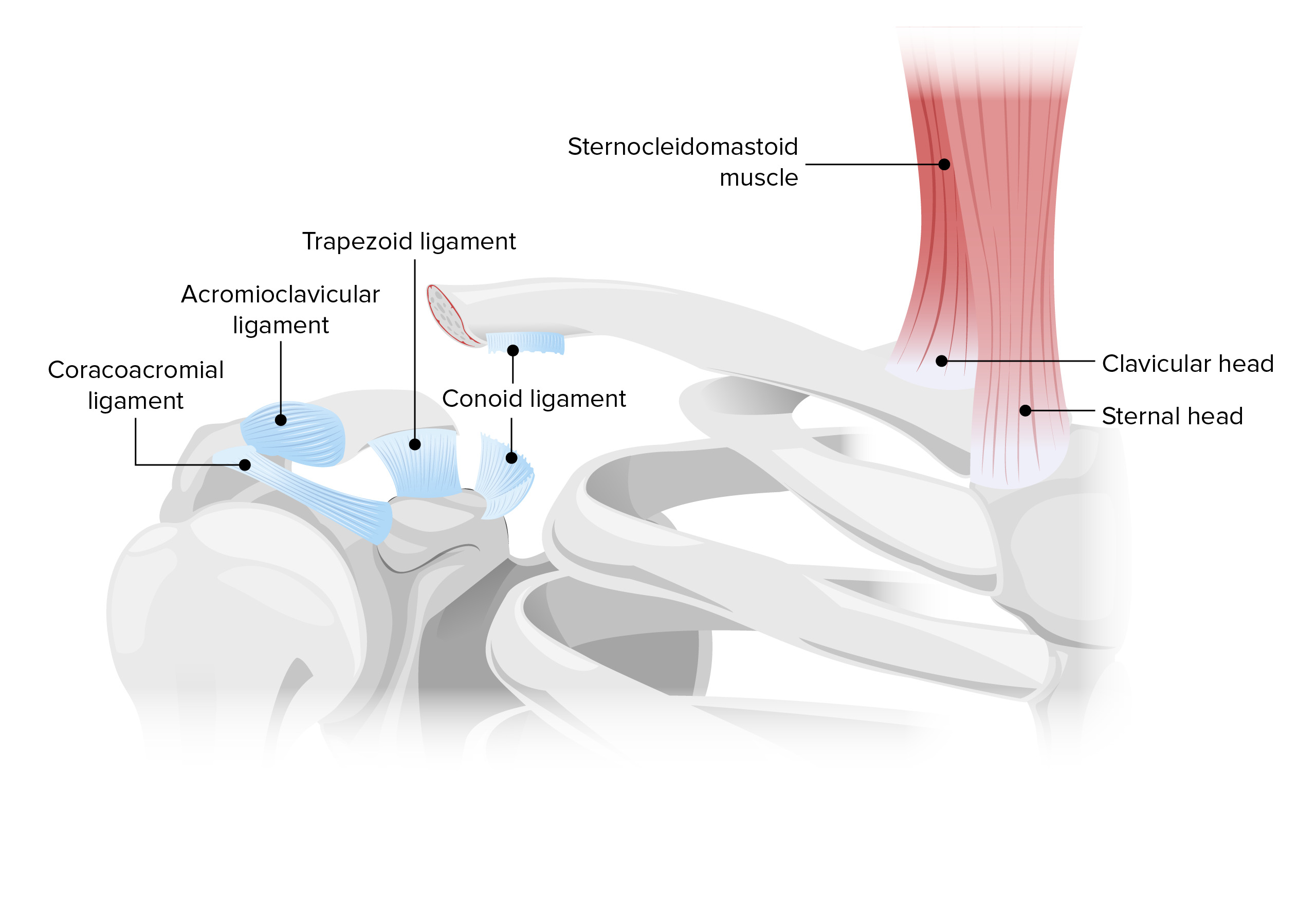
With a displaced fractureFractureA fracture is a disruption of the cortex of any bone and periosteum and is commonly due to mechanical stress after an injury or accident. Open fractures due to trauma can be a medical emergency. Fractures are frequently associated with automobile accidents, workplace injuries, and trauma.Overview of Bone Fractures, the proximal fragment is almost always displaced superiorly by the pull of the sternocleidomastoidSternocleidomastoidMuscles of the Neck: Anatomy muscle
Note the bony and ligamentous attachments. Image by BioDigital, edited by Lecturio
Displacement pattern of a typical clavicle fracture: Note the proximal portion of the clavicle being displaced superiorly by the sternocleidomastoid muscle.
Direct fall onto the lateral shoulder (the vast majority)
Fall onto an outstretched handHandThe hand constitutes the distal part of the upper limb and provides the fine, precise movements needed in activities of daily living. It consists of 5 metacarpal bones and 14 phalanges, as well as numerous muscles innervated by the median and ulnar nerves. Hand: Anatomy
Least common type of clavicle fractureFractureA fracture is a disruption of the cortex of any bone and periosteum and is commonly due to mechanical stress after an injury or accident. Open fractures due to trauma can be a medical emergency. Fractures are frequently associated with automobile accidents, workplace injuries, and trauma.Overview of Bone Fractures but most often associated with a serious injury
Associated with high-energy trauma and chest, head, and neckNeckThe part of a human or animal body connecting the head to the rest of the body.Peritonsillar Abscess injuries
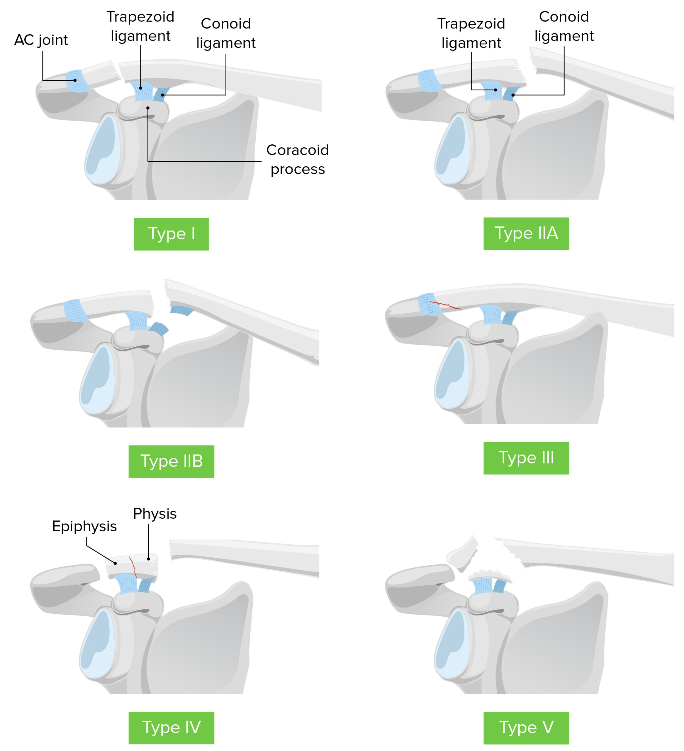
Distal third clavicle fractures (group II)
May be confused with acromioclavicular injuries
Classified based on location of the fractureFractureA fracture is a disruption of the cortex of any bone and periosteum and is commonly due to mechanical stress after an injury or accident. Open fractures due to trauma can be a medical emergency. Fractures are frequently associated with automobile accidents, workplace injuries, and trauma.Overview of Bone Fractures and ligamentous stability
Neer’s classification of distal end clavicle fracture AC: acromioclavicular
The examination of a clavicular fractureClavicular FractureAcute Shoulder Pain depends on the clinical situation, although the majority of clavicular fractures present with a classic midshaft, group I fractureFractureA fracture is a disruption of the cortex of any bone and periosteum and is commonly due to mechanical stress after an injury or accident. Open fractures due to trauma can be a medical emergency. Fractures are frequently associated with automobile accidents, workplace injuries, and trauma.Overview of Bone Fractures. If the fractureFractureA fracture is a disruption of the cortex of any bone and periosteum and is commonly due to mechanical stress after an injury or accident. Open fractures due to trauma can be a medical emergency. Fractures are frequently associated with automobile accidents, workplace injuries, and trauma.Overview of Bone Fractures occurred in the context of high-energy trauma, patientsPatientsIndividuals participating in the health care system for the purpose of receiving therapeutic, diagnostic, or preventive procedures.Clinician–Patient Relationship may require simultaneous examination and management following the Advanced Trauma Life Support (ATLS) method (most commonly seen with group III fractures).
History
PatientsPatientsIndividuals participating in the health care system for the purpose of receiving therapeutic, diagnostic, or preventive procedures.Clinician–Patient Relationship, or first responders, will report trauma:
Recent
High-energy blunt or penetrating trauma
Vehicle accident
Fall from height
High-impact sports injury
Attack with a blunt or sharp object
The clinicianClinicianA physician, nurse practitioner, physician assistant, or another health professional who is directly involved in patient care and has a professional relationship with patients.Clinician–Patient Relationship must assess the mechanism of trauma and injury mechanism details:
Vehicle accidents: types of restraints, airbags, patient position in the vehicle, status of other passengers
Fall mechanism
In elderly patientsPatientsIndividuals participating in the health care system for the purpose of receiving therapeutic, diagnostic, or preventive procedures.Clinician–Patient Relationship, emphasis is on comorbiditiesComorbiditiesThe presence of co-existing or additional diseases with reference to an initial diagnosis or with reference to the index condition that is the subject of study. Comorbidity may affect the ability of affected individuals to function and also their survival; it may be used as a prognostic indicator for length of hospital stay, cost factors, and outcome or survival.St. Louis Encephalitis Virus and fall risk:
Factors that increase risk of syncopeSyncopeSyncope is a short-term loss of consciousness and loss of postural stability followed by spontaneous return of consciousness to the previous neurologic baseline without the need for resuscitation. The condition is caused by transient interruption of cerebral blood flow that may be benign or related to a underlying life-threatening condition. Syncope:
Stroke
MIMIMI is ischemia and death of an area of myocardial tissue due to insufficient blood flow and oxygenation, usually from thrombus formation on a ruptured atherosclerotic plaque in the epicardial arteries. Clinical presentation is most commonly with chest pain, but women and patients with diabetes may have atypical symptoms.Myocardial Infarction
Arrhythmia
Factors that increase risk of fall:
ArthritisArthritisAcute or chronic inflammation of joints.Osteoarthritis in weight-bearing joints
PainPainAn unpleasant sensation induced by noxious stimuli which are detected by nerve endings of nociceptive neurons.Pain: Types and Pathways at location of the clavicle fractureFractureA fracture is a disruption of the cortex of any bone and periosteum and is commonly due to mechanical stress after an injury or accident. Open fractures due to trauma can be a medical emergency. Fractures are frequently associated with automobile accidents, workplace injuries, and trauma.Overview of Bone Fractures, exacerbated with motion or palpationPalpationApplication of fingers with light pressure to the surface of the body to determine consistency of parts beneath in physical diagnosis; includes palpation for determining the outlines of organs.Dermatologic Examination
Visible/palpable deformityDeformityExamination of the Upper Limbs over the fractureFractureA fracture is a disruption of the cortex of any bone and periosteum and is commonly due to mechanical stress after an injury or accident. Open fractures due to trauma can be a medical emergency. Fractures are frequently associated with automobile accidents, workplace injuries, and trauma.Overview of Bone Fractures site:
Tenting of skinSkinThe skin, also referred to as the integumentary system, is the largest organ of the body. The skin is primarily composed of the epidermis (outer layer) and dermis (deep layer). The epidermis is primarily composed of keratinocytes that undergo rapid turnover, while the dermis contains dense layers of connective tissue.Skin: Structure and Functions may occur.
EcchymosisEcchymosisExtravasation of blood into the skin, resulting in a nonelevated, rounded or irregular, blue or purplish patch, larger than a petechia.Orbital Fractures/hematomaHematomaA collection of blood outside the blood vessels. Hematoma can be localized in an organ, space, or tissue.Intussusception may be present.
DisplacementDisplacementThe process by which an emotional or behavioral response that is appropriate for one situation appears in another situation for which it is inappropriate.Defense Mechanisms:
Lateral fragment may be displaced inferiorly and medially because of unopposed action of pectoralis major muscle.
Assess for possible concomitant fractures/dislocations/subluxations:
Fractures:
RibsRibsA set of twelve curved bones which connect to the vertebral column posteriorly, and terminate anteriorly as costal cartilage. Together, they form a protective cage around the internal thoracic organs.Chest Wall: Anatomy
SternumSternumA long, narrow, and flat bone commonly known as breastbone occurring in the midsection of the anterior thoracic segment or chest region, which stabilizes the rib cage and serves as the point of origin for several muscles that move the arms, head, and neck.Chest Wall: Anatomy
HumerusHumerusBone in humans and primates extending from the shoulder joint to the elbow joint.Arm: Anatomy
Injury to the subclavian artery → decreased pulses/pallor in affected limb
Injury to the brachial plexusBrachial PlexusThe large network of nerve fibers which distributes the innervation of the upper extremity. The brachial plexus extends from the neck into the axilla. In humans, the nerves of the plexus usually originate from the lower cervical and the first thoracic spinal cord segments (c5-c8 and T1), but variations are not uncommon.Peripheral Nerve Injuries in the Cervicothoracic Region → motorMotorNeurons which send impulses peripherally to activate muscles or secretory cells.Nervous System: Histology/sensorySensoryNeurons which conduct nerve impulses to the central nervous system.Nervous System: Histology abnormalities in affected limb
Assess for pulmonary injury/pneumothoraxPneumothoraxA pneumothorax is a life-threatening condition in which air collects in the pleural space, causing partial or full collapse of the lung. A pneumothorax can be traumatic or spontaneous. Patients present with a sudden onset of sharp chest pain, dyspnea, and diminished breath sounds on exam.Pneumothorax:
DyspneaDyspneaDyspnea is the subjective sensation of breathing discomfort. Dyspnea is a normal manifestation of heavy physical or psychological exertion, but also may be caused by underlying conditions (both pulmonary and extrapulmonary). Dyspnea
Decreased lung sounds and lung excursion
Hyperresonance to percussionPercussionAct of striking a part with short, sharp blows as an aid in diagnosing the condition beneath the sound obtained.Pulmonary Examination
Diagnosis
Diagnosis is initially made clinically and confirmed with diagnostic imaging (X-rayX-rayPenetrating electromagnetic radiation emitted when the inner orbital electrons of an atom are excited and release radiant energy. X-ray wavelengths range from 1 pm to 10 nm. Hard x-rays are the higher energy, shorter wavelength x-rays. Soft x-rays or grenz rays are less energetic and longer in wavelength. The short wavelength end of the x-ray spectrum overlaps the gamma rays wavelength range. The distinction between gamma rays and x-rays is based on their radiation source.Pulmonary Function Tests).
X-rayX-rayPenetrating electromagnetic radiation emitted when the inner orbital electrons of an atom are excited and release radiant energy. X-ray wavelengths range from 1 pm to 10 nm. Hard x-rays are the higher energy, shorter wavelength x-rays. Soft x-rays or grenz rays are less energetic and longer in wavelength. The short wavelength end of the x-ray spectrum overlaps the gamma rays wavelength range. The distinction between gamma rays and x-rays is based on their radiation source.Pulmonary Function Tests
Anteroposterior (AP) view usually sufficient for diagnosis and classification
Chest X-rayX-rayPenetrating electromagnetic radiation emitted when the inner orbital electrons of an atom are excited and release radiant energy. X-ray wavelengths range from 1 pm to 10 nm. Hard x-rays are the higher energy, shorter wavelength x-rays. Soft x-rays or grenz rays are less energetic and longer in wavelength. The short wavelength end of the x-ray spectrum overlaps the gamma rays wavelength range. The distinction between gamma rays and x-rays is based on their radiation source.Pulmonary Function Tests is indicated if clinical suspicion of:
PneumothoraxPneumothoraxA pneumothorax is a life-threatening condition in which air collects in the pleural space, causing partial or full collapse of the lung. A pneumothorax can be traumatic or spontaneous. Patients present with a sudden onset of sharp chest pain, dyspnea, and diminished breath sounds on exam.Pneumothorax
Sternal fractureFractureA fracture is a disruption of the cortex of any bone and periosteum and is commonly due to mechanical stress after an injury or accident. Open fractures due to trauma can be a medical emergency. Fractures are frequently associated with automobile accidents, workplace injuries, and trauma.Overview of Bone Fractures
Shoulder X-raysX-raysX-rays are high-energy particles of electromagnetic radiation used in the medical field for the generation of anatomical images. X-rays are projected through the body of a patient and onto a film, and this technique is called conventional or projectional radiography. X-rays indicated if clinical suspicion of:
Glenoid or scapular fractureFractureA fracture is a disruption of the cortex of any bone and periosteum and is commonly due to mechanical stress after an injury or accident. Open fractures due to trauma can be a medical emergency. Fractures are frequently associated with automobile accidents, workplace injuries, and trauma.Overview of Bone Fractures
Humeral fractureFractureA fracture is a disruption of the cortex of any bone and periosteum and is commonly due to mechanical stress after an injury or accident. Open fractures due to trauma can be a medical emergency. Fractures are frequently associated with automobile accidents, workplace injuries, and trauma.Overview of Bone Fractures (proximal)
Cervical X-raysX-raysX-rays are high-energy particles of electromagnetic radiation used in the medical field for the generation of anatomical images. X-rays are projected through the body of a patient and onto a film, and this technique is called conventional or projectional radiography. X-rays if clinical suspicion of vertebral fractureFractureA fracture is a disruption of the cortex of any bone and periosteum and is commonly due to mechanical stress after an injury or accident. Open fractures due to trauma can be a medical emergency. Fractures are frequently associated with automobile accidents, workplace injuries, and trauma.Overview of Bone Fractures
FractureFractureA fracture is a disruption of the cortex of any bone and periosteum and is commonly due to mechanical stress after an injury or accident. Open fractures due to trauma can be a medical emergency. Fractures are frequently associated with automobile accidents, workplace injuries, and trauma.Overview of Bone Fractures description:
Location of fractureFractureA fracture is a disruption of the cortex of any bone and periosteum and is commonly due to mechanical stress after an injury or accident. Open fractures due to trauma can be a medical emergency. Fractures are frequently associated with automobile accidents, workplace injuries, and trauma.Overview of Bone Fractures (Allman classification):
Increased opacityOpacityImaging of the Lungs and Pleura of soft tissues (inflammationInflammationInflammation is a complex set of responses to infection and injury involving leukocytes as the principal cellular mediators in the body’s defense against pathogenic organisms. Inflammation is also seen as a response to tissue injury in the process of wound healing. The 5 cardinal signs of inflammation are pain, heat, redness, swelling, and loss of function. Inflammation and edemaEdemaEdema is a condition in which excess serous fluid accumulates in the body cavity or interstitial space of connective tissues. Edema is a symptom observed in several medical conditions. It can be categorized into 2 types, namely, peripheral (in the extremities) and internal (in an organ or body cavity). Edema)
Periosteal reaction/callus formation (older fractureFractureA fracture is a disruption of the cortex of any bone and periosteum and is commonly due to mechanical stress after an injury or accident. Open fractures due to trauma can be a medical emergency. Fractures are frequently associated with automobile accidents, workplace injuries, and trauma.Overview of Bone Fractures)
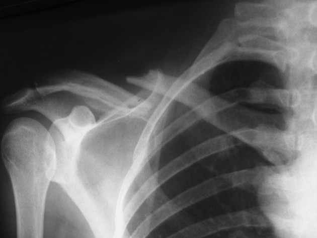
X-ray image of middle third, or midshaft, comminuted fracture of the clavicle
Image: “Comminuted Midshaft Clavicular Fracture” by Keihan Shokouh H et al. License: CC BY 3.0
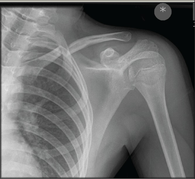
X-ray image of middle third, or midshaft, fracture of the clavicle associated with acute brachial plexus injury and subclavian artery compression
Image: “Plain x-ray showing midshaft clavicle fracture” by Gill I et al. License: CC BY 3.0
CT
May be needed to guide or plan definitive management in complex or uncommon cases
Considered in the evaluation of medial third fractures with posterior displacementDisplacementThe process by which an emotional or behavioral response that is appropriate for one situation appears in another situation for which it is inappropriate.Defense Mechanisms
Considered if concomitant cervical/thoracic vertebral fractureFractureA fracture is a disruption of the cortex of any bone and periosteum and is commonly due to mechanical stress after an injury or accident. Open fractures due to trauma can be a medical emergency. Fractures are frequently associated with automobile accidents, workplace injuries, and trauma.Overview of Bone Fractures suspected
Ultrasonography is a consideration if possible arterial injuryArterial InjuryHemothorax is suspected (subclavian artery).
Management
The vast majority of clavicle fractures will heal without operative intervention. However, there remain challenges secondary to the occurrence of complications and nonunionNonunionHip Fractures of the fractureFractureA fracture is a disruption of the cortex of any bone and periosteum and is commonly due to mechanical stress after an injury or accident. Open fractures due to trauma can be a medical emergency. Fractures are frequently associated with automobile accidents, workplace injuries, and trauma.Overview of Bone Fractures. Definitive management depends on the type of fractureFractureA fracture is a disruption of the cortex of any bone and periosteum and is commonly due to mechanical stress after an injury or accident. Open fractures due to trauma can be a medical emergency. Fractures are frequently associated with automobile accidents, workplace injuries, and trauma.Overview of Bone Fractures and is often done in consultation with an orthopedic surgeon. A thoracic or vascular surgeryVascular surgeryVascular surgery is the specialized field of medicine that focuses on the surgical management of the pathologies of the peripheral circulation. The main goal of most vascular procedures is to restore circulatory function to the affected vessels by relieving occlusions or by redirecting blood flow (e.g., bypass).Vascular Surgery consult may be indicated in a small number of clavicle fractures with associated pneumothoraxPneumothoraxA pneumothorax is a life-threatening condition in which air collects in the pleural space, causing partial or full collapse of the lung. A pneumothorax can be traumatic or spontaneous. Patients present with a sudden onset of sharp chest pain, dyspnea, and diminished breath sounds on exam.Pneumothorax or vascular injury.
Indications for orthopedic surgical referral
Complete fractureComplete FractureOverview of Bone FracturesdisplacementDisplacementThe process by which an emotional or behavioral response that is appropriate for one situation appears in another situation for which it is inappropriate.Defense Mechanisms (displacementDisplacementThe process by which an emotional or behavioral response that is appropriate for one situation appears in another situation for which it is inappropriate.Defense Mechanisms > 1 boneBoneBone is a compact type of hardened connective tissue composed of bone cells, membranes, an extracellular mineralized matrix, and central bone marrow. The 2 primary types of bone are compact and spongy. Bones: Structure and Types width)
Longitudinal shortening ≥ 2 cm, severe tenting of the skinSkinThe skin, also referred to as the integumentary system, is the largest organ of the body. The skin is primarily composed of the epidermis (outer layer) and dermis (deep layer). The epidermis is primarily composed of keratinocytes that undergo rapid turnover, while the dermis contains dense layers of connective tissue.Skin: Structure and Functions with risk of puncture
Comminuted fractures
Open fractures
Displaced medial clavicular fractures
Type II distal clavicular fractures
Neurovascular compromise
Associated glenoid fractureFractureA fracture is a disruption of the cortex of any bone and periosteum and is commonly due to mechanical stress after an injury or accident. Open fractures due to trauma can be a medical emergency. Fractures are frequently associated with automobile accidents, workplace injuries, and trauma.Overview of Bone Fractures (floating shoulder)
Management depends on displacementDisplacementThe process by which an emotional or behavioral response that is appropriate for one situation appears in another situation for which it is inappropriate.Defense Mechanisms, age, and level of activity
The focus remains on nonoperative management, although recently, there is more support for surgical management for some clavicle fractures
Nonoperative:
Sling versus figure 8 brace: literature shows no real difference in outcomes
Healing time 6–8 weeks in adults
Surgical intervention:
Increasing evidence for operative treatment of displaced clavicle fractures
Surgical options:
Intramedullary fixation
Plate-and-screw fixation
Suggested indications:
Complicated fractures of the middle third
Fractures overlapped > 2 cm
Severe tenting of the skinSkinThe skin, also referred to as the integumentary system, is the largest organ of the body. The skin is primarily composed of the epidermis (outer layer) and dermis (deep layer). The epidermis is primarily composed of keratinocytes that undergo rapid turnover, while the dermis contains dense layers of connective tissue.Skin: Structure and Functions
Orthopedic referral for possible surgical intervention
High incidenceIncidenceThe number of new cases of a given disease during a given period in a specified population. It also is used for the rate at which new events occur in a defined population. It is differentiated from prevalence, which refers to all cases in the population at a given time.Measures of Disease Frequency of nonunionNonunionHip Fractures
Generally, type II fractures of the distal clavicle (level of the coracoclavicular ligaments) are treated surgically
Group III fractures (medial third):
Generally treated conservatively
Essential to assess for other associated thorax and neckNeckThe part of a human or animal body connecting the head to the rest of the body.Peritonsillar Abscess injuries
A posterior sternoclavicular dislocationSternoclavicular DislocationAcute Shoulder Pain needs emergent evaluation because of the potential for great-vessel injury and other intrathoracic injuries
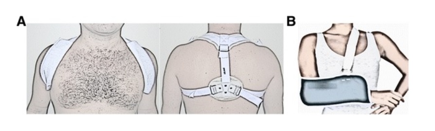
Conservative management
The most widely used devices are the sling, sling and swathe, and figure 8 brace
Range of motionRange of motionThe distance and direction to which a bone joint can be extended. Range of motion is a function of the condition of the joints, muscles, and connective tissues involved. Joint flexibility can be improved through appropriate muscle strength exercises.Examination of the Upper Limbs (ROM) exercises of the elbow and wrist are started within 2–3 days of injury.
Fewer complications and a faster recovery in nondisplaced midshaft fractures
NonunionNonunionHip Fractures: occurs when fractureFractureA fracture is a disruption of the cortex of any bone and periosteum and is commonly due to mechanical stress after an injury or accident. Open fractures due to trauma can be a medical emergency. Fractures are frequently associated with automobile accidents, workplace injuries, and trauma.Overview of Bone Fractures has not healed in 6 months
Conservative management of clavicle fractures: A. Figure 8 bandage B. Simple arm sling
Image: “Conservative management of clavicle fractures” by Lenza M et al. License: CC BY 4.0
Surgical management
Recent literature has challenged the belief that all midshaft fractures heal without difficulty.
Open reduction and internal fixation of the fractureFractureA fracture is a disruption of the cortex of any bone and periosteum and is commonly due to mechanical stress after an injury or accident. Open fractures due to trauma can be a medical emergency. Fractures are frequently associated with automobile accidents, workplace injuries, and trauma.Overview of Bone Fractures with either:
Plate fixation
Intramedullary pin fixation
Surgical complicationsSurgical complicationsSurgical complications are conditions, disorders, or adverse events that occur following surgical procedures. The most common general surgical complications include bleeding, infections, injury to the surrounding organs, venous thromboembolic events, and complications from anesthesia.Surgical Complications:
Infection
Hardware failure or migration
SkinSkinThe skin, also referred to as the integumentary system, is the largest organ of the body. The skin is primarily composed of the epidermis (outer layer) and dermis (deep layer). The epidermis is primarily composed of keratinocytes that undergo rapid turnover, while the dermis contains dense layers of connective tissue.Skin: Structure and Functions breakdown
Brachial plexusBrachial PlexusThe large network of nerve fibers which distributes the innervation of the upper extremity. The brachial plexus extends from the neck into the axilla. In humans, the nerves of the plexus usually originate from the lower cervical and the first thoracic spinal cord segments (c5-c8 and T1), but variations are not uncommon.Peripheral Nerve Injuries in the Cervicothoracic Region injury
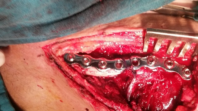
Surgical repair of clavicular fracture: Open reduction and plate osteosynthesis
Image: “Open reduction and plate osteosynthesis of the clavicle fracture” by Redouane H et al. License: CC BY 2.0
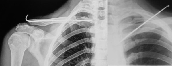
Postoperative clavicle X-ray: K-wire fixation on middle third clavicle fracture
Image: “Xray shows a K-wire fixation on middle third clavicle fracture and the migration of K-wire” by Paladini P et al. License: CC BY 2.5
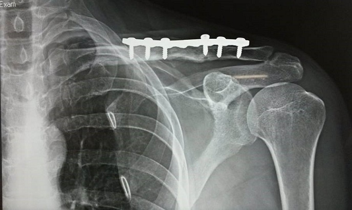
Postoperative clavicle X-ray: Callus formation of the fractured clavicle fixed with plate and screws
Image: “Chest radiographs showing callus formation of the fractured clavicle 14 months after the injury with no recurrence of pneumothorax” by Hani R et al. License: CC BY 2.0
Rehabilitation
As soon as tolerated, ROM exercises begin with the elbow and wrist with gentle pendulum exercise for the shoulder.
2–4 months required for athletes in contact sports and evidence of radiologic healing
Complications related to a clavicle fractureFractureA fracture is a disruption of the cortex of any bone and periosteum and is commonly due to mechanical stress after an injury or accident. Open fractures due to trauma can be a medical emergency. Fractures are frequently associated with automobile accidents, workplace injuries, and trauma.Overview of Bone Fractures
Direct injury to the subclavian artery or brachial plexusBrachial PlexusThe large network of nerve fibers which distributes the innervation of the upper extremity. The brachial plexus extends from the neck into the axilla. In humans, the nerves of the plexus usually originate from the lower cervical and the first thoracic spinal cord segments (c5-c8 and T1), but variations are not uncommon.Peripheral Nerve Injuries in the Cervicothoracic Region
PneumothoraxPneumothoraxA pneumothorax is a life-threatening condition in which air collects in the pleural space, causing partial or full collapse of the lung. A pneumothorax can be traumatic or spontaneous. Patients present with a sudden onset of sharp chest pain, dyspnea, and diminished breath sounds on exam.Pneumothorax due to injury to the lung apex
NonunionNonunionHip Fractures of the fractureFractureA fracture is a disruption of the cortex of any bone and periosteum and is commonly due to mechanical stress after an injury or accident. Open fractures due to trauma can be a medical emergency. Fractures are frequently associated with automobile accidents, workplace injuries, and trauma.Overview of Bone Fractures
Posttraumatic arthritisArthritisAcute or chronic inflammation of joints.Osteoarthritis (acromioclavicular (AC) or sternoclavicular (SC) joint)
PrognosisPrognosisA prediction of the probable outcome of a disease based on a individual’s condition and the usual course of the disease as seen in similar situations.Non-Hodgkin Lymphomas
Most clavicular fractures are managed conservatively with good results.
Support for surgical treatment of middle third fractures is increasing.
Serious complications are uncommon; most common complication is a malunionMalunionHip Fractures.
PatientsPatientsIndividuals participating in the health care system for the purpose of receiving therapeutic, diagnostic, or preventive procedures.Clinician–Patient Relationship return to sports/work based on:
FractureFractureA fracture is a disruption of the cortex of any bone and periosteum and is commonly due to mechanical stress after an injury or accident. Open fractures due to trauma can be a medical emergency. Fractures are frequently associated with automobile accidents, workplace injuries, and trauma.Overview of Bone Fractures location
Degree of healing
Activity requirements
Majority of patientsPatientsIndividuals participating in the health care system for the purpose of receiving therapeutic, diagnostic, or preventive procedures.Clinician–Patient Relationship return to normal activities in 6–8 weeks.
Clinical Relevance
Other common diagnoses related to shoulder trauma
Rotator cuff tendon tearRotator Cuff Tendon TearChronic Shoulder Pain: injury to the tendons of the muscles that make up the rotator cuff. Rotator cuff tendon tearRotator Cuff Tendon TearChronic Shoulder Pain can occur as a result of acute trauma, falls, repetitive motion, or tendon degeneration. The tendon of the supraspinatus muscle is the most commonly torn.
Glenohumeral dislocationGlenohumeral DislocationAcute Shoulder Pain: dislocation of the humeral headHumeral headThe upper rounded extremity of the humerus fitting into the glenoid cavity of the scapula.Arm: Anatomy from the glenoid fossa. The glenohumeral joint is the most commonly dislocated joint, and > 90% of shoulder dislocations are anterior–inferior.
Acromioclavicular jointAcromioclavicular jointThe gliding joint formed by the outer extremity of the clavicle and the inner margin of the acromion process of the scapula.Examination of the Upper Limbs injury: common injury in adults. Acromioclavicular jointAcromioclavicular jointThe gliding joint formed by the outer extremity of the clavicle and the inner margin of the acromion process of the scapula.Examination of the Upper Limbs injury is caused by a fall on the lateral shoulder with the armArmThe arm, or “upper arm” in common usage, is the region of the upper limb that extends from the shoulder to the elbow joint and connects inferiorly to the forearm through the cubital fossa. It is divided into 2 fascial compartments (anterior and posterior).Arm: Anatomy adducted.
Sternoclavicular jointSternoclavicular JointExamination of the Upper Limbs injury: This type of joint injury may take the form of sprains or dislocations, and dislocations can be anterior or posterior. Posterior dislocations require emergent care because of the potential for injury to mediastinal structures.
Clinically relevant topics related to clavicle fractures and falls
PneumothoraxPneumothoraxA pneumothorax is a life-threatening condition in which air collects in the pleural space, causing partial or full collapse of the lung. A pneumothorax can be traumatic or spontaneous. Patients present with a sudden onset of sharp chest pain, dyspnea, and diminished breath sounds on exam.Pneumothorax: accumulation of air within the pleural spacePleural spaceThe thin serous membrane enveloping the lungs (lung) and lining the thoracic cavity. Pleura consist of two layers, the inner visceral pleura lying next to the pulmonary parenchyma and the outer parietal pleura. Between the two layers is the pleural cavity which contains a thin film of liquid.Pleuritis (between the parietalParietalOne of a pair of irregularly shaped quadrilateral bones situated between the frontal bone and occipital bone, which together form the sides of the cranium.Skull: Anatomy and visceral pleuraVisceral pleuraPleura: Anatomy), which can be open (in communicationCommunicationThe exchange or transmission of ideas, attitudes, or beliefs between individuals or groups.Decision-making Capacity and Legal Competence with the atmosphere) or under tension (without an opening in the chest wallChest wallThe chest wall consists of skin, fat, muscles, bones, and cartilage. The bony structure of the chest wall is composed of the ribs, sternum, and thoracic vertebrae. The chest wall serves as armor for the vital intrathoracic organs and provides the stability necessary for the movement of the shoulders and arms. Chest Wall: Anatomy). PneumothoraxPneumothoraxA pneumothorax is a life-threatening condition in which air collects in the pleural space, causing partial or full collapse of the lung. A pneumothorax can be traumatic or spontaneous. Patients present with a sudden onset of sharp chest pain, dyspnea, and diminished breath sounds on exam.Pneumothorax can occur in clavicular fractures because of injury to the lung apex.
SyncopeSyncopeSyncope is a short-term loss of consciousness and loss of postural stability followed by spontaneous return of consciousness to the previous neurologic baseline without the need for resuscitation. The condition is caused by transient interruption of cerebral blood flow that may be benign or related to a underlying life-threatening condition. Syncope: self-limited, transient loss of consciousness caused by inadequate cerebral perfusionCerebral PerfusionSyncope, most often the result of an abrupt drop of systemic BP. Syncopal episodes are brief (8–10 seconds) and are classified as cardiogenic, orthostatic, or neurally mediated.
Stroke: Stroke is an injury to brainBrainThe part of central nervous system that is contained within the skull (cranium). Arising from the neural tube, the embryonic brain is comprised of three major parts including prosencephalon (the forebrain); mesencephalon (the midbrain); and rhombencephalon (the hindbrain). The developed brain consists of cerebrum; cerebellum; and other structures in the brain stem.Nervous System: Anatomy, Structure, and Classification tissue after interruption of blood flowBlood flowBlood flow refers to the movement of a certain volume of blood through the vasculature over a given unit of time (e.g., mL per minute).Vascular Resistance, Flow, and Mean Arterial Pressure (ischemic strokeIschemic StrokeAn ischemic stroke (also known as cerebrovascular accident) is an acute neurologic injury that occurs as a result of brain ischemia; this condition may be due to cerebral blood vessel occlusion by thrombosis or embolism, or rarely due to systemic hypoperfusion. Ischemic Stroke) or active hemorrhage (hemorrhagic strokeHemorrhagic strokeStroke due to rupture of a weakened blood vessel in the brain (e.g., cerebral hemispheres; cerebellum; subarachnoid space).Subarachnoid Hemorrhage), which has characteristic neurologic clinical features.
MIMIMI is ischemia and death of an area of myocardial tissue due to insufficient blood flow and oxygenation, usually from thrombus formation on a ruptured atherosclerotic plaque in the epicardial arteries. Clinical presentation is most commonly with chest pain, but women and patients with diabetes may have atypical symptoms.Myocardial Infarction: injury to the myocardiumMyocardiumThe muscle tissue of the heart. It is composed of striated, involuntary muscle cells connected to form the contractile pump to generate blood flow.Heart: Anatomy due to ischemiaIschemiaA hypoperfusion of the blood through an organ or tissue caused by a pathologic constriction or obstruction of its blood vessels, or an absence of blood circulation.Ischemic Cell Damage. MIMIMI is ischemia and death of an area of myocardial tissue due to insufficient blood flow and oxygenation, usually from thrombus formation on a ruptured atherosclerotic plaque in the epicardial arteries. Clinical presentation is most commonly with chest pain, but women and patients with diabetes may have atypical symptoms.Myocardial Infarction is characterized by an increase in cardiac enzymesEnzymesEnzymes are complex protein biocatalysts that accelerate chemical reactions without being consumed by them. Due to the body’s constant metabolic needs, the absence of enzymes would make life unsustainable, as reactions would occur too slowly without these molecules. Basics of Enzymes (especially troponin T), ECGECGAn electrocardiogram (ECG) is a graphic representation of the electrical activity of the heart plotted against time. Adhesive electrodes are affixed to the skin surface allowing measurement of cardiac impulses from many angles. The ECG provides 3-dimensional information about the conduction system of the heart, the myocardium, and other cardiac structures. Electrocardiogram (ECG) changes suggestive of ischemiaIschemiaA hypoperfusion of the blood through an organ or tissue caused by a pathologic constriction or obstruction of its blood vessels, or an absence of blood circulation.Ischemic Cell Damage in 2 contiguous leads, and chest painPainAn unpleasant sensation induced by noxious stimuli which are detected by nerve endings of nociceptive neurons.Pain: Types and Pathways.
References
Browner, B., Jupiter, J., Krettek, C., Anderson, P. (2020). Skeletal Trauma: Basic Science, Management, and Reconstruction. Philadelphia: Elsevier.