Labor is defined as regularRegularInsulin, effective uterine contractions resulting in cervical changes that culminate in expulsion of the fetus and products of conception. Complications may arise during childbirth that necessitate prompt recognition and management by the delivering team. Four important complications/topics related to the moments surrounding delivery include episiotomy and lacerations, operative vaginal deliveries (forcepsForcepsSurgical Instruments and Sutures and vacuum-assisted deliveries), shoulder dystocia, and amniotic fluidAmniotic fluidA clear, yellowish liquid that envelopes the fetus inside the sac of amnion. In the first trimester, it is likely a transudate of maternal or fetal plasma. In the second trimester, amniotic fluid derives primarily from fetal lung and kidney. Cells or substances in this fluid can be removed for prenatal diagnostic tests (amniocentesis).Placenta, Umbilical Cord, and Amniotic Cavity embolism.
The perineumPerineumThe body region lying between the genital area and the anus on the surface of the trunk, and to the shallow compartment lying deep to this area that is inferior to the pelvic diaphragm. The surface area is between the vulva and the anus in the female, and between the scrotum and the anus in the male.Vagina, Vulva, and Pelvic Floor: Anatomy is the space between the vaginal and anal orifices.
Thick, circular, striated muscleStriated muscleOne of two types of muscle in the body, characterized by the array of bands observed under microscope. Striated muscles can be divided into two subtypes: the cardiac muscle and the skeletal muscle.Muscle Tissue: Histology surrounding the anal orifice
Responsible for solid, liquid, and gas continence at rest and during rectal distention
Thin condensation of the smooth muscle of the distal colonColonThe large intestines constitute the last portion of the digestive system. The large intestine consists of the cecum, appendix, colon (with ascending, transverse, descending, and sigmoid segments), rectum, and anal canal. The primary function of the colon is to remove water and compact the stool prior to expulsion from the body via the rectum and anal canal. Colon, Cecum, and Appendix: Anatomy submucosa
Responsible for continence at rest
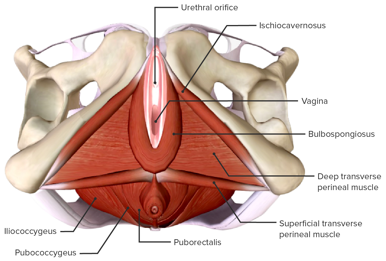
Muscular anatomy of the perineum and pelvic floor, external view
Lacerations are spontaneous tears that occur due to the trauma of the infant passing through the vaginal canal during delivery.
Epidemiology:
Up to 80% of women will sustain some type of lacerationLacerationTorn, ragged, mangled wounds.Blunt Chest Trauma at vaginal delivery.
Most are 1st- and 2nd-degree tears.
Location:
Perineal (most common)
Periclitoral
Periurethral
Labial
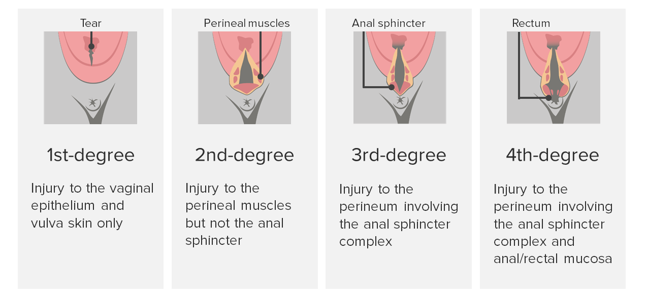
Classifications of perineal lacerations:
1st-degree:
Tear extends to the vaginal epitheliumEpitheliumThe epithelium is a complex of specialized cellular organizations arranged into sheets and lining cavities and covering the surfaces of the body. The cells exhibit polarity, having an apical and a basal pole. Structures important for the epithelial integrity and function involve the basement membrane, the semipermeable sheet on which the cells rest, and interdigitations, as well as cellular junctions. Surface Epithelium: Histology and vulvaVulvaThe vulva is the external genitalia of the female and includes the mons pubis, labia majora, labia minora, clitoris, vestibule, vestibular bulb, and greater vestibular glands. Vagina, Vulva, and Pelvic Floor: AnatomyskinSkinThe skin, also referred to as the integumentary system, is the largest organ of the body. The skin is primarily composed of the epidermis (outer layer) and dermis (deep layer). The epidermis is primarily composed of keratinocytes that undergo rapid turnover, while the dermis contains dense layers of connective tissue.Skin: Structure and Functions only.
Pelvic organ prolapsePelvic Organ ProlapsePelvic organ prolapse (POP) is a general term that refers to herniation of 1 or more pelvic organs (e.g., bladder, uterus, rectum) into the vaginal canal, and potentially all the way through the introitus. Weakness and insufficiency of the pelvic floor may result in POP.Pelvic Organ Prolapse
Rectovaginal fistulas
PainPainAn unpleasant sensation induced by noxious stimuli which are detected by nerve endings of nociceptive neurons.Pain: Types and Pathways, including dyspareuniaDyspareuniaRecurrent genital pain occurring during, before, or after sexual intercourse in either the male or the female.Primary Ovarian Insufficiency
Degrees of perineal lacerations
Image by Lecturio.
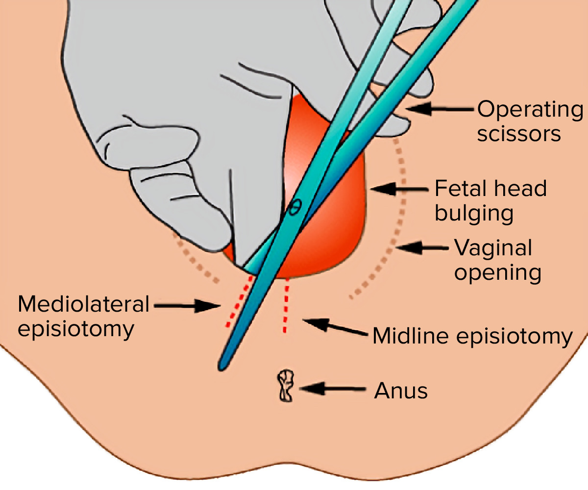
Episiotomy
An episiotomyis an intentional 3–5-cm incision made by the delivering provider to enlarge the vaginal opening at the time of delivery.
Not recommended for routine deliveries
Indications:
Woman at high risk for a 3rd- or 4th-degree lacerationLacerationTorn, ragged, mangled wounds.Blunt Chest Trauma.
Nonreassuring fetal heart rateHeart rateThe number of times the heart ventricles contract per unit of time, usually per minute.Cardiac Physiology tracing that warrants hastening vaginal delivery
Create space for the provider to:
Perform internal maneuvers to relieve a shoulder dystocia
3rd- or 4th-degree lacerations in subsequent deliveries
Greater blood loss
Infection and dehiscence
DyspareuniaDyspareuniaRecurrent genital pain occurring during, before, or after sexual intercourse in either the male or the female.Primary Ovarian Insufficiency at 1 year postpartum
Midline versus mediolateral episiotomy
Image: “Medio-lateral-episiotomy” by Jeremykemp. License: Public Domain, edited by Lecturio.
Overall incidenceIncidenceThe number of new cases of a given disease during a given period in a specified population. It also is used for the rate at which new events occur in a defined population. It is differentiated from prevalence, which refers to all cases in the population at a given time.Measures of Disease Frequency: approximately 3% of deliveries
Fetal distress in the 2nd stage of labor2nd stage of laborThe period of obstetric labor that is from the complete dilatation of the cervix uteri to the expulsion of the fetus.Normal and Abnormal Labor (2nd stage: time from complete dilation through delivery of the fetus; “pushing”)
Prolonged or arrested 2nd stage of labor2nd stage of laborThe period of obstetric labor that is from the complete dilatation of the cervix uteri to the expulsion of the fetus.Normal and Abnormal Labor
Shortening of the 2nd stage of labor2nd stage of laborThe period of obstetric labor that is from the complete dilatation of the cervix uteri to the expulsion of the fetus.Normal and Abnormal Labor for maternal medical indications (conditions that prevent safe maternal pushing):
Maternal cardiac disease (e.g., heart failureHeart FailureA heterogeneous condition in which the heart is unable to pump out sufficient blood to meet the metabolic need of the body. Heart failure can be caused by structural defects, functional abnormalities (ventricular dysfunction), or a sudden overload beyond its capacity. Chronic heart failure is more common than acute heart failure which results from sudden insult to cardiac function, such as myocardial infarction.Total Anomalous Pulmonary Venous Return (TAPVR))
At least +2 station (fetal head is 2 cm below the maternal ischial spineSpineThe human spine, or vertebral column, is the most important anatomical and functional axis of the human body. It consists of 7 cervical vertebrae, 12 thoracic vertebrae, and 5 lumbar vertebrae and is limited cranially by the skull and caudally by the sacrum.Vertebral Column: Anatomy)
Knowledge of the fetal positionFetal positionDirection of the fetal head in relation to the maternal pelvis in vertex presentationsNormal and Abnormal Labor (e.g., occiput anterior versus posterior)
Adequate anesthesiaAnesthesiaA state characterized by loss of feeling or sensation. This depression of nerve function is usually the result of pharmacologic action and is induced to allow performance of surgery or other painful procedures.Anesthesiology: History and Basic Concepts
Empty maternal bladderBladderA musculomembranous sac along the urinary tract. Urine flows from the kidneys into the bladder via the ureters, and is held there until urination.Pyelonephritis and Perinephric Abscess
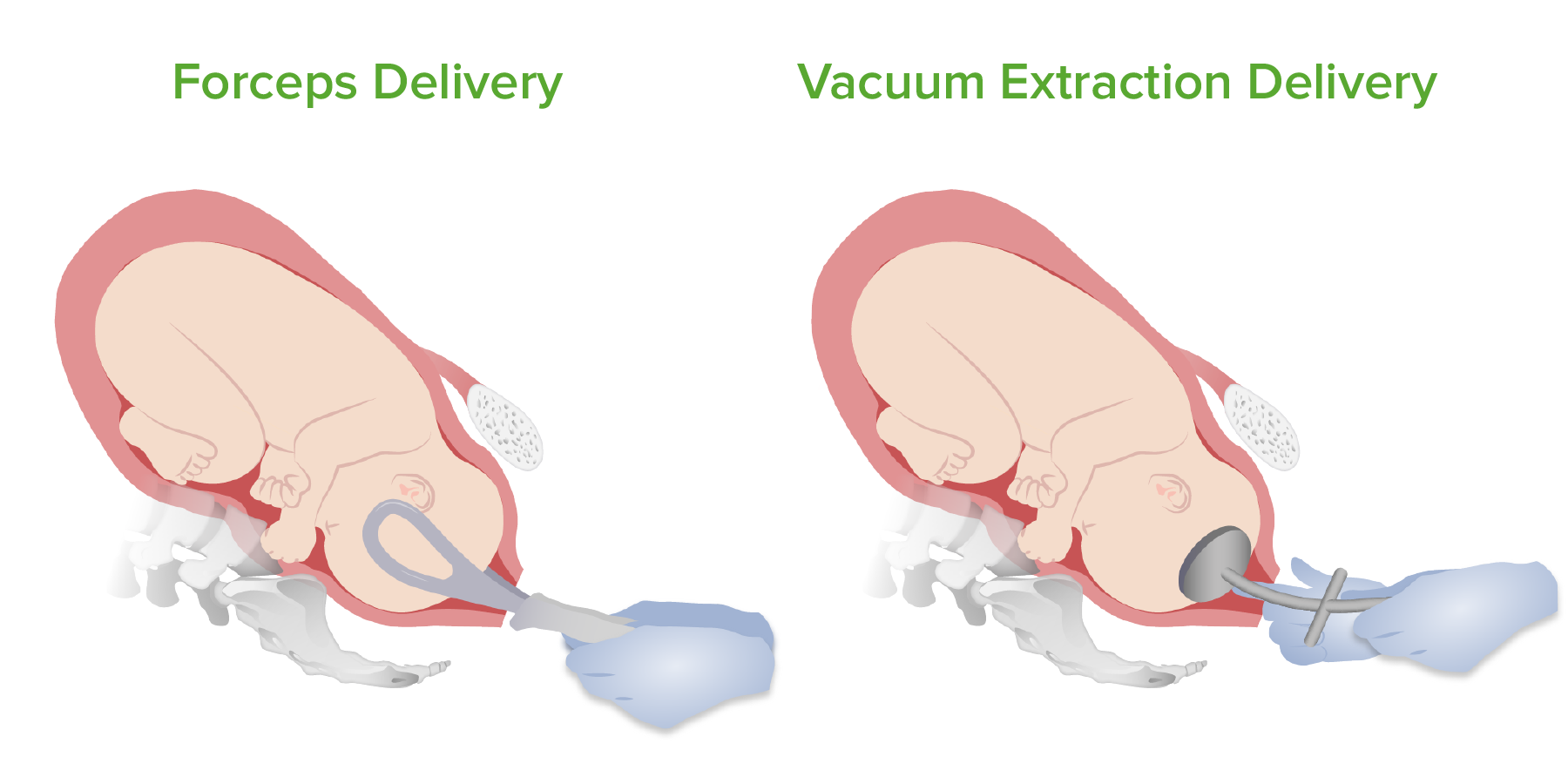
Forceps and vacuum deliveries
Image by Lecturio.
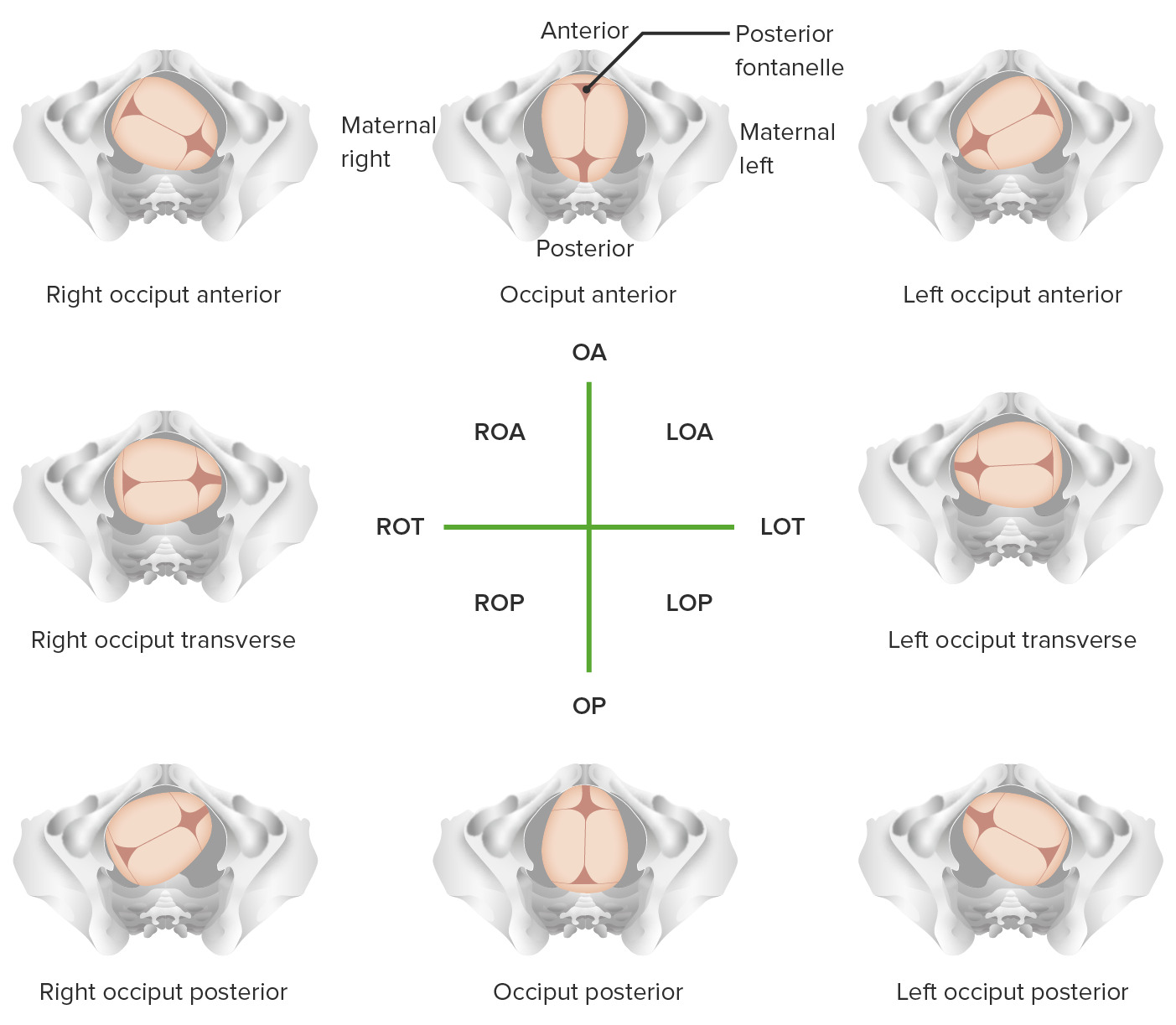
Fetal head position: Position is determined by the location of the fetal occiput (posterior portion of the head located behind the smaller triangular shaped fontanelle) relative to the maternal pelvis (anterior/posterior and right/left). For example, ROA stands for right occiput anterior and means that the fetal occiput is against the right anterior portion of the maternal pelvis.
Image by Lecturio.
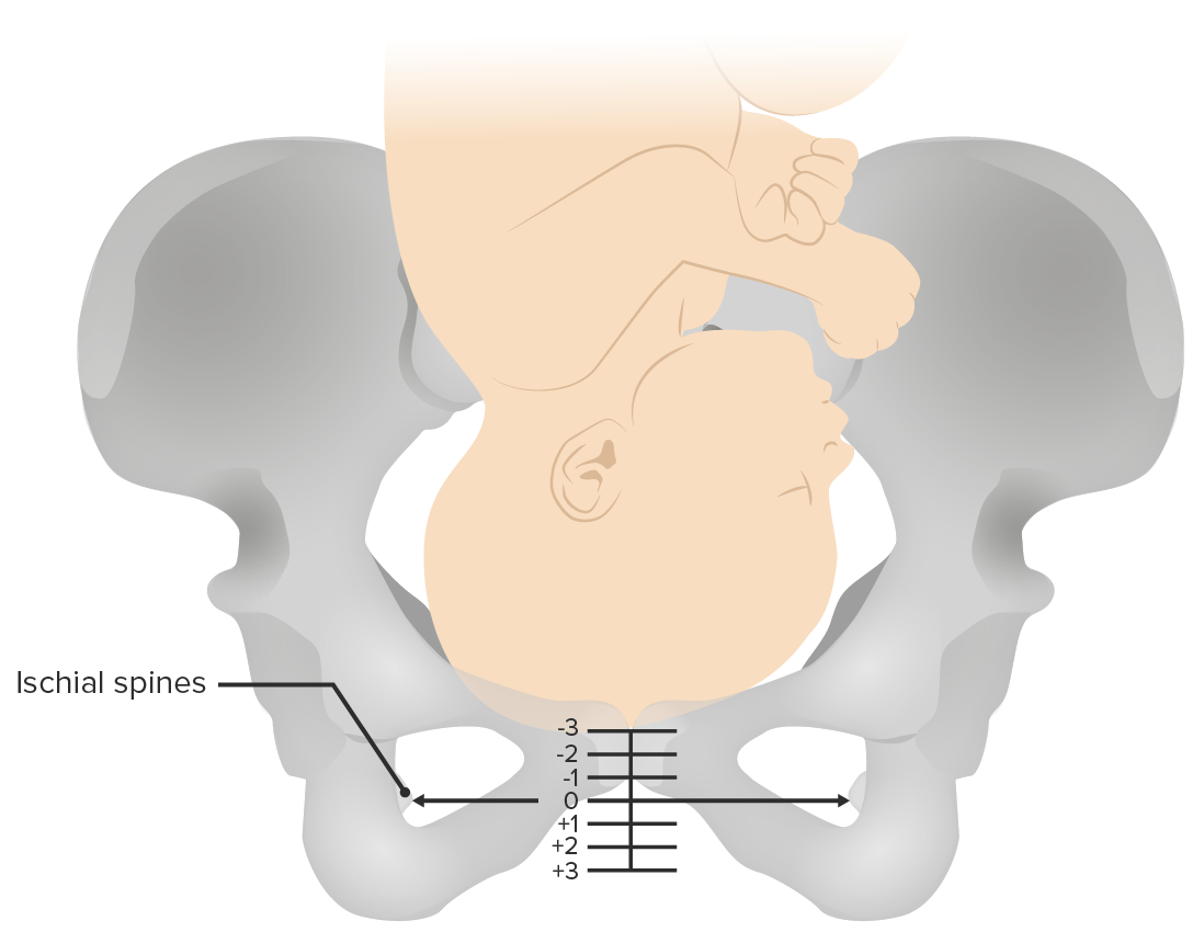
Fetal station: The presenting fetal part is measured in centimeters from the ischial spine. Negative numbers denote a higher fetal station, while positive numbers denote a lower fetal station as the fetus descends in the birth canal.
Image by Lecturio.
Vacuum-assisted delivery
Technique:
A vacuum extractor is applied to the fetal vertex.
Steady downward traction is applied during contractions with maternal pushing.
Maternal complications
2-fold ↑ risk of 3rd- and 4th-degree perineal lacerations (involves the anal sphincter)
Vulvar and vaginal hematomas
Urinary tractUrinary tractThe urinary tract is located in the abdomen and pelvis and consists of the kidneys, ureters, urinary bladder, and urethra. The structures permit the excretion of urine from the body. Urine flows from the kidneys through the ureters to the urinary bladder and out through the urethra.Urinary Tract: Anatomy injury
CephalohematomaCephalohematomaVon Willebrand Disease → hyperbilirubinemiaHyperbilirubinemiaA condition characterized by an abnormal increase of bilirubin in the blood, which may result in jaundice. Bilirubin, a breakdown product of heme, is normally excreted in the bile or further catabolized before excretion in the urine.Jaundice
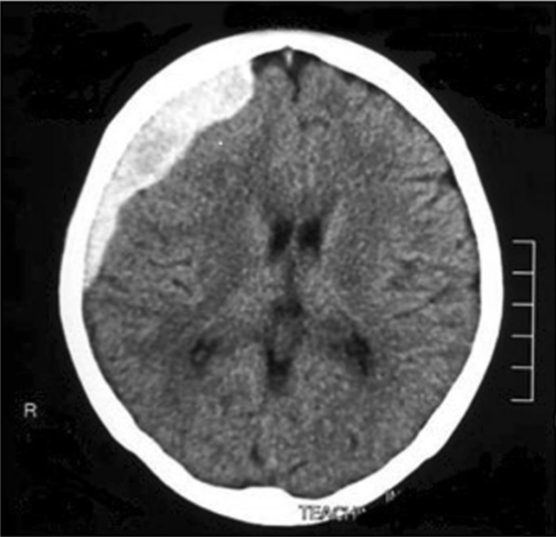
Intracranial hemorrhageIntracranial hemorrhageSubarachnoid hemorrhage (SAH) is a type of cerebrovascular accident (stroke) resulting from intracranial hemorrhage into the subarachnoid space between the arachnoid and the pia mater layers of the meninges surrounding the brain. Most sahs originate from a saccular aneurysm in the circle of willis but may also occur as a result of trauma, uncontrolled hypertension, vasculitis, anticoagulant use, or stimulant use.Subarachnoid Hemorrhage
CT scan of fetal head demonstrating an intracranial hemorrhage: This is a potential complication of operative vaginal deliveries.
Image: “Image of computed tomography scan of brain on postpartum day 23” by University Obstetrics Unit, De Soysa Hospital for Women, Colombo, Sri Lanka. License: CC BY 4.0
Steady downward traction is applied during contractions with maternal pushing.
Maternal complications:
6-fold ↑ risk of 3rd- and 4th-degree perineal lacerations
Vulvar and vaginal hematomas
Urinary tractUrinary tractThe urinary tract is located in the abdomen and pelvis and consists of the kidneys, ureters, urinary bladder, and urethra. The structures permit the excretion of urine from the body. Urine flows from the kidneys through the ureters to the urinary bladder and out through the urethra.Urinary Tract: Anatomy injury
SkullSkullThe skull (cranium) is the skeletal structure of the head supporting the face and forming a protective cavity for the brain. The skull consists of 22 bones divided into the viscerocranium (facial skeleton) and the neurocranium.Skull: AnatomyfractureFractureA fracture is a disruption of the cortex of any bone and periosteum and is commonly due to mechanical stress after an injury or accident. Open fractures due to trauma can be a medical emergency. Fractures are frequently associated with automobile accidents, workplace injuries, and trauma.Overview of Bone Fractures
Intracranial hemorrhageIntracranial hemorrhageSubarachnoid hemorrhage (SAH) is a type of cerebrovascular accident (stroke) resulting from intracranial hemorrhage into the subarachnoid space between the arachnoid and the pia mater layers of the meninges surrounding the brain. Most sahs originate from a saccular aneurysm in the circle of willis but may also occur as a result of trauma, uncontrolled hypertension, vasculitis, anticoagulant use, or stimulant use.Subarachnoid Hemorrhage
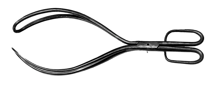
Obstetric forceps
Image: “Obstetric forceps” by Wellcome Collection gallery. License: CC BY 4.0
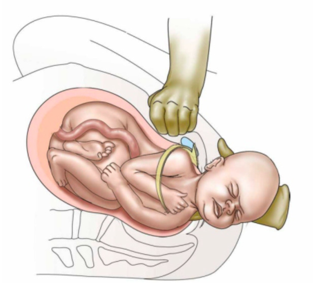
Shoulder dystocia is when the baby’s anterior shoulder becomes impacted behind the maternal pubic symphysisPubic SymphysisA slightly movable cartilaginous joint which occurs between the pubic bones.Vagina, Vulva, and Pelvic Floor: Anatomy, preventing delivery of the fetal body. Shoulder dystocia is a true obstetric emergency.
Definition:
Failure to deliver the fetal shoulders with gentle downward traction, where additional maneuvers are required to deliver the baby
This is a bone-on-bone obstruction → episiotomy will not relieve shoulder dystocia
During the dystocia, the fetus is not getting any oxygen.
Epidemiology:
IncidenceIncidenceThe number of new cases of a given disease during a given period in a specified population. It also is used for the rate at which new events occur in a defined population. It is differentiated from prevalence, which refers to all cases in the population at a given time.Measures of Disease Frequency: 0.2%–3% of vaginal births in the vertex presentation
Risk of recurrence: 10%–16%
Unpredictable and can occur in any laboring woman
Risk factors (though shoulder dystocia often occurs in the absence of risk factors):
Fetal macrosomiaFetal macrosomiaA condition of fetal overgrowth leading to a large-for-gestational-age fetus. It is defined as birth weight greater than 4, 000 grams or above the 90th percentile for population and sex-specific growth curves. It is commonly seen in gestational diabetes; prolonged pregnancy; and pregnancies complicated by pre-existing diabetes mellitus.Wilms Tumor
Maternal diabetesDiabetesDiabetes mellitus (DM) is a metabolic disease characterized by hyperglycemia and dysfunction of the regulation of glucose metabolism by insulin. Type 1 DM is diagnosed mostly in children and young adults as the result of autoimmune destruction of β cells in the pancreas and the resulting lack of insulin. Type 2 DM has a significant association with obesity and is characterized by insulin resistance.Diabetes Mellitus mellitus (pregestational or gestational)
Previous shoulder dystocia
Maternal obesityObesityObesity is a condition associated with excess body weight, specifically with the deposition of excessive adipose tissue. Obesity is considered a global epidemic. Major influences come from the western diet and sedentary lifestyles, but the exact mechanisms likely include a mixture of genetic and environmental factors. Obesity
Operative vaginal delivery
Prolonged 2nd stage of labor2nd stage of laborThe period of obstetric labor that is from the complete dilatation of the cervix uteri to the expulsion of the fetus.Normal and Abnormal Labor
Clinical presentation:
Failure of the fetal shoulders to deliver with gentle downward traction on the fetal head
Turtle sign: retraction of the fetal head tightly against the maternal perineumPerineumThe body region lying between the genital area and the anus on the surface of the trunk, and to the shallow compartment lying deep to this area that is inferior to the pelvic diaphragm. The surface area is between the vulva and the anus in the female, and between the scrotum and the anus in the male.Vagina, Vulva, and Pelvic Floor: Anatomy
Abnormal progression of the 2nd stage of labor2nd stage of laborThe period of obstetric labor that is from the complete dilatation of the cervix uteri to the expulsion of the fetus.Normal and Abnormal Labor
Causes cephalad rotationRotationMotion of an object in which either one or more points on a line are fixed. It is also the motion of a particle about a fixed point.X-rays of the pubic symphysisPubic SymphysisA slightly movable cartilaginous joint which occurs between the pubic bones.Vagina, Vulva, and Pelvic Floor: Anatomy and flattens the lumbar lordosis → maximizes the pelvic diameter
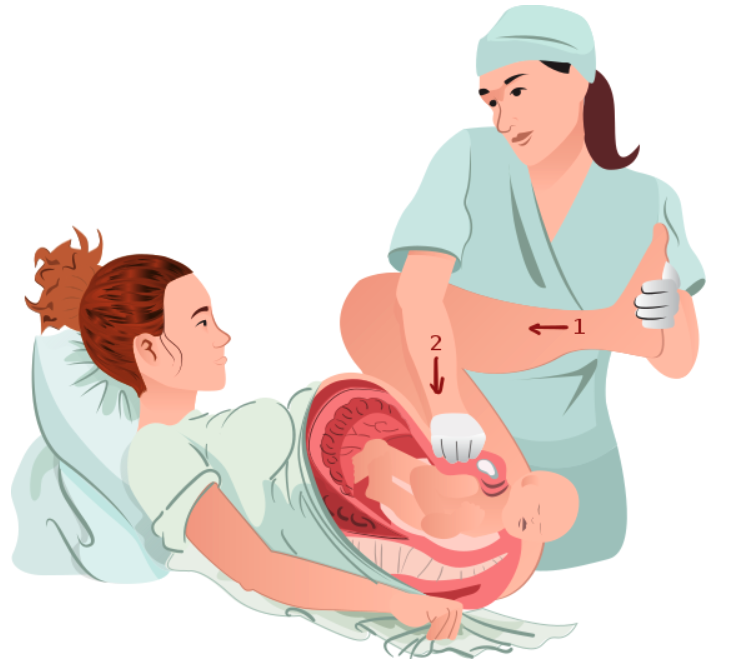
Suprapubic pressure:
Attempts to push the shoulder down and inward
Slightly rotates the fetus and dislodges the impacted shoulder
Avoid fundal pressure, which may make the dystocia worse.
Manual delivery of the posterior armArmThe arm, or “upper arm” in common usage, is the region of the upper limb that extends from the shoulder to the elbow joint and connects inferiorly to the forearm through the cubital fossa. It is divided into 2 fascial compartments (anterior and posterior).Arm: Anatomy
Reduces the bisacromial diameter
↑ Risk of clavicular and/or humerusHumerusBone in humans and primates extending from the shoulder joint to the elbow joint.Arm: AnatomyfractureFractureA fracture is a disruption of the cortex of any bone and periosteum and is commonly due to mechanical stress after an injury or accident. Open fractures due to trauma can be a medical emergency. Fractures are frequently associated with automobile accidents, workplace injuries, and trauma.Overview of Bone Fractures
95% of shoulder dystocias will be relieved with the management described.
Suprapubic pressure being used to dislodge and internally rotate an impacted shoulder in shoulder dystocia
Image: “Suprapubic-pressureforSD” by Henry Lerner. License: CC BY 4.0
McRoberts position with suprapubic pressure
Image: “McRoberts maneuver” by geraldbaeck. License: CC0 1.0
Additional maneuvers
Additional maneuvers to attempt if the dystocia persists include:
Rotational maneuvers:
Attempt to manually rotate the shoulders
Rubin’s maneuver: rotate the anterior or posterior fetal shoulder anteriorly toward the fetal face
Woods screw maneuver: rotate the posterior fetal shoulder backward
Gaskin maneuver:
Have mother assume a position on her hands and knees.
Pop the anterior clavicleClavicleA bone on the ventral side of the shoulder girdle, which in humans is commonly called the collar bone.Clavicle Fracture outward to ↓ the bisacromial diameter.
↑ Risk of injury to vasculature and pulmonary structures
Less morbidityMorbidityThe proportion of patients with a particular disease during a given year per given unit of population.Measures of Health Status than procedures of last resort
Episiotomy:
Episiotomy will not relieve shoulder dystocia.
Consider cutting one to allow space to adequately perform the maneuvers.
Repeat all above maneuvers several times before moving on to procedures of last resort.
Procedures of last resort:
Zavanelli maneuver:
Replace the fetal head in the abdomen by reversing the cardinal movements of labor and perform an urgent cesarean deliveryCesarean DeliveryCesarean delivery (CD) is the operative delivery of ≥ 1 infants through a surgical incision in the maternal abdomen and uterus. Cesarean deliveries may be indicated for a number of either maternal or fetal reasons, most commonly including fetal intolerance to labor, arrest of labor, a history of prior uterine surgery, fetal malpresentation, and placental abnormalities. Cesarean Delivery.
Make a hysterotomy, manually reduce the impacted shoulder, and deliver vaginally.
Done when the head is unable to be manually replaced during attempted Zavanelli maneuver
Symphysiotomy:
Surgical division of the cartilageCartilageCartilage is a type of connective tissue derived from embryonic mesenchyme that is responsible for structural support, resilience, and the smoothness of physical actions. Perichondrium (connective tissue membrane surrounding cartilage) compensates for the absence of vasculature in cartilage by providing nutrition and support. Cartilage: Histology of the pubic symphysisPubic SymphysisA slightly movable cartilaginous joint which occurs between the pubic bones.Vagina, Vulva, and Pelvic Floor: Anatomy
Only used when an OR is unavailable
To remember the management of a shoulder dystocia, remember HELPERR:
Call for Help.
Consider an Episiotomy (to allow space for maneuvers).
Elevate the Legs.
Apply suprapubic Pressure.
Enter the vaginaVaginaThe vagina is the female genital canal, extending from the vulva externally to the cervix uteri internally. The structures have sexual, reproductive, and urinary functions and a rich blood supply, mainly arising from the internal iliac artery.Vagina, Vulva, and Pelvic Floor: Anatomy for internal rotational maneuvers.
Reliever the posterior armArmThe arm, or “upper arm” in common usage, is the region of the upper limb that extends from the shoulder to the elbow joint and connects inferiorly to the forearm through the cubital fossa. It is divided into 2 fascial compartments (anterior and posterior).Arm: Anatomy.
Rotate the woman to hands-and-knees position.
Complications
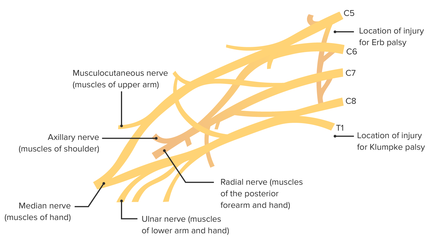
Brachial plexusBrachial PlexusThe large network of nerve fibers which distributes the innervation of the upper extremity. The brachial plexus extends from the neck into the axilla. In humans, the nerves of the plexus usually originate from the lower cervical and the first thoracic spinal cord segments (c5-c8 and T1), but variations are not uncommon.Peripheral Nerve Injuries in the Cervicothoracic Region injury
Stretching of the C5 and C6 nerves from continuous downward traction on the head
Often reversible
75% of brachial plexusBrachial PlexusThe large network of nerve fibers which distributes the innervation of the upper extremity. The brachial plexus extends from the neck into the axilla. In humans, the nerves of the plexus usually originate from the lower cervical and the first thoracic spinal cord segments (c5-c8 and T1), but variations are not uncommon.Peripheral Nerve Injuries in the Cervicothoracic Region injury
Klumpke palsyPalsyparalysis of an area of the body, thus incapable of voluntary movementCranial Nerve Palsies: stretching of C8 and T1 nerves
HumerusHumerusBone in humans and primates extending from the shoulder joint to the elbow joint.Arm: AnatomyfractureFractureA fracture is a disruption of the cortex of any bone and periosteum and is commonly due to mechanical stress after an injury or accident. Open fractures due to trauma can be a medical emergency. Fractures are frequently associated with automobile accidents, workplace injuries, and trauma.Overview of Bone Fractures
Fetal asphyxiaAsphyxiaA pathological condition caused by lack of oxygen, manifested in impending or actual cessation of life.Drowning
Contusions and lacerations
Brachial plexus injuries associated with shoulder dystocia
Amniotic fluidAmniotic fluidA clear, yellowish liquid that envelopes the fetus inside the sac of amnion. In the first trimester, it is likely a transudate of maternal or fetal plasma. In the second trimester, amniotic fluid derives primarily from fetal lung and kidney. Cells or substances in this fluid can be removed for prenatal diagnostic tests (amniocentesis).Placenta, Umbilical Cord, and Amniotic Cavity embolism (AFE) is a complication of labor affecting the mother in the immediate postpartum periodPostpartum periodIn females, the period that is shortly after giving birth (parturition).Postpartum Complications.
Caused by entry of amniotic fluidAmniotic fluidA clear, yellowish liquid that envelopes the fetus inside the sac of amnion. In the first trimester, it is likely a transudate of maternal or fetal plasma. In the second trimester, amniotic fluid derives primarily from fetal lung and kidney. Cells or substances in this fluid can be removed for prenatal diagnostic tests (amniocentesis).Placenta, Umbilical Cord, and Amniotic Cavity into the maternal circulationCirculationThe movement of the blood as it is pumped through the cardiovascular system.ABCDE Assessment by:
Placental tears
Uterine vein rupture
IncidenceIncidenceThe number of new cases of a given disease during a given period in a specified population. It also is used for the rate at which new events occur in a defined population. It is differentiated from prevalence, which refers to all cases in the population at a given time.Measures of Disease Frequency: 1 in 40,000 deliveries
AFE causes 10% of maternal deaths in developed countries.
Risk factors
Cesarean deliveryCesarean DeliveryCesarean delivery (CD) is the operative delivery of ≥ 1 infants through a surgical incision in the maternal abdomen and uterus. Cesarean deliveries may be indicated for a number of either maternal or fetal reasons, most commonly including fetal intolerance to labor, arrest of labor, a history of prior uterine surgery, fetal malpresentation, and placental abnormalities. Cesarean Delivery
Operative vaginal deliveries
Placental abnormalitiesPlacental abnormalitiesNormal placental structure and function are essential for a healthy pregnancy. Some of the most common placental abnormalities include structural anomalies (such as a succenturiate lobe or velamentous cord insertion), implantation anomalies (such as placenta accreta and placenta previa), and functional anomalies (such as placental insufficiency). Placental Abnormalities (e.g., placenta previaPlacenta PreviaAbnormal placentation in which the placenta implants in the lower segment of the uterus (the zone of dilation) and may cover part or all of the opening of the cervix. It is often associated with serious antepartum bleeding and premature labor.Placental Abnormalities)
PreeclampsiaPreeclampsiaA complication of pregnancy, characterized by a complex of symptoms including maternal hypertension and proteinuria with or without pathological edema. Symptoms may range between mild and severe. Pre-eclampsia usually occurs after the 20th week of gestation, but may develop before this time in the presence of trophoblastic disease.Hypertensive Pregnancy Disorders/eclampsiaEclampsiaOnset of hyperreflexia; seizures; or coma in a previously diagnosed pre-eclamptic patient (pre-eclampsia).Hypertensive Pregnancy Disorders
Pathogenesis
Unclear
Amniotic fluidAmniotic fluidA clear, yellowish liquid that envelopes the fetus inside the sac of amnion. In the first trimester, it is likely a transudate of maternal or fetal plasma. In the second trimester, amniotic fluid derives primarily from fetal lung and kidney. Cells or substances in this fluid can be removed for prenatal diagnostic tests (amniocentesis).Placenta, Umbilical Cord, and Amniotic Cavity enters maternal circulationCirculationThe movement of the blood as it is pumped through the cardiovascular system.ABCDE Assessment and triggers:
↑ Pulmonary pressure → right ventricular failure → systemic hypotensionHypotensionHypotension is defined as low blood pressure, specifically < 90/60 mm Hg, and is most commonly a physiologic response. Hypotension may be mild, serious, or life threatening, depending on the cause. Hypotension
Pulmonary edemaPulmonary edemaPulmonary edema is a condition caused by excess fluid within the lung parenchyma and alveoli as a consequence of a disease process. Based on etiology, pulmonary edema is classified as cardiogenic or noncardiogenic. Patients may present with progressive dyspnea, orthopnea, cough, or respiratory failure.Pulmonary Edema
An abnormal immune response:
Intense inflammatory response (similar to SIRS) is activated.
Inflammatory mediators activate the coagulation cascadeCoagulation cascadeThe coagulation cascade is a series of reactions that ultimately generates a strong, cross-linked fibrin clot.Hemostasis systemically → DICDICDisseminated intravascular coagulation (DIC) is a condition characterized by systemic bodywide activation of the coagulation cascade. This cascade results in both widespread microvascular thrombi contributing to multiple organ dysfunction and consumption of clotting factors and platelets, leading to hemorrhage. Disseminated Intravascular Coagulation
DICDICDisseminated intravascular coagulation (DIC) is a condition characterized by systemic bodywide activation of the coagulation cascade. This cascade results in both widespread microvascular thrombi contributing to multiple organ dysfunction and consumption of clotting factors and platelets, leading to hemorrhage. Disseminated Intravascular Coagulation leads to:
Hemorrhage → further hemodynamic instability
Ischemic multiorgan failure
Mechanical obstructionMechanical ObstructionAny impairment, arrest, or reversal of the normal flow of intestinal contents toward the anal canal.Imaging of the Intestines from amniotic fluidAmniotic fluidA clear, yellowish liquid that envelopes the fetus inside the sac of amnion. In the first trimester, it is likely a transudate of maternal or fetal plasma. In the second trimester, amniotic fluid derives primarily from fetal lung and kidney. Cells or substances in this fluid can be removed for prenatal diagnostic tests (amniocentesis).Placenta, Umbilical Cord, and Amniotic Cavity debris likely does not play a significant role.
Clinical presentation
Amniotic fluidAmniotic fluidA clear, yellowish liquid that envelopes the fetus inside the sac of amnion. In the first trimester, it is likely a transudate of maternal or fetal plasma. In the second trimester, amniotic fluid derives primarily from fetal lung and kidney. Cells or substances in this fluid can be removed for prenatal diagnostic tests (amniocentesis).Placenta, Umbilical Cord, and Amniotic Cavity embolism typically presents dramatically, as sudden onset cardiopulmonary collapse occurring during labor or within 30 minutes after delivery.
Signs:
Cardiopulmonary collapse: loss of breathing and pulse
DyspneaDyspneaDyspnea is the subjective sensation of breathing discomfort. Dyspnea is a normal manifestation of heavy physical or psychological exertion, but also may be caused by underlying conditions (both pulmonary and extrapulmonary). Dyspnea
HypotensionHypotensionHypotension is defined as low blood pressure, specifically < 90/60 mm Hg, and is most commonly a physiologic response. Hypotension may be mild, serious, or life threatening, depending on the cause. Hypotension
TachycardiaTachycardiaAbnormally rapid heartbeat, usually with a heart rate above 100 beats per minute for adults. Tachycardia accompanied by disturbance in the cardiac depolarization (cardiac arrhythmia) is called tachyarrhythmia.Sepsis in Children
Other symptoms:
NauseaNauseaAn unpleasant sensation in the stomach usually accompanied by the urge to vomit. Common causes are early pregnancy, sea and motion sickness, emotional stress, intense pain, food poisoning, and various enteroviruses.Antiemetics and vomitingVomitingThe forcible expulsion of the contents of the stomach through the mouth.Hypokalemia
Mental status changes
Seizure
DICDICDisseminated intravascular coagulation (DIC) is a condition characterized by systemic bodywide activation of the coagulation cascade. This cascade results in both widespread microvascular thrombi contributing to multiple organ dysfunction and consumption of clotting factors and platelets, leading to hemorrhage. Disseminated Intravascular Coagulation:
Usually develops shortly after an AFE
Leads to obstetric hemorrhage
Bleeding from catheter sites and mucosal surfaces
Fetal heart rateHeart rateThe number of times the heart ventricles contract per unit of time, usually per minute.Cardiac Physiology abnormalities indicating distress (e.g., late decelerations, terminal bradycardiaBradycardiaBradyarrhythmia is a rhythm in which the heart rate is less than 60/min. Bradyarrhythmia can be physiologic, without symptoms or hemodynamic change. Pathologic bradyarrhythmia results in reduced cardiac output and hemodynamic instability causing syncope, dizziness, or dyspnea.Bradyarrhythmias) if still pregnant
Diagnosis
Amniotic fluidAmniotic fluidA clear, yellowish liquid that envelopes the fetus inside the sac of amnion. In the first trimester, it is likely a transudate of maternal or fetal plasma. In the second trimester, amniotic fluid derives primarily from fetal lung and kidney. Cells or substances in this fluid can be removed for prenatal diagnostic tests (amniocentesis).Placenta, Umbilical Cord, and Amniotic Cavity embolism is a clinical diagnosis based on presentation.
↑ Prothrombin timeProthrombin timeClotting time of plasma recalcified in the presence of excess tissue thromboplastin. Factors measured are fibrinogen; prothrombin; factor V; factor VII; and factor X.Hemostasis
↓ FibrinogenFibrinogenPlasma glycoprotein clotted by thrombin, composed of a dimer of three non-identical pairs of polypeptide chains (alpha, beta, gamma) held together by disulfide bonds. Fibrinogen clotting is a sol-gel change involving complex molecular arrangements: whereas fibrinogen is cleaved by thrombin to form polypeptides a and b, the proteolytic action of other enzymes yields different fibrinogen degradation products.Hemostasis
AnemiaAnemiaAnemia is a condition in which individuals have low Hb levels, which can arise from various causes. Anemia is accompanied by a reduced number of RBCs and may manifest with fatigue, shortness of breath, pallor, and weakness. Subtypes are classified by the size of RBCs, chronicity, and etiology. Anemia: Overview and Types
ThrombocytopeniaThrombocytopeniaThrombocytopenia occurs when the platelet count is < 150,000 per microliter. The normal range for platelets is usually 150,000-450,000/µL of whole blood. Thrombocytopenia can be a result of decreased production, increased destruction, or splenic sequestration of platelets. Patients are often asymptomatic until platelet counts are < 50,000/µL. Thrombocytopenia
AcidosisAcidosisA pathologic condition of acid accumulation or depletion of base in the body. The two main types are respiratory acidosis and metabolic acidosis, due to metabolic acid build up.Respiratory Acidosis (both respiratory and metabolic)
Imaging (once woman is stable enough):
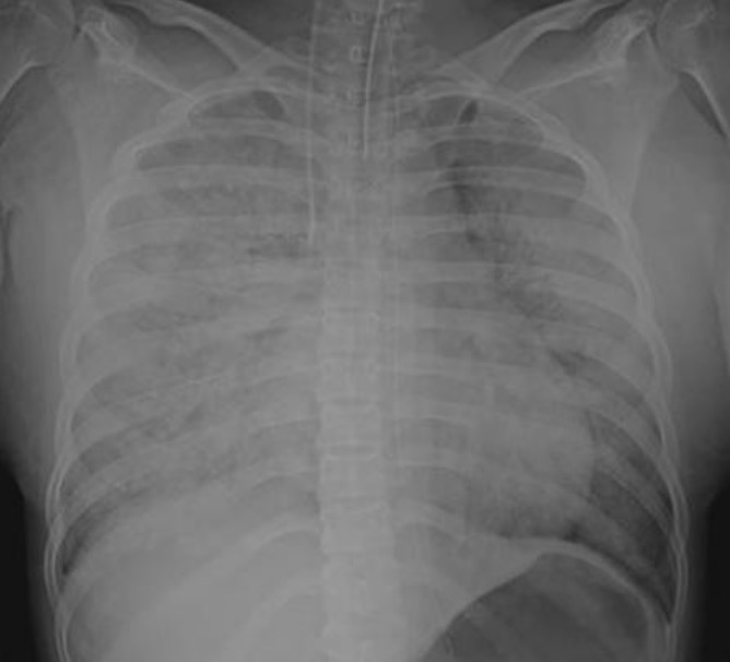
Chest radiography: bilateral diffuse infiltrates
EchocardiographyEchocardiographyUltrasonic recording of the size, motion, and composition of the heart and surrounding tissues. The standard approach is transthoracic.Tricuspid Valve Atresia (TVA) to assess cardiac function, which may have become compromised
Chest x-ray in a woman with amniotic fluid embolism: Diffuse infiltration is evident throughout the lungs.
Image: “X-ray” by Department of Emergency and Critical Care, The University of Tokushima Graduate School, Kuramoto Tokushima, 770-8503, Japan. License: CC BY 2.0
CirculationCirculationThe movement of the blood as it is pumped through the cardiovascular system.ABCDE Assessment:
High-quality cardiopulmonary resuscitationResuscitationThe restoration to life or consciousness of one apparently dead. .Neonatal Respiratory Distress Syndrome (CPRCPRThe artificial substitution of heart and lung action as indicated for heart arrest resulting from electric shock, drowning, respiratory arrest, or other causes. The two major components of cardiopulmonary resuscitation are artificial ventilation and closed-chest cardiac massage.Cardiac Arrest)
Transfuse to combat DICDICDisseminated intravascular coagulation (DIC) is a condition characterized by systemic bodywide activation of the coagulation cascade. This cascade results in both widespread microvascular thrombi contributing to multiple organ dysfunction and consumption of clotting factors and platelets, leading to hemorrhage. Disseminated Intravascular Coagulation; typically a 1:1:1 ratio of:
Hematologic: DICDICDisseminated intravascular coagulation (DIC) is a condition characterized by systemic bodywide activation of the coagulation cascade. This cascade results in both widespread microvascular thrombi contributing to multiple organ dysfunction and consumption of clotting factors and platelets, leading to hemorrhage. Disseminated Intravascular Coagulation
Cardiovascular: hemorrhage and cardiac arrestCardiac arrestCardiac arrest is the sudden, complete cessation of cardiac output with hemodynamic collapse. Patients present as pulseless, unresponsive, and apneic. Rhythms associated with cardiac arrest are ventricular fibrillation/tachycardia, asystole, or pulseless electrical activity. Cardiac Arrest
Pulmonary-related: pulmonary edemaPulmonary edemaPulmonary edema is a condition caused by excess fluid within the lung parenchyma and alveoli as a consequence of a disease process. Based on etiology, pulmonary edema is classified as cardiogenic or noncardiogenic. Patients may present with progressive dyspnea, orthopnea, cough, or respiratory failure.Pulmonary Edema and ARDS